. 2023 Apr 7;9(4):452.
doi: 10.3390/jof9040452.

Pneumocystis Exacerbates Inflammation and Mucus Hypersecretion in a Murine, Elastase-Induced-COPD Model

Affiliations

Pneumocystis Exacerbates Inflammation and Mucus Hypersecretion in a Murine, Elastase-Induced-COPD Model

Diego A Rojas et al. J Fungi (Basel). .

Abstract

Inflammation and mucus hypersecretion are frequent pathology features of chronic respiratory diseases such as asthma and COPD. Selected bacteria, viruses and fungi may synergize as co-factors in aggravating disease by activating pathways that are able to induce airway pathology. Pneumocystis infection induces inflammation and mucus hypersecretion in immune competent and compromised humans and animals. This fungus is a frequent colonizer in patients with COPD. Therefore, it becomes essential to identify whether it has a role in aggravating COPD severity. This work used an elastase-induced COPD model to evaluate the role of Pneumocystis in the exacerbation of pathology, including COPD-like lung lesions, inflammation and mucus hypersecretion. Animals infected with Pneumocystis developed increased histology features of COPD, inflammatory cuffs around airways and lung vasculature plus mucus hypersecretion. Pneumocystis induced a synergic increment in levels of inflammation markers (Cxcl2, IL6, IL8 and IL10) and mucins (Muc5ac/Muc5b). Levels of STAT6-dependent transcription factors Gata3, FoxA3 and Spdef were also synergically increased in Pneumocystis infected animals and elastase-induced COPD, while the levels of the mucous cell-hyperplasia transcription factor FoxA2 were decreased compared to the other groups. Results document that Pneumocystis is a co-factor for disease severity in this elastase-induced-COPD model and highlight the relevance of STAT6 pathway in Pneumocystis pathogenesis.

Keywords: COPD; Muc5ac; Muc5b; Pneumocystis; hypersecretion; inflammation; mucins; mucus.


Conflict of interest statement

The authors declare no conflict of interest.

Figures

Figure 1
Figure 1
Evaluation of elastase-induced lung lesions. (A) Scheme of the animal model indicating the four experimental groups: Saline, Elastase (ELT), Pneumocystis infection (Pc), and Elastase-Pneumocystis infection (ELT-Pc). The experiment took 14 weeks total in all experimental groups. This time included the one-week of cohabitation with Pneumocystis-seeding rats starting 4 weeks after elastase instillation in the Pneumocystis groups. (B) Alveolar spaces were evaluated in lung sections from rats of the four groups using hematoxylin-eosin (H/E) staining. Microscopic analyses were performed using 40× magnification. (C) Pneumocystis infection was evaluated in the animals using PCR amplification of the mitochondrial large subunit (mtLSU) rRNA. Amplification of actin was used as internal control.
Figure 2
Figure 2
Evaluation of peribronchiolar and perivascular inflammation cuffs in the elastase-induced COPD model infected with Pneumocystis. Lung sections of rats from the four experimental groups were stained with hematoxylin-eosin and the presence of inflammation cuffs were identified by microscopic evaluation in images with 100× magnification. (A) Peribronchiolar and (B) perivascular inflammation cuffs were identified in the images. Quantification of the cuffs observed in the images was determined using a score described in the Materials and Methods section. Quantification of (C) peribronchiolar and (D) perivascular inflammatory cuffs is indicated. Data are expressed as mean ± SD and analyzed by ANOVA. ns = nonsignificant; *** = p < 0.001; **** = p < 0.0001.
Figure 3
Figure 3
Characterization of cytokine profile in the elastase-induced COPD animal model infected with Pneumocystis. (A) Tnf, (B) Cxcl2, (C) Il6, (D) Il8, (E) Il10 and (F) GM-CSF cytokines mRNA levels were evaluated by qPCR. Data were determined using the 2−DDCt, actin levels as internal control and normalized to saline control animal group. Data are expressed as mean ± SD and analyzed by ANOVA. ns = non-significant; * = p < 0.05; ** = p < 0.01; *** = p < 0.001; **** = p < 0.0001.
Figure 4
Figure 4
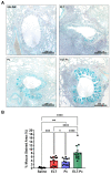
Evaluation of mucus secretion in the elastase-induced COPD animal model infected with Pneumocystis. (A) Representative images of lung sections from the four experimental animal groups stained with alcian-blue/periodic acid Schiff (AB/PAS). Microscopic magnification was 100×. (B) The stained area of the airway epithelium was quantified as described in the Materials and Methods section. Data are expressed as mean ± SD and analyzed by ANOVA. ns = non significant; ** = p < 0.01; *** = p < 0.001; **** = p < 0.0001.
Figure 5
Figure 5
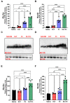
Evaluation of the mucin levels in the elastase-induced COPD animal model infected with Pneumocystis. mRNA levels of (A) Muc5ac and (B) Muc5b were evaluated by qPCR. Data were determined using the 2−DDCt, actin levels as internal control and normalized to saline control animal group. Data are expressed as mean ± SD and analyzed by ANOVA. ns = nonsignificant; * = p < 0.05; ** = p < 0.01; **** = p < 0.0001. Protein levels of the mucins were evaluated by western blot. Representative images of western blot to evaluate (C) Muc5ac and (D) Muc5b are indicated in the figure. Actin was used as internal control. Quantification of (E) Muc5ac and (F) Muc5b are indicated. Data are expressed as mean ± SD and analyzed by ANOVA. ns = nonsignificant; ** = p < 0.01; **** = p < 0.0001.
Figure 6
Figure 6
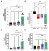
Evaluation of the expression level of STAT6-dependent transcription factor in the elastase-induced COPD animal model infected with Pneumocystis. mRNA levels of (A) Gata3, (B) FoxA2, (C) FoxA3 and (D) Spdef were evaluated by qPCR. Data were determined using the 2−DDCt method, actin levels as internal control and normalized to saline control animal group. Data are expressed as mean ± SD and analyzed by ANOVA. ns = nonsignificant; * = p < 0.05; ** = p < 0.01; *** = p < 0.001; **** = p < 0.0001.

References

    1. Kim V., Criner G.J. Chronic Bronchitis and Chronic Obstructive Pulmonary Disease. Am. J. Respir. Crit. Care Med. 2013;187:228–237. doi: 10.1164/rccm.201210-1843CI. - DOI - PMC - PubMed
    1. McDonough J.E., Yuan R., Suzuki M., Seyednejad N., Elliott W.M., Sanchez P.G., Wright A.C., Gefter W.B., Litzky L., Coxson H.O., et al. Small-Airway Obstruction and Emphysema in Chronic Obstructive Pulmonary Disease. N. Engl. J. Med. 2011;365:1567–1575. doi: 10.1056/NEJMoa1106955. - DOI - PMC - PubMed
    1. Mio T., Romberger D.J., Thompson A.B., Robbins R.A., Heires A., Rennard S.I. Cigarette Smoke Induces Interleukin-8 Release from Human Bronchial Epithelial Cells. Am. J. Respir. Crit. Care Med. 1997;155:1770–1776. doi: 10.1164/ajrccm.155.5.9154890. - DOI - PubMed
    1. Retamales I., Elliott W.M., Meshi B., Coxson H.O., Pare P.D., Sciurba F.C., Rogers R.M., Hayashi S., Hogg J.C. Amplification of Inflammation in Emphysema and Its Association with Latent Adenoviral Infection. Am. J. Respir. Crit. Care Med. 2001;164:469–473. doi: 10.1164/ajrccm.164.3.2007149. - DOI - PubMed
    1. Hellermann G.R., Nagy S.B., Kong X., Lockey R.F., Mohapatra S.S. Mechanism of Cigarette Smoke Condensate-Induced Acute Inflammatory Response in Human Bronchial Epithelial Cells. Respir. Res. 2002;3:15. doi: 10.1186/rr172. - DOI - PMC - PubMed