Case Reports
. 2023 Apr 6;13(4):637.
doi: 10.3390/jpm13040637.

Preserving Ambulation in a Gene Therapy-Treated Girl Affected by Metachromatic Leukodystrophy: A Case Report

Affiliations
Case Reports

Preserving Ambulation in a Gene Therapy-Treated Girl Affected by Metachromatic Leukodystrophy: A Case Report

Silvia Faccioli et al. J Pers Med. .

Abstract

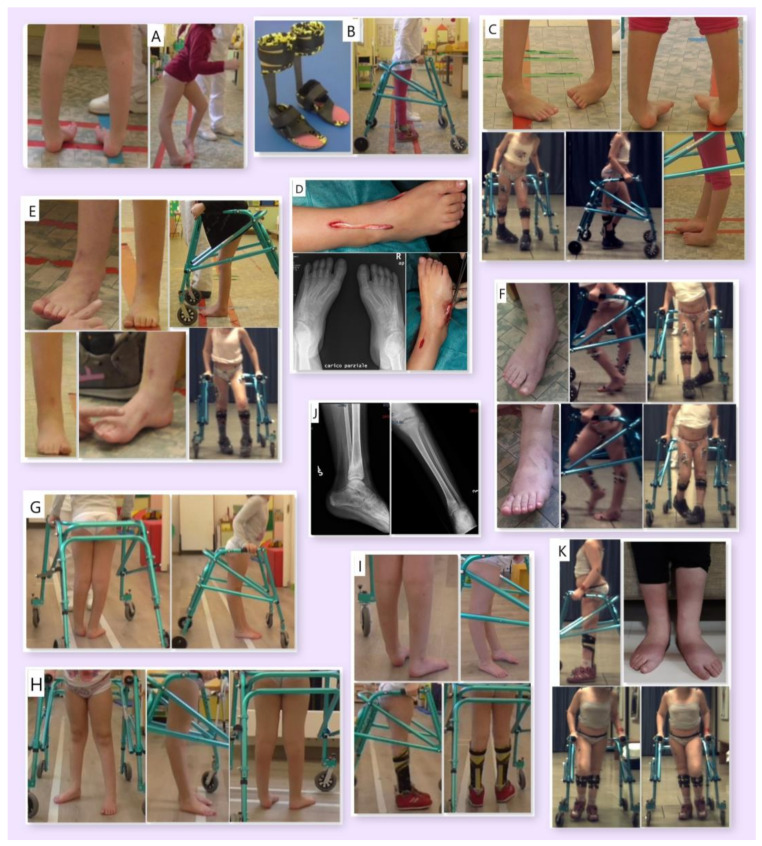
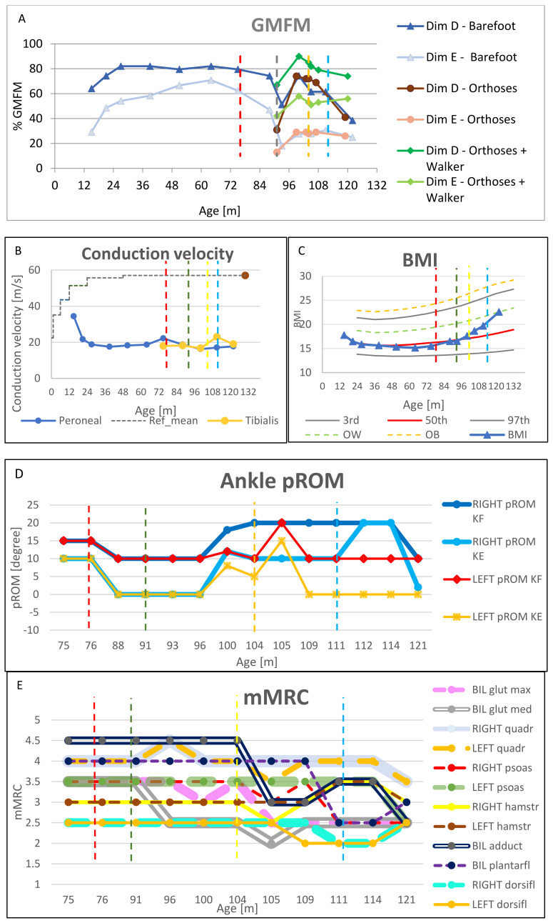
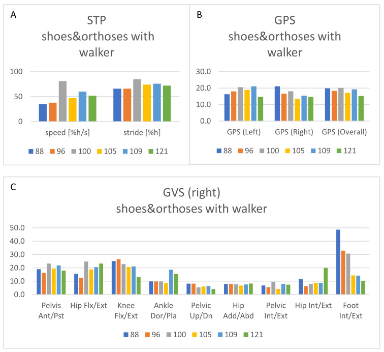
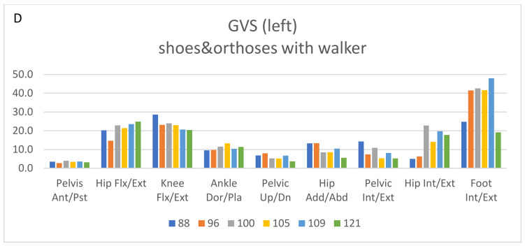
(1) Background: Atidarsagene autotemcel is a hematopoietic stem and progenitor cell gene therapy (HSPC-GT) approved to treat early-onset metachromatic leukodystrophy (MLD). The purpose of this case report is to describe the long-term management of residual gait impairment of a child with late infantile MLD treated with HSPC-GT. (2) Methods: Assessment included Gross Motor Function Measure-88, nerve conduction study, body mass index (BMI), Modified Tardieu Scale, passive range of motion, modified Medical Research Council scale, and gait analysis. Interventions included orthoses, a walker, orthopedic surgery, physiotherapy, and botulinum. (3) Results: Orthoses and a walker were fundamental to maintaining ambulation. Orthopedic surgery positively influenced gait by reducing equinovarus. Nonetheless, unilateral recurrence of varo-supination was observed, attributable to spasticity and muscle imbalance. Botulinum improved foot alignment but induced transient overall weakness. A significant increase in BMI occurred. Finally, a shift to bilateral valgopronation was observed, more easily managed with orthoses. (4) Conclusions: HSPC-GT preserved survival and locomotor abilities. Rehabilitation was then considered fundamental as a complementary treatment. Muscle imbalance and increased BMI contributed to gait deterioration in the growing phase. Caution is recommended when considering botulinum in similar subjects, as the risk of inducing overall weakness can outweigh the benefits of spasticity reduction.

Keywords: assistive devices; botulinum toxins; disabled children; gait analysis; muscle spasticity; nerve conduction studies; orthopedics; rehabilitation.


Conflict of interest statement

S.F., S.S. (Silvia Sassi), D.P., C.B. (Corrado Borghi), V.M., M.S., S.S. (Stefano Scarparo), and C.B. (Carla Butera) certify that they have no conflict of interest with any financial organization regarding the material discussed in the manuscript. The patient presented in the manuscript had previously been enrolled in the clinical trial ClinicalTrials.gov Identifier: NCT01560182. The trial was initially funded by Telethon Foundation. Subsequently, MLD gene therapy was licensed to GlaxoSmithKline (GSK) in 2014 and then to Orchard Therapeutics, the current license holder of the investigational medicinal product OTL-200, in 2018. AA, VC, and FF are investigators of HSPC-GT clinical trials for MLD sponsored by Orchard Therapeutics, the license holder of investigational medicinal product OTL-200. FF and VC have acted as ad hoc consultants for Orchard Therapeutic advisory boards.

Figures

Figure 1
Figure 1
Images of the patient: at first access to the rehabilitation unit ((A), age 75 months); AFOs and walker ((B), age 76 months); pre-surgery ((C), age 88 months); intraoperative ((D), age 91 months); 4 and 9 months after surgery, respectively ((E), age 96 months; (F), age 100 months); pre-botulinum toxin injection ((G), age 104 months); 1 month after botulinum toxin injection ((H), age 105 months); 4 months after botulinum toxin injection ((I), age 109 months); left tibial fracture ((J), age 111 months); last assessment ((K), age 121 months).
Figure 2
Figure 2
Clinical and instrumental assessments performed over the follow-up period, at different months of age. Time at which interventions and fracture occurred is represented with vertical dot lines: respectively, AFO in red, surgery in green, botulinum in yellow, and tibial fracture in light blue. (A): Gross Motor Function-88 (GMFM-88) dimensions D and E, in different conditions: barefoot, with orthoses, with orthoses and walker. (B): Conduction velocity relative to the right leg. (C): BMI of the patient, with Italian growth norms for females [14], related to the 3rd, 50th, and 97th percentile and extra-centiles for overweight (OW) and obesity (OB). (D): Lower limb passive range of motion (pROM), according to Modified Tardieu Scale: slow pROM to assess muscle stiffness and contractures, fast pROM to assess spastic reaction (which identified the range at which the catch was evoked). (E): Lower limb strength assessment by means of modified Medical Research Council (mMRC) scale.
Figure 3
Figure 3
Results from the 3DGA over the follow-up period at 88, 96, 100, 105, 109, and 121 months of age. (A): Spatiotemporal parameters (speed and stride length, both normalized to the patient’s height), referring to gait analyses acquired with orthoses and walker. (B): Gait profile score (GPS) for the left and right side and overall, referring to gait analyses acquired with orthoses and walker. (C): Gait variable score (GVS) for the right side, referring to gait analyses acquired with orthoses and walker. (D): Gait variable score (GVS) for the left side, referring to gait analyses acquired with orthoses and walker.
Figure 3
Figure 3
Results from the 3DGA over the follow-up period at 88, 96, 100, 105, 109, and 121 months of age. (A): Spatiotemporal parameters (speed and stride length, both normalized to the patient’s height), referring to gait analyses acquired with orthoses and walker. (B): Gait profile score (GPS) for the left and right side and overall, referring to gait analyses acquired with orthoses and walker. (C): Gait variable score (GVS) for the right side, referring to gait analyses acquired with orthoses and walker. (D): Gait variable score (GVS) for the left side, referring to gait analyses acquired with orthoses and walker.

References

    1. Van Rappard D.F., Boelens J.J., Wolf N.I. Metachromatic leukodystrophy: Disease spectrum and approaches for treatment. Best Pract. Res. Clin. Endocrinol. Metab. 2015;29:261–273. doi: 10.1016/j.beem.2014.10.001. - DOI - PubMed
    1. Kehrer C., Elgün S., Raabe C., Böhringer J., Beck-Wödl S., Bevot A., Kaiser N., Schöls L., Krägeloh-Mann I., Groeschel S. Association of Age at Onset and First Symptoms With Disease Progression in Patients With Metachromatic Leukodystrophy. Neurology. 2020;96:e255–e266. doi: 10.1212/WNL.0000000000011047. - DOI - PubMed
    1. Fumagalli F., Zambon A.A., Rancoita P.M.V., Baldoli C., Canale S., Spiga I., Medaglini S., Penati R., Facchini M., Ciotti F., et al. Metachromatic leukodystrophy: A single-center longitudinal study of 45 patients. J. Inherit. Metab. Dis. 2021;44:1151–1164. doi: 10.1002/jimd.12388. - DOI - PubMed
    1. Artigalás O., Lagranha V.L., Saraiva-Pereira M.L., Burin M.G., Lourenço C.M., van der Linden H., Jr., Santos M.L.F., Rosemberg S., Steiner C.E., Kok F., et al. Clinical and biochemical study of 29 Brazilian patients with metachromatic leukodystrophy. J. Inherit. Metab. Dis. 2010;33((Suppl. 3)):S257–S262. doi: 10.1007/s10545-010-9140-4. - DOI - PubMed
    1. Groeschel S., Kehrer C., Engel C., Dali C., Bley A., Steinfeld R., Grodd W., Krägeloh-Mann I. Metachromatic leukodystrophy: Natural course of cerebral MRI changes in relation to clinical course. J. Inherit. Metab. Dis. 2011;34:1095–1102. doi: 10.1007/s10545-011-9361-1. - DOI - PubMed

Publication types

LinkOut - more resources