

Chlorogenic Acid Attenuates Doxorubicin-Induced Oxidative Stress and Markers of Apoptosis in Cardiomyocytes via Nrf2/HO-1 and Dityrosine Signaling
- PMID: 37109035
- PMCID: PMC10140899
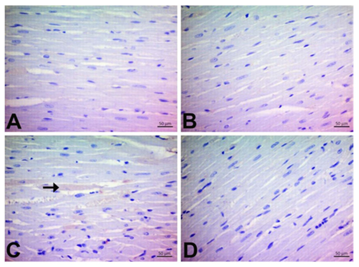
- DOI: 10.3390/jpm13040649
Chlorogenic Acid Attenuates Doxorubicin-Induced Oxidative Stress and Markers of Apoptosis in Cardiomyocytes via Nrf2/HO-1 and Dityrosine Signaling
Abstract
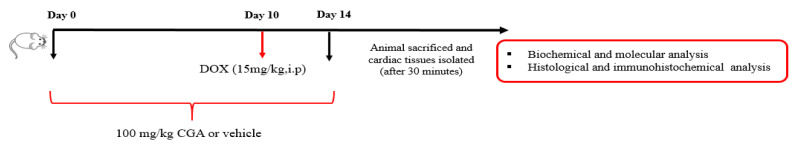
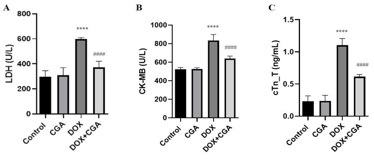
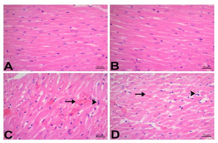
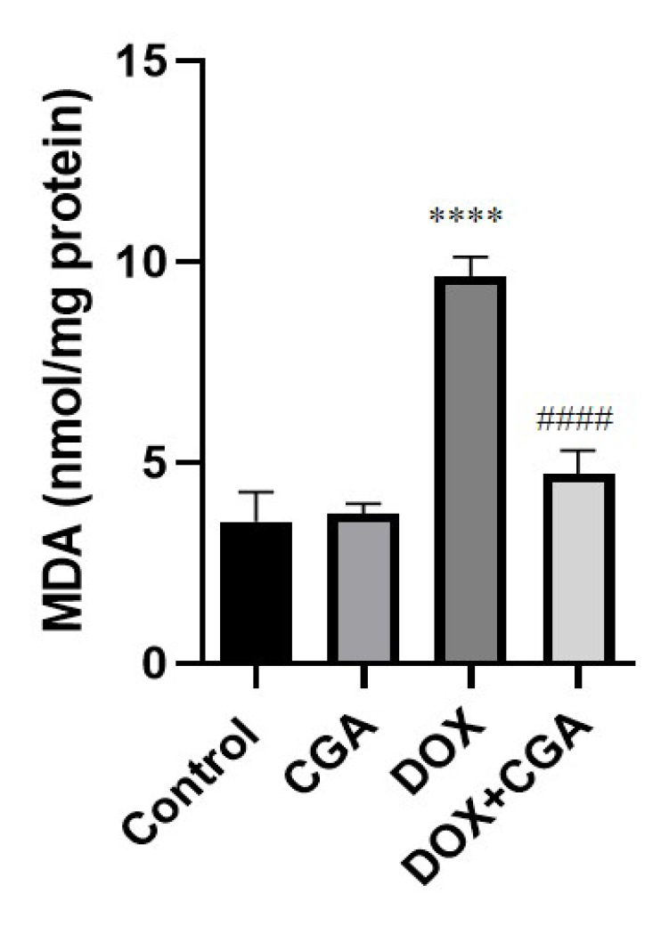
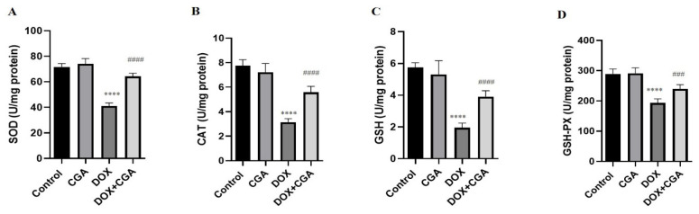
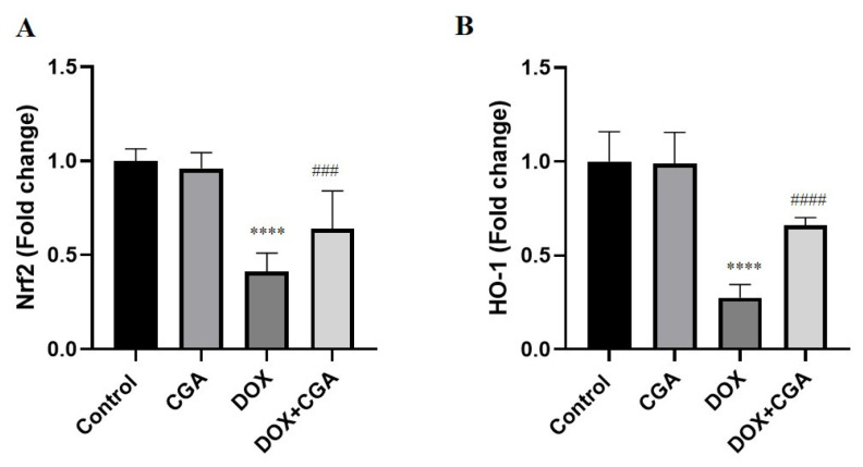
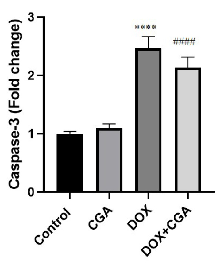
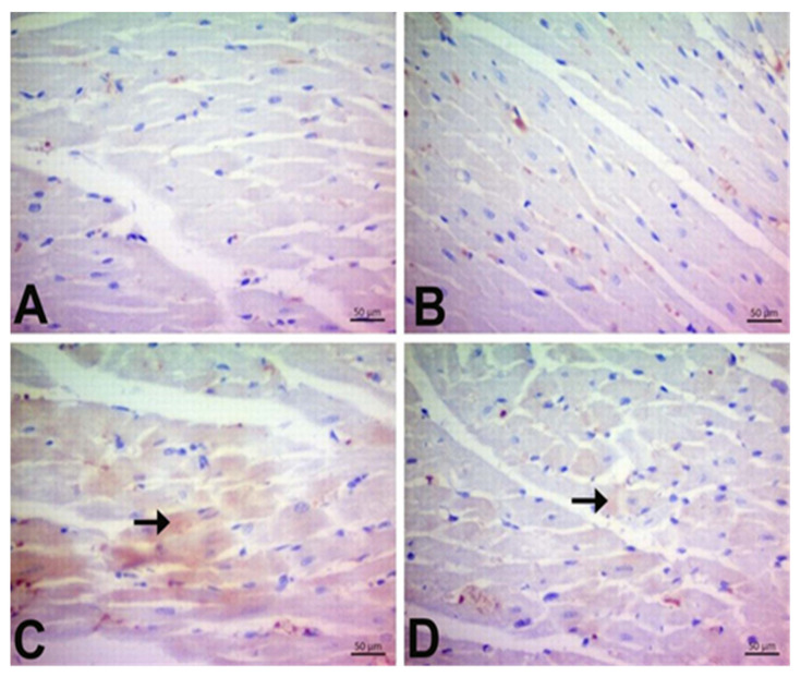
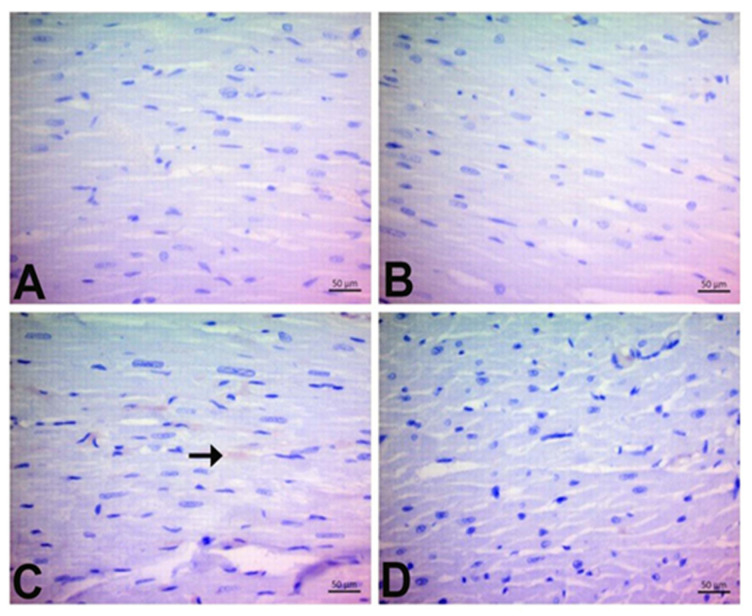
(1) Background: Doxorubicin (DOX) is extensively used for cancer treatments; however, its clinical application is limited because of its cardiotoxic adverse effects. A combination of DOX and agents with cardioprotective properties is an effective strategy to ameliorate DOX-related cardiotoxicity. Polyphenolic compounds are ideal for the investigation of novel cardioprotective agents. Chlorogenic acid (CGA), an essential dietary polyphenol found in plants, has been previously reported to exert antioxidant, cardioprotective, and antiapoptotic properties. The current research evaluated CGA's in vivo cardioprotective properties in DOX-induced cardiotoxicity and the probable mechanisms underlying this protection. (2) Methods: CGA's cardioprotective properties were investigated in rats that were treated with CGA (100 mg/kg, p.o.) for fourteen days. The experimental model of cardiotoxicity was induced with a single intraperitoneal (15 mg/kg i.p.) injection of DOX on the 10th day. (3) Results: Treatment with CGA significantly improved the DOX-caused altered cardiac damage markers (LDH, CK-MB, and cTn-T), and a marked improvement in cardiac histopathological features accompanied this. DOX downregulated the expression of Nrf2/HO-1 signaling pathways, and the CGA reversed this effect. Consistently, caspase-3, an apoptotic-related marker, and dityrosine expression were suppressed, while Nrf2 and HO-1 expressions were elevated in the cardiac tissues of DOX-treated rats after treatment with the CGA. Furthermore, the recovery was confirmed by the downregulation of 8-OHdG and dityrosine (DT) expressions in immunohistochemical findings. (4) Conclusions: CGA demonstrated a considerable cardioprotective effect against DOX-induced cardiotoxicity. One of the possible mechanisms for these protective properties was the upregulation of the Nrf2/HO-1-dependent pathway and the downregulation of DT, which may ameliorate oxidative stress and cardiomyocyte apoptosis. These findings suggest that CGA may be cardioprotective, particularly in patients receiving DOX-based chemotherapy.
Keywords: Nrf2/HO-1; cardiotoxicity; chlorogenic acid; doxorubicin; oxidative stress.
Conflict of interest statement
Konstantinos Tsarouhas is a member of the Editorial Board of the Journal of Personalized Medicine, but has no personal involvement in the reviewing process or any influence in terms of adjudicating the final decision for this article. The other authors declare that they have no conflict of interest.
Figures
References
-
- Sun L., Wang H., Dan X., Yu S., Zhang L., Li X. Lapatinib induces mitochondrial dysfunction to enhance oxidative stress and ferroptosis in doxorubicin-induced cardiomyocytes via inhibition of PI3K/AKT signaling pathway. Bioengineered. 2022;13:48–60. doi: 10.1080/21655979.2021.2004980. - DOI - PMC - PubMed
LinkOut - more resources
Full Text Sources
Research Materials
Miscellaneous
