

Texture Analysis for the Bone Age Assessment from MRI Images of Adolescent Wrists in Boys
- PMID: 37109098
- PMCID: PMC10141677
- DOI: 10.3390/jcm12082762
Texture Analysis for the Bone Age Assessment from MRI Images of Adolescent Wrists in Boys
Abstract
Currently, bone age is assessed by X-rays. It enables the evaluation of the child's development and is an important diagnostic factor. However, it is not sufficient to diagnose a specific disease because the diagnoses and prognoses may arise depending on how much the given case differs from the norms of bone age.
Background: The use of magnetic resonance images (MRI) to assess the age of the patient would extend diagnostic possibilities. The bone age test could then become a routine screening test. Changing the method of determining the bone age would also prevent the patient from taking a dose of ionizing radiation, making the test less invasive.
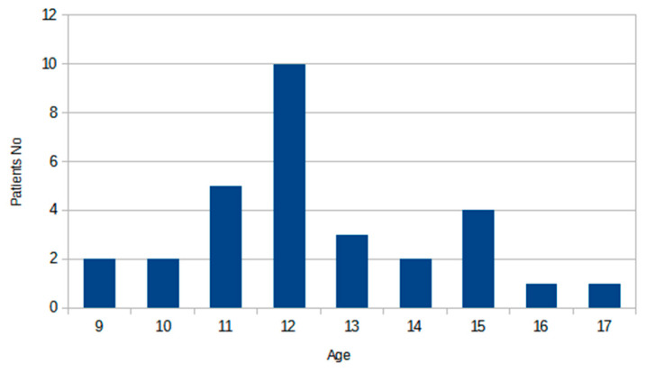
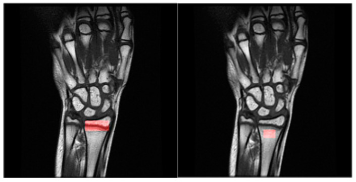
Methods: The regions of interest containing the wrist area and the epiphyses of the radius are marked on the magnetic resonance imaging of the non-dominant hand of boys aged 9 to 17 years. Textural features are computed for these regions, as it is assumed that the texture of the wrist image contains information about bone age.
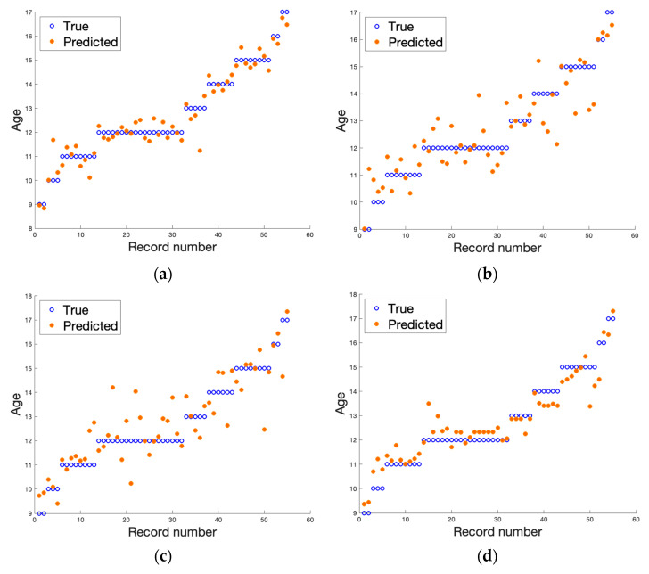
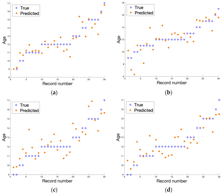
Results: The regression analysis revealed that there is a high correlation between the bone age of a patient and the MRI-derived textural features derived from MRI. For DICOM T1-weighted data, the best scores reached 0.94 R2, 0.46 RMSE, 0.21 MSE, and 0.33 MAE.
Conclusions: The experiments performed have shown that using the MRI images gives reliable results in the assessment of bone age while not exposing the patient to ionizing radiation.
Keywords: bone MRI; bone age; image texture; pediatric radiology; regression analysis.
Conflict of interest statement
The authors declare no conflict of interest.
Figures
References
-
- Schmitt A. Forensic Anthropology and Medicine: Complementary Sciences from Recovery to Cause of Death. Humana; Paramus, NJ, USA: 2006. pp. 212–219.
-
- Abdelbary M.H., Abdelkawi M.M., Nasr M.A. Age determination by MR imaging of the wrist in Egyptian male foot ballplayers How far is it reliable? Egypt. J. Radiol. Nucl. Med. 2018;49:146–151. doi: 10.1016/j.ejrnm.2017.12.005. - DOI
-
- Dekhne M.S., Kocher I.D., Hussain Z.B., Feroe A.G., Sankarankutty S., Williams K.A., Heyworth B.E., Milewski M.D., Kocher M.S. Tibial tubercle apophyseal stage to determine skeletal age in pediatric patients undergoing ACL Reconstruction: A validation and reliability study. Orthop. J. Sports Med. 2021;9:23259671211036897. doi: 10.1177/23259671211036897. - DOI - PMC - PubMed
LinkOut - more resources
Full Text Sources
