. 2023 Apr 13;12(8):2853.
doi: 10.3390/jcm12082853.

Reoperation in Chiari-1 Malformations

Affiliations

Reoperation in Chiari-1 Malformations

Giuseppe Talamonti et al. J Clin Med. .

Abstract

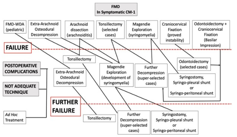
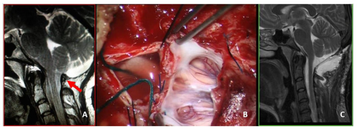
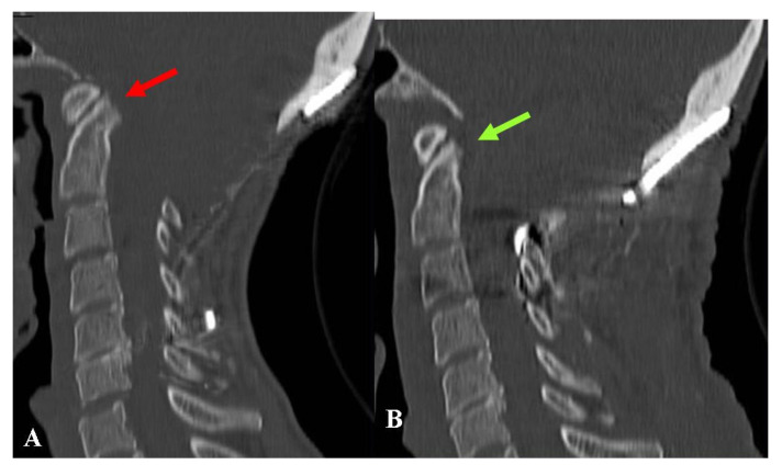
(1) Background: The issue of unsuccessful surgery for Chiari-1 malformation (CM-1), as well as its potential causes and possible solutions, remains poorly documented and studied. (2) Methods: From a retrospective review of a personal series of 98 patients undergoing treatment for CM-1 during the past 10 years, we created two study groups. Group 1: 8 patients (8.1%) requiring additional surgeries owing to postoperative complications (7 cerebrospinal fluid leakage, 1 extradural hematoma); 7 patients (7.1%) undergoing reoperations for failed decompression during the follow-up. Group 2: During the same period, we also managed 19 patients who had previously been operated on elsewhere: 8 patients who required adequate CM-1 treatment following extradural section of the filum terminale; 11 patients requiring reoperations for failed decompression. Failed decompression was managed by adequate osteodural decompression, which was associated with tonsillectomy (6 cases), subarachnoid exploration (8 cases), graft substitution (6 cases), and occipito-cervical fixation/revision (1 case). (3) Results: There was no mortality or surgical morbidity in Group 1. However, one patient's condition worsened due to untreatable syrinx. In Group 2, there were two cases of mortality, and surgical morbidity was represented by functional limitation and pain in the patient who needed revision of the occipitocervical fixation. Twenty patients improved (58.8%), 6 remained unchanged (32.3%), 1 worsened (2.9%) and 2 died (5.9%). (4) Conclusions: The rate of complications remains high in CM-1 treatment. Unfortunately, a certain rate of treatment failure is unavoidable, but it appears that a significant number of re-operations could have been avoided using proper indications and careful technique.

Keywords: CSF-leak; Chiari malformation; complications; foramen magnum decompression; reoperation; syringomyelia.


Conflict of interest statement

The authors declare no conflict of interest.

Figures

Figure 1
Figure 1
Schematic drawing of treatment for CM-1. The first line shows the initial surgical plan. The following lines describe the management of FMD failures. Postoperative complications and improper techniques were treated “ad hoc”. In the event of postoperative instability, craniocervical fixation was also performed. FMD = foramen magnum decompression; FMD-WDA = foramen magnum decompression without dural augmentation.
Figure 2
Figure 2
This 48-year-old man complained of an invalidating Valsalva-related headache. (A) Preoperative MRI, T2-weight, sagittal view showing a CM-1.5. Osteodural decompression was performed, and the arachnoid was accidentally violated. The dural sac was repaired using biological graft and fibrin glue. (B) Postoperative MRI, T2-weight, sagittal view showing a deep extradural pseudomeningocele that was not evident at skin level. Conservative treatment was attempted at first. The headache continued to worsen, especially in orthostatism, and 4 months later, he underwent reoperation for duraplasty using autologous fascia lata. (C) Follow-up MRI, T2-weight, sagittal view obtained 9 months after reoperation. There was no evidence of CSF collection; however, the orthostatic pain improved, but the headache did not yet resolve. Adhesions may have developed between the autologous graft and the cerebellum (red arrow). A further surgical procedure was offered, but was refused by the patient. The final CCOS score was 12.
Figure 3
Figure 3
This 41-year-old woman had undergone bone decompression alone. Over the next 8 years, her headache recurred and progressively worsened. She also complained of dizziness and bouts of diplopia. (A) Preoperative MRI, T2-weight, sagittal view showing the bone decompression with scarce CSF film around the CM-1. (B) Intraoperative picture showing a thick and hard extradural fibrous scar that was maintaining the compression. (C) Intraoperative picture following dural opening with intact arachnoid. The dural sac was repaired using biologic graft. (D) Follow-up MRI, T2-weight, sagittal view obtained 6 months later showing the CM-1 was well bathed in CSF. Preoperative symptoms were almost completely resolved. The final CCOS score was 14.
Figure 4
Figure 4
This 64-year-old woman had been treated with osteodural decompression, leaving C1 intact. One year later, she was referred to us for severe nocturnal breathing disturbances and mild quadriparesis. (A) Preoperative MRI, T2-weight, sagittal view, showing a small craniectomy with C1 still maintaining a significant obliteration of the arachnoid space (red arrow). A wide syringomyelia was evident. You will notice a marked skin retraction over the craniectomy (yellow arrow). (B) Intraoperative picture showing an “arc” incision and the skin retraction. The reoperation consisted of C1 laminectomy, tonsillectomy, and dural sac augmentation. (C) Postoperative MRI, T2-weight, sagittal view showing a relatively large subarachnoid space and initial syrinx shrinkage. There was mild local cerebellar edema owing to the tonsillectomy. An asymptomatic pseudomeningocele was also evident, which resolved spontaneously within a couple of weeks. Since the first postoperative period, breathing disturbances have significantly improved, whereas quadriparesis improvement has been slower. The final CCOS score was 10.
Figure 5
Figure 5
This 29-year-old woman had undergone osteodural decompression without laminectomy. One year later, she was admitted to our department with an untreatable persistent hiccup responsible for difficult alimentation, invalidating headache, dizziness, tinnitus, fever, rigor nucalis, and diffuse asthenia. An infection was suspected, but was ruled out. (A) Preoperative MRI, T2-weight, sagittal view showing a relatively wide craniectomy with C1 (red arrow) still compressing the arachnoid space, and a scarce presence of CSF around the CM-1. She underwent laminectomy and dural sac augmentation. (B) Intraoperative picture depicting a diffuse, tight adherence between the dural graft and the parenchyma. To replace the graft, a meticulous dissection was necessary. Finally, the dural sac was repaired with a synthetic graft. (C) Postoperative MRI, T2-weight, sagittal view showing a well-bathed CM-1. An asymptomatic pseudomeningocele was detected. This remained completely asymptomatic, even though it was still evident (but reduced) 6 months later. One year after the reoperation, the patient reported sporadic headache bouts, but had no other symptoms. The final CCOS score was 13.
Figure 6
Figure 6
This 43-year-old woman had undergone osteodural decompression and craniocervical fixation. Six months later, she was referred to us due to progressively worsening swallowing disturbances, severe weight loss, asthenia, and marked continuous occipital pain. (A) Preoperative CT-scan, sagittal reconstruction showing a relatively wide bone decompression and a basilar impression with a quite acute clivo-axial angle (red arrow). Under neurophysiologic and radiologic monitoring, the patient underwent the removal of the fixation device and cranial traction. The basilar impression could be reduced, which made the planned procedure of odontoidectomy unnecessary. New craniocervical fixation devices were placed to maintain the new position. (B) Follow-up CT-scan, sagittal reconstruction, obtained one year later, showing resolution of the basilar impression (green arrow). The swallowing problems almost completely went away, and the headache got better, but the asthenia stayed. The final CCOS score was 10.

References

    1. Milhorat T.H., Chou M.W., Trinidad E.M., Kula R.W., Mandell M., Wolpert C., Speer M.C. Chiari I Malformation Redefined: Clinical and Radiographic Findings for 364 Symptomatic Patients. Neurosurgery. 1999;44:1005–1017. doi: 10.1097/00006123-199905000-00042. - DOI - PubMed
    1. Meadows J., Kraut M., Guarnieri M., Haroun R.I., Carson B.S. Asymptomatic Chiari Type I malformations identified on magnetic resonance imaging. J. Neurosurg. 2000;92:920–926. doi: 10.3171/jns.2000.92.6.0920. - DOI - PubMed
    1. Menezes A.H., Greenlee J., Donovan K.A. Honored guest presentation: Lifetime experiences and where we are going: Chiari I with syringohydromyelia--controversies and development of decision trees. Clin. Neurosurg. 2005;52:297–305. - PubMed
    1. Talamonti G., Ferrari E., D’Aliberti G. Chiari malformation type 1: Are we doing less with more? Illustrative case. J. Neurosurg. Case Lessons. 2021;1:CASE20145. doi: 10.3171/CASE20145. - DOI - PMC - PubMed
    1. CreveCoeur T.S., Yahanda A.T., Maher C.O., Johnson G.W., Ackerman L.L., Adelson P.D., Ahmed R., Albert G.W., Aldana P.R., Alden T.D., et al. Occipital-Cervical Fusion and Ventral Decompression in the Surgical Management of Chiari-1 Malformation and Syringomyelia: Analysis of Data from the Park-Reeves Syringomyelia Research Consortium. Neurosurgery. 2021;88:332–341. doi: 10.1093/neuros/nyaa460. - DOI - PMC - PubMed

LinkOut - more resources