Review
. 2023 Apr 17;12(8):2919.
doi: 10.3390/jcm12082919.

Continuing Challenges in the Definitive Diagnosis of Cushing's Disease: A Structured Review Focusing on Molecular Imaging and a Proposal for Diagnostic Work-Up

Affiliations
Review

Continuing Challenges in the Definitive Diagnosis of Cushing's Disease: A Structured Review Focusing on Molecular Imaging and a Proposal for Diagnostic Work-Up

Tessa N A Slagboom et al. J Clin Med. .

Abstract

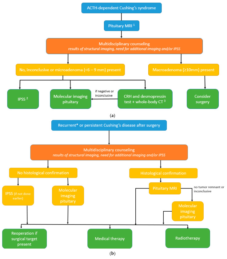
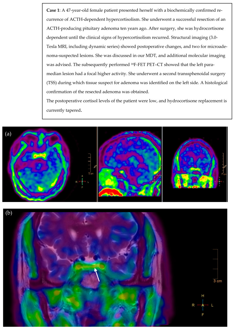
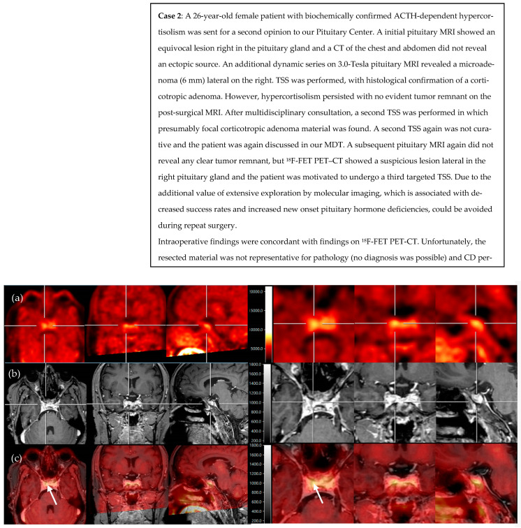
The definitive diagnosis of Cushing's disease (CD) in the presence of pituitary microadenoma remains a continuous challenge. Novel available pituitary imaging techniques are emerging. This study aimed to provide a structured analysis of the diagnostic accuracy as well as the clinical use of molecular imaging in patients with ACTH-dependent Cushing's syndrome (CS). We also discuss the role of multidisciplinary counseling in decision making. Additionally, we propose a complementary diagnostic algorithm for both de novo and recurrent or persistent CD. A structured literature search was conducted and two illustrative CD cases discussed at our Pituitary Center are presented. A total of 14 CD (n = 201) and 30 ectopic CS (n = 301) articles were included. MRI was negative or inconclusive in a quarter of CD patients. 11C-Met showed higher pituitary adenoma detection than 18F-FDG PET-CT (87% versus 49%). Up to 100% detection rates were found for 18F-FET, 68Ga-DOTA-TATE, and 68Ga-DOTA-CRH, but were based on single studies. The use of molecular imaging modalities in the detection of pituitary microadenoma in ACTH-dependent CS is of added and complementary value, serving as one of the available tools in the diagnostic work-up. In selected CD cases, it seems justified to even refrain from IPSS.

Keywords: Cushing’s disease; PET; diagnosis; hypercortisolism; molecular imaging; pituitary.


Conflict of interest statement

The authors declare no conflict of interest.

Figures

Figure 3
Figure 3
(a) Diagnostic algorithm for de novo diagnosis of Cushing’s syndrome.1 optimized as proposed by Bashari et al. [17]: Step 1a = conventional MRI: spin echo (SE) pre- and post-gadolinium, if no clear (micro)adenoma >, Step 1b = subsequent MRI sequences: gadolinium-enhanced 3D-spoiled gradient (recalled) echo (3D-SGE/3D-GRE) or gadolinium-enhanced dynamic (dMRI), if still not clear >, Step 2 = subsequent MRI sequences: fluid-attenuated inversion recovery (FLAIR) with gadolinium contrast, constructive interference in steady state (CISS) or isotropic 3D-fast turbo SE and/or use of the ultra-high field (7.0-Tesla) MRI. 2 optimally performed with CRH stimulation. 3 Alternative: whole-body 68Gallium-somatostatin receptor PET–CT. (b) Algorithm for persistent or recurrent Cushing’s disease. * In selected cases of Cushing’s disease with the equivocal biochemical response after surgery.
Figure 1
Figure 1
18F-FET PET scan and fusion of PET with T2 MR images reveal asymmetrical focal high uptake (in orange) in a pituitary adenoma left paramedian, as shown with the crosshair (a) and white arrow (b). (a) axial, sagittal, and coronal view of PET scan after infusion of 219 MBq 18F-FET, (b) fusion of PET/MRI (T2-weighted coronal view, pre-gadolinium). L = left, R = right, H = head, F = feet.
Figure 2
Figure 2
18F-FET PET and fusion of PET with post-contrast T1 MR images reveal asymmetrical focal high uptake (in yellow) lateral in the right pituitary, as centered with the crosshair (ac) and shown with the white arrows (c). (a) axial, coronal, and sagittal view of PET scan after infusion of 192 MBq 18F-FET, (b) axial, coronal, and sagittal view of 3D T1-weighted, post-gadolinium MRI, and (c) axial, coronal, and sagittal view of PET/MRI fusion. Scans on the right side present the same lesion but in more detail.

References

    1. Boscaro M., Barzon L., Fallo F., Sonino N. Cushing’s syndrome. Lancet. 2001;357:783–791. doi: 10.1016/S0140-6736(00)04172-6. - DOI - PubMed
    1. Grober Y., Grober H., Wintermark M., Jane J.A., Oldfield E.H. Comparison of MRI techniques for detecting microadenomas in Cushing’s disease. J. Neurosurg. JNS. 2018;128:1051–1057. doi: 10.3171/2017.3.JNS163122. - DOI - PubMed
    1. Invitti C., Giraldi F.P., De Martin M., Cavagnini F. Diagnosis and Management of Cushing’s Syndrome: Results of an Italian Multicentre Study1. J. Clin. Endocrinol. Metab. 1999;84:440–448. doi: 10.1210/jcem.84.2.5465. - DOI - PubMed
    1. Buchfelder M., Nistor R., Fahlbusch R., Huk W.J. The accuracy of CT and MR evaluation of the sella turcica for detection of adrenocorticotropic hormone-secreting adenomas in Cushing disease. Am. J. Neuroradiol. 1993;14:1183–1190. - PMC - PubMed
    1. Arnaldi G., Angeli A., Atkinson A.B., Bertagna X., Cavagnini F., Chrousos G.P., Fava G.A., Findling J.W., Gaillard R.C., Grossman A.B., et al. Diagnosis and Complications of Cushing’s Syndrome: A Consensus Statement. J. Clin. Endocrinol. Metab. 2003;88:5593–5602. doi: 10.1210/jc.2003-030871. - DOI - PubMed

LinkOut - more resources