

WNT16 Regulation of the Articular Chondrocyte Phenotype in Mice
- PMID: 37109407
- PMCID: PMC10145094
- DOI: 10.3390/life13040878
WNT16 Regulation of the Articular Chondrocyte Phenotype in Mice
Abstract
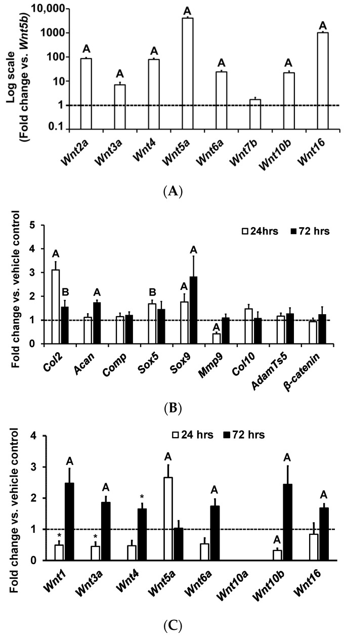
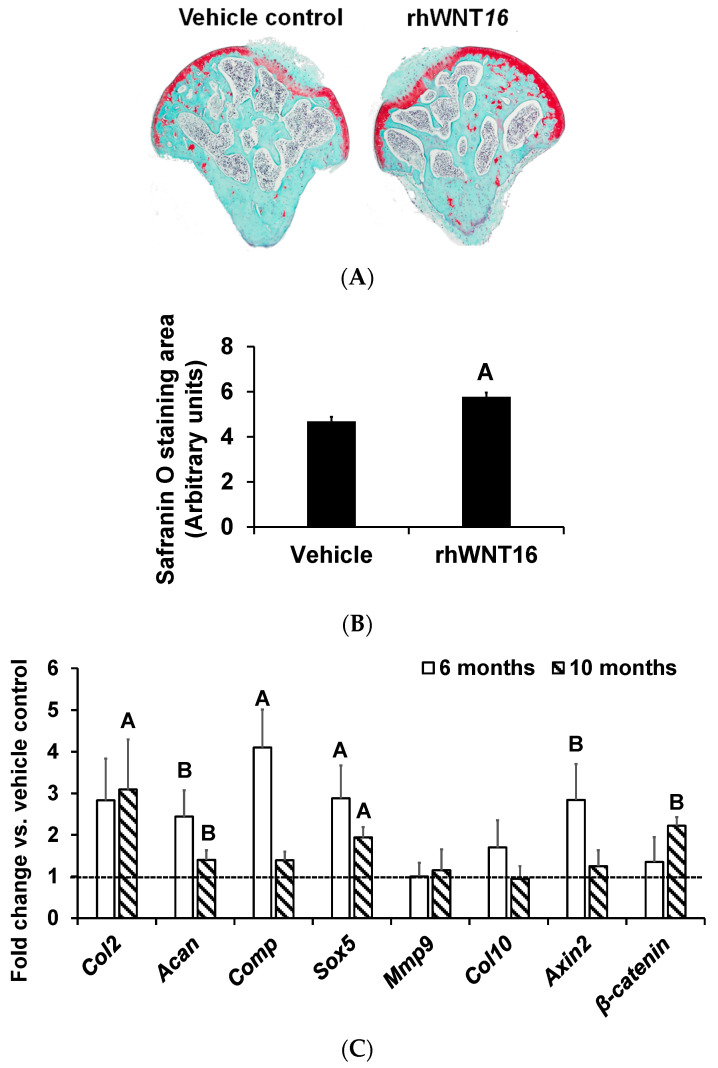
The anabolic effects of WNT16 on osteoblasts are well established, however, little is known regarding the role of WNT16 in chondrocytes. In this study, we evaluated Wnt16 expression and its biological effects on mouse articular chondrocytes (ACs), since these cells are key to the development of osteoarthritis. While ACs derived from the long bone epiphysis of 7-day old C57BL/6J mice express multiple Wnts, Wnt5b and Wnt16 represent the two most highly expressed Wnts (expressed at several-fold higher levels than other Wnts). Treatment of serum-free AC cultures, with 100 ng/mL of recombinant human (rh) WNT16 for 24 h (hrs), increased proliferation (20%, p < 0.05) and expression levels of makers (Sox9 and Col2) of immature chondrocytes at both 24 h and 72 h, while Acan increased at 72 h. Expression of Mmp9, a marker of mature chondrocytes was decreased at 24 h. Additionally, WNT16 treatment regulated expression levels of Wnt ligands in a biphasic manner, inhibiting its expression at 24 h, while stimulating expression at 72 h. To determine whether WNT16 exerted anabolic effects on the AC phenotype, ex vivo cultures of tibial epiphyses were treated with rhWNT16 or vehicle for 9 days, and the articular cartilage phenotype was evaluated by safranin O cartilage staining and expression of articular cartilage marker genes. Both articular cartilage area and expression levels of AC markers were increased after rhWNT16 treatment. Our data suggest that Wnt16 expressed in ACs may play a role in regulating joint cartilage homeostasis via its direct effect, as well as through modulating the expression of other Wnt ligands.
Keywords: WNT16; cell culture; chondrocytes; chondrogenesis; mice; osteoarthritis.
Conflict of interest statement
The authors declare no conflict of interest.
Figures
References
-
- Buckwalter J.A., Brown T.D. Joint injury, repair, and remodeling: Roles in post-traumatic osteoarthritis. Clin. Orthop. Relat. Res. 2004;426:7–16. doi: 10.1097/01.blo.0000131638.81519.de. - DOI - PubMed
Grants and funding
LinkOut - more resources
Full Text Sources
Research Materials
Miscellaneous
