. 2023 Apr 4;13(4):949.
doi: 10.3390/life13040949.

Molecular Mechanism of Sirtuin 1 Inhibition by Human Immunodeficiency Virus 1 Tat Protein

Affiliations

Molecular Mechanism of Sirtuin 1 Inhibition by Human Immunodeficiency Virus 1 Tat Protein

Ramona S Adolph et al. Life (Basel). .

Abstract

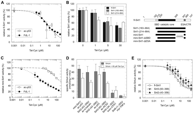
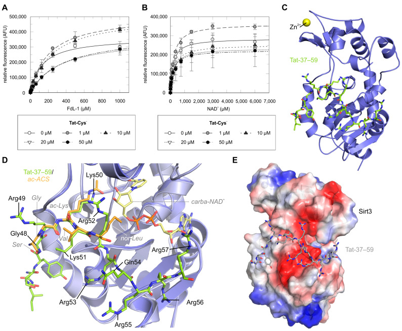
Sirtuins are NAD+-dependent protein lysine deacylases implicated in metabolic regulation and aging-related dysfunctions. The nuclear isoform Sirt1 deacetylates histones and transcription factors and contributes, e.g., to brain and immune cell functions. Upon infection by human immunodeficiency virus 1 (HIV1), Sirt1 deacetylates the viral transactivator of transcription (Tat) protein to promote the expression of the viral genome. Tat, in turn, inhibits Sirt1, leading to the T cell hyperactivation associated with HIV infection. Here, we describe the molecular mechanism of Tat-dependent sirtuin inhibition. Using Tat-derived peptides and recombinant Tat protein, we mapped the inhibitory activity to Tat residues 34-59, comprising Tat core and basic regions and including the Sirt1 deacetylation site Lys50. Tat binds to the sirtuin catalytic core and inhibits Sirt1, Sirt2, and Sirt3 with comparable potencies. Biochemical data and crystal structures of sirtuin complexes with Tat peptides reveal that Tat exploits its intrinsically extended basic region for binding to the sirtuin substrate binding cleft through substrate-like β-strand interactions, supported by charge complementarity. Tat Lys50 is positioned in the sirtuin substrate lysine pocket, although binding and inhibition do not require prior acetylation and rely on subtle differences to the binding of regular substrates. Our results provide mechanistic insights into sirtuin regulation by Tat, improving our understanding of physiological sirtuin regulation and the role of this interaction during HIV1 infection.

Keywords: HIV; Sirt1; crystal structure; deacetylase; regulation.


Conflict of interest statement

The authors declare no conflict of interest.

Figures

Figure 1
Figure 1
Sirt1 inhibition by Tat and mapping of the interaction sites. (A) Titration of fl-Sirt1 with Tat-Cys in a coupled enzymatic assay at 100 µM substrate (○) and an FdL assay at substrate Km (●) (error bars: s.d., n = 2 in coupled assay, n = 3 in FdL assay). (B) Dose-dependent inhibition of various Sirt1 constructs by Tat-Cys at 100 µM FdL-1 (black: full-length Sirt1; gray: Sirt1-183–664; white: Sirt1-214–664). Sirt1 regions included in the respective construct are illustrated in the scheme on the right (error bars: s.d., n = 2). (C) Inhibition of mini-Sirt1 by Tat-Cys at substrate Km in a coupled-enzyme assay (□) and an FdL assay (●), respectively, (error bars: s.d., n = 3–5). (D) Deacylation activity of human Sirtuin isoforms at substrate Km in absence of Tat-Cys (gray) or in presence of 65 μM Tat-Cys (white). Sirt1,2,3 tested against acetylated peptides, Sirt5 against succinylated, and Sirt6 against myristoylated peptides (error bars: s.d., n = 3). (E) Tat-Cys titration of fl-Sirt1 (○), Sirt2 (▲), and Sirt3 (▼) in an FdL assay at substrate Km (error bars: s.d., n = 3).
Figure 2
Figure 2
Mapping and characterization of the Tat region required for sirtuin inhibition. (A) Comparison of 2D-(1H, 15N)-HSQC spectra of 58 µM Tat-Cys before (●) and after (●) addition of mini-Sirt1 (2-fold molar excess). Remaining signals of backbone amides are labeled by sequence numbers. Signals with * denote a second amino acid conformation, signals connected via a horizontal line refer to the sidechain-NH2 of Gln/Asn. (B) Dose-dependent inhibition of fl-Sirt1 by the truncated Tat variants Tat-Cys-(1–63) (gray) and Tat-Cys-(21–63) (white) in an FdL assay at 100 µM FdL-1 (error bars: s.d., n = 2). (C) Dose-dependent inhibition of fl-Sirt1 by Tat-46–54 in a coupled-enzyme assay at 100 µM ac-p53 (error bars: s.d., n = 2). (D) Michaelis–Menten kinetics for deacetylation of ac-p53-Lys382 (○) and ac-Tat-46–54 (●) peptides by mini-Sirt1 (error bars: s.d., n = 3). (E) Dose-dependent increase of fl-Sirt1 activity by ac-Tat-46–54, whose addition increases the total substrate concentration, in a coupled-enzyme assay at 100 µM ac-p53 as the basal substrate (error bars: s.d., n = 2). (F) Dose-dependent inhibition of mini-Sirt1 by synthetic Tat peptides in an FdL assay at substrate Km. Control refers to Tat addition after stopping the reaction. Tat regions covered by peptides are indicated at the bottom (error bars: s.d., n = 3). (G) Titration of mini-Sirt1 with Tat-31–49, Tat-49–60, and Tat-31–60 in an FdL assay at substrate Km (error bars: s.d., n = 3). (H) Titration of mini-Sirt1 with Tat-34–54, Tat-34–59, and Tat-37–59 in an FdL assay at substrate Km (error bars: s.d., n = 3).
Scheme 1
Scheme 1
Schematic summary of the effects of different Tat fragments, with or without Lys50 acetylation, on the deacetylase activity of human Sirt1.
Figure 3
Figure 3
Mechanistic features of Tat-dependent sirtuin inhibition. (A) Michaelis–Menten kinetics for deacetylation of FdL-1 substrate by mini-Sirt1 in presence of increasing concentrations of Tat-Cys (error bars: s.d., n = 3). (B) Michaelis–Menten kinetics for the mini-Sirt1 cosubstrate NAD+ in presence of increasing concentrations of Tat-Cys (error bars: s.d., n = 3–6). (C) Sirt3 (blue) in complex with Tat-37–59 (lime). (D) Superposition of Sirt3/Tat-37–59 (blue/lime) with a Sirt3/ac-ACS/carba-NAD+ complex (light blue/orange; PDB ID 4FVT, RMSD = 0.5 Å for 242 Cα atoms). Residues of ac-ACS2 are labeled in italics. (E) Sirt3/Tat-37–59 with Sirt3 shown as surface colored according to electrostatic potential, from −62.5 kbT/ec (red) to +62.5 kbT/ec (blue).
Figure 4
Figure 4
Structural basis of Tat deacetylation and Tat-dependent sirtuin inhibition. (A) MS2 spectrum of DSSO-interlinked peptides of mini-Sirt1 and Tat-Cys (parent 798.24 m/z). Selected ions for MS3 analysis are highlighted (red line). Tat Arg78 (purple) is deaminated. (B) Visualization of the crosslink between Tat-Lys71 and Sirt1-Lys238 (both Lys as sticks). An NMR structure of wt-Tat (dark green; PDB ID 1TBC) was superpositioned on Sirt3/Tat-37–59 and in turn overlaid with a mini-Sirt1/ac-p53/carba-NAD+/STAC complex (gray; PDB ID 4ZZJ). For clarity, only Sirt1 and Tat are shown. (C) Superposition of Sirt3/ac-Tat-46–54 (teal) with a mini-Sirt1/ac-p53/carba-NAD+/STAC complex (gray; PDB ID 4ZZJ, RMSD 1.2 Å for 208 Cα atoms). The ligands ac-Tat-46–54 (salmon), ac-p53 (light orange), carba-NAD+ (light yellow), and STAC (brown) are shown as sticks. (D) Interactions of ac-Tat-46–54 (salmon) with Sirt3 (teal), and Sirt1 overlaid in gray with their respective residues labeled in italics. Hydrogen bonds are shown as orange dotted lines. (E) Superposition of Sirt3/Tat-37–59 (blue) with Sirt3/ac-Tat-46–54 (teal, RMSD = 0.3 Å for 247 Cα atoms) and a mini-Sirt1/ac-p53/carba-NAD+/STAC complex (gray; PDB ID 4ZZJ, RMSD = 1.2 Å for 210 Cα atoms). Hydrogen bonds (orange dotted lines) and hydrophobic interactions of Tat-37–59 (lime) and ac-Tat-46–54 (salmon) with Sirt3 and the putatively involved residues in Sirt1 are labeled in italics.

References

    1. Morris B.J. Seven sirtuins for seven deadly diseases of aging. Free. Radic. Biol. Med. 2013;56:133–171. doi: 10.1016/j.freeradbiomed.2012.10.525. - DOI - PubMed
    1. Klatzmann D., Champagne E., Chamaret S., Gruest J., Guetard D., Hercend T., Gluckman J.C., Montagnier L. T-lymphocyte T4 molecule behaves as the receptor for human retrovirus LAV. Nature. 1984;312:767–768. doi: 10.1038/312767a0. - DOI - PubMed
    1. Barre-Sinoussi F., Chermann J.C., Rey F., Nugeyre M.T., Chamaret S., Gruest J., Dauguet C., Axler-Blin C., Vezinet-Brun F., Rouzioux C., et al. Isolation of a T-lymphotropic retrovirus from a patient at risk for acquired immune deficiency syndrome (AIDS) Science. 1983;220:868–871. doi: 10.1126/science.6189183. - DOI - PubMed
    1. Dayton A.I., Sodroski J.G., Rosen C.A., Goh W.C., Haseltine W.A. The trans-activator gene of the human T cell lymphotropic virus type III is required for replication. Cell. 1986;44:941–947. doi: 10.1016/0092-8674(86)90017-6. - DOI - PubMed
    1. Clark E., Nava B., Caputi M. Tat is a multifunctional viral protein that modulates cellular gene expression and functions. Oncotarget. 2017;8:27569–27581. doi: 10.18632/oncotarget.15174. - DOI - PMC - PubMed

LinkOut - more resources