. 2023 Apr 11;28(8):3363.
doi: 10.3390/molecules28083363.

Computational Insights into the Dynamic Structural Features and Binding Characteristics of Recombinase UvsX Compared with RecA

Affiliations

Computational Insights into the Dynamic Structural Features and Binding Characteristics of Recombinase UvsX Compared with RecA

Yue Pan et al. Molecules. .

Abstract

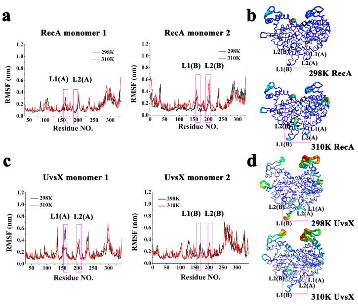
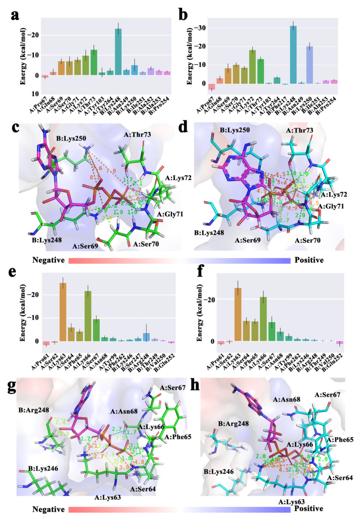
RecA family recombinases are the core enzymes in the process of homologous recombination, and their normal operation ensures the stability of the genome and the healthy development of organisms. The UvsX protein from bacteriophage T4 is a member of the RecA family recombinases and plays a central role in T4 phage DNA repair and replication, which provides an important model for the biochemistry and genetics of DNA metabolism. UvsX shares a high degree of structural similarity and function with RecA, which is the most deeply studied member of the RecA family. However, the detailed molecular mechanism of UvsX has not been resolved. In this study, a comprehensive all-atom molecular dynamics simulation of the UvsX protein dimer complex was carried out in order to investigate the conformational and binding properties of UvsX in combination with ATP and DNA, and the simulation of RecA was synchronized with the property comparison learning for UvsX. This study confirmed the highly conserved molecular structure characteristics and catalytic centers of RecA and UvsX, and also discovered differences in regional conformation, volatility and the ability to bind DNA between the two proteins at different temperatures, which would be helpful for the subsequent understanding and application of related recombinases.

Keywords: DNA recombinases; RecA; UvsX; homologous modelling; molecular dynamics simulations.


Conflict of interest statement

The authors declare no conflict of interest.

Figures

Figure 1
Figure 1
(a) Sequence alignment of RecA and UvsX. Regions with the same amino acid sequence are filled with red background, and regions with similar amino acids are filled with red letters. (b) Overview of initial structures included simulated initial structure of RecA, ATP structure, dsDNA structure and simulated initial structure of UvsX.
Figure 2
Figure 2
(a) RMSF curves of RecA monomers; (b) Visualizations of the backbone flexibility of RecA; (c) RMSF curves of UvsX monomers; (d) Visualizations of the backbone flexibility of UvsX. (The DNA binding domains were framed with pink rectangles.)
Figure 3
Figure 3
Cross-correlation matrices between residues. (a) 298 K RecA; (b) 310 K RecA; (c) 298 K UvsX; (d) 310 K UvsX.
Figure 4
Figure 4
Free energy landscape and lowest energy conformations (a) 298 K RecA; (b) 310 K RecA; (c) 298 K UvsX; (d) 310 K UvsX. Comparing the lowest energy conformation of each protein at 298 K and 310 K, the regions with obvious differences are circled in red, and these parts of the protein are colored by secondary structure, with cyan as helix and the magentas as loop.
Figure 5
Figure 5
Energy contribution of ATP binding residues in RecA (a) 298 K, (b) 310 K. Schematic diagram of ATP interacting with RecA (c) 298 K, (d) 310 K. Energy contribution of ATP binding residues in UvsX (e) 298 K, (f) 310 K; Schematic diagram of ATP interacting with UvsX (g) at 298 K, (h) at 310 K. (The dashed green lines represent conventional hydrogen bond interactions, the lemon lines represent carbon hydrogen bonds, and the orange lines represent electrostatic interactions and bond lengths in Å.).
Figure 6
Figure 6
Schematic diagram of DNA binding region (a) 298 K RecA; (b) 310 K RecA; (c) 298 K UvsX; (d) 310 K UvsX. (The colors of the dashed lines represent the interactions as shown in Figure 5).
Figure 7
Figure 7
(a) DNA rise parameter; (b) Diagram of DNA in four systems and the helical axis of the systems. (Cyan for 298 K RecA, purple for 310 K RecA, pink for 298 K UvsX and yellow for 310 K UvsX).
Figure 8
Figure 8
ATP-DNA communication pathway. (a) 298 K RecA; (b) 310 K RecA; (c) 298 K UvsX; (d) 310 K UvsX.

Similar articles

Cited by

References

    1. Bianco P.R., Tracy R.B., Kowalczykowski S.C. DNA strand exchange proteins: A biochemical and physical comparison. Front. Biosci. 1998;3:D570–D603. doi: 10.2741/a304. - DOI - PubMed
    1. West S.C. Molecular views of recombination proteins and their control. Nat. Rev. Mol. Cell Biol. 2003;4:435–445. doi: 10.1038/nrm1127. - DOI - PubMed
    1. Pierce A.J., Stark J.M., Araujo F.D., Moynahan M.E., Berwick M., Jasin M. Double-strand breaks and tumorigenesis. Trends Cell Biol. 2001;11:S52–S59. doi: 10.1016/S0962-8924(01)02149-3. - DOI - PubMed
    1. Jasin M. Homologous repair of DNA damage and tumorigenesis: The BRCA connection. Oncogene. 2002;21:8981–8993. doi: 10.1038/sj.onc.1206176. - DOI - PubMed
    1. Symington L.S. Role of RAD52 epistasis group genes in homologous recombination and double-strand break repair. Microbiol. Mol. Biol. Rev. 2002;66:630–670. doi: 10.1128/MMBR.66.4.630-670.2002. - DOI - PMC - PubMed

MeSH terms

LinkOut - more resources