. 2023 Apr 21;28(8):3619.
doi: 10.3390/molecules28083619.

Suppressing Kaposi's Sarcoma-Associated Herpesvirus Lytic Gene Expression and Replication by RNase P Ribozyme

Affiliations

Suppressing Kaposi's Sarcoma-Associated Herpesvirus Lytic Gene Expression and Replication by RNase P Ribozyme

Yujun Liu et al. Molecules. .

Abstract

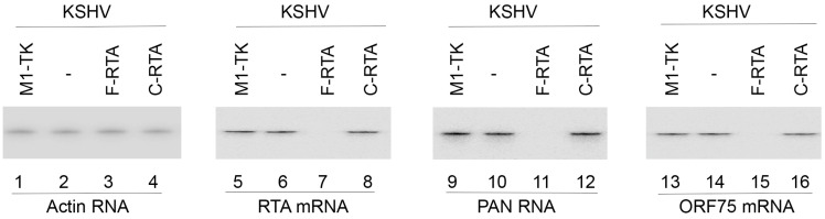
Kaposi's sarcoma, an AIDS-defining illness, is caused by Kaposi's sarcoma-associated herpesvirus (KSHV), an oncogenic virus. In this study, we engineered ribozymes derived from ribonuclease P (RNase P) catalytic RNA with targeting against the mRNA encoding KSHV immediate early replication and transcription activator (RTA), which is vital for KSHV gene expression. The functional ribozyme F-RTA efficiently sliced the RTA mRNA sequence in vitro. In cells, KSHV production was suppressed with ribozyme F-RTA expression by 250-fold, and RTA expression was suppressed by 92-94%. In contrast, expression of control ribozymes hardly affected RTA expression or viral production. Further studies revealed both overall KSHV early and late gene expression and viral growth decreased because of F-RTA-facilitated suppression of RTA expression. Our results indicate the first instance of RNase P ribozymes having potential for use in anti-KSHV therapy.

Keywords: Kaposi sarcoma-associated herpesvirus; RNase P; antiviral; catalytic RNA; gene therapy; herpesvirus; ribozyme.


Conflict of interest statement

The authors declare no conflict of interest.

Figures

Figure 1
Figure 1
(A,B) Substrates for RNase P and M1 RNA. (C) Substrate for M1GS ribozyme. (D) rta39 substrate in this study. (E) The mutated positions in ribozyme C-RTA (circled and in red), compared to F-RTA, shown in the secondary structure of M1 RNA [13,14]. The cleavage site is marked by an arrow.
Figure 2
Figure 2
Northern blot analysis of cellular ribozyme expression. RNAs (30 µg) from BCBL-1 cells transfected with empty LXSN vector with no ribozyme sequences (-) and cells with different ribozymes (F-RTA, C-RTA, and M1-TK) were hybridized with probes against human H1 RNA (loading control) (lanes 1–4) and M1GS RNAs (lanes 5–8).
Figure 3
Figure 3
Expression of viral RNAs detected by Northern blot analysis. RNAs (40 µg) from BCBL-1 cells with no ribozyme sequences (-) and cells with different ribozymes (F-RTA, C-RTA, and M1-TK) were hybridized with probes for actin mRNA (lanes 1–4), RTA mRNA (lanes 5–8), PAN RNA (lanes 9–12), and ORF75 mRNA (lanes 13–16).
Figure 4
Figure 4
Expression of KSHV proteins detected by Western blot analysis. Proteins (35 µg) from BCBL-1 cells with no ribozyme sequences (-) and cells with different ribozymes (F-RTA, C-RTA, and M1-TK) were allowed to bind to antibodies specifically against human actin (loading control), RTA protein, ORF59 protein, and ORF26 protein.
Figure 5
Figure 5
Levels of KSHV DNAs in supernatants of the cell cultures by qPCR. DNAs were purified from supernatants of cell cultures treated with or without TPA and collected at different time points. We employed qPCR to determine viral DNA levels, following procedures described in Materials and Methods. The values show the increase in virion DNA level in the samples, compared to that of virion DNA level in BCBL-1 cells with empty pLXSN vector in the absence of TPA (BCBL-1 no TPA). The averages of three independent experiments are shown and bars indicate the standard deviations.
Figure 6
Figure 6
KSHV latent genome DNA levels in cells by qPCR. We harvested DNA samples from cells without TPA induction and performed qPCR assays. The values show the relative percentage of viral genome DNA levels in samples, as compared with the level of KSHV genome DNA in the BCBL-1 cells transfected with empty pLXSN vector and no ribozyme sequence (BCBL-1). The averages of three independent experiments are shown and bars indicate the standard deviations. NS, not significant.

Similar articles

Cited by

References

    1. Roizman B., Pellett P.E. The family herpesviridae: A brief introduction. In: Knipe D.M., Howley P.M., editors. Fields Virology. Volume 2. Lippincott-William & Wilkins; Philadelphia, PA, USA: 2001. pp. 2381–2398.
    1. Ganem D. Kaposi’s sarcoma-associated herpesvirus. In: Knipe D.M., Howley P.M., Griffin D.E., Martin M.A., Lamb R.A., Roizman B., Straus S.E., editors. Fields Virology. Lippincott-William & Wilkins; Philadelphia, PA, USA: 2007. pp. 2820–2845.
    1. Chang Y., Cesarman E., Pessin M.S., Lee F., Culpepper J., Knowles D.M., Moore P.S. Identification of herpesvirus-like DNA sequences in AIDS-associated Kaposi’s sarcoma. Science. 1994;266:1865–1869. doi: 10.1126/science.7997879. - DOI - PubMed
    1. Arvanitakis L., Geras-Raaka E., Varma A., Gershengorn M.C., Cesarman E. Human herpesvirus KSHV encodes a constitutively active G-protein-coupled receptor linked to cell proliferation. Nature. 1997;385:347–350. doi: 10.1038/385347a0. - DOI - PubMed
    1. Boshoff C., Endo Y., Collins P.D., Takeuchi Y., Reeves J.D., Schweickart V.L., Siani M.A., Sasaki T., Williams T.J., Gray P.W., et al. Angiogenic and HIV-inhibitory functions of KSHV-encoded chemokines. Science. 1997;278:290–294. doi: 10.1126/science.278.5336.290. - DOI - PubMed

MeSH terms

LinkOut - more resources