. 2023 Apr 13;15(8):1882.
doi: 10.3390/nu15081882.

Antidiabetic Activity of Potential Probiotics Limosilactobacillus spp., Levilactobacillus spp., and Lacticaseibacillus spp. Isolated from Fermented Sugarcane Juice: A Comprehensive In Vitro and In Silico Study

Affiliations

Antidiabetic Activity of Potential Probiotics Limosilactobacillus spp., Levilactobacillus spp., and Lacticaseibacillus spp. Isolated from Fermented Sugarcane Juice: A Comprehensive In Vitro and In Silico Study

Chandana Kumari V B et al. Nutrients. .

Abstract

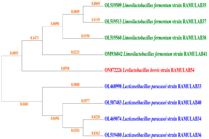
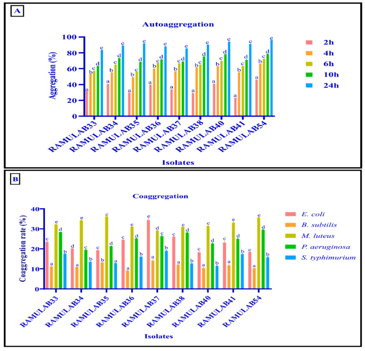
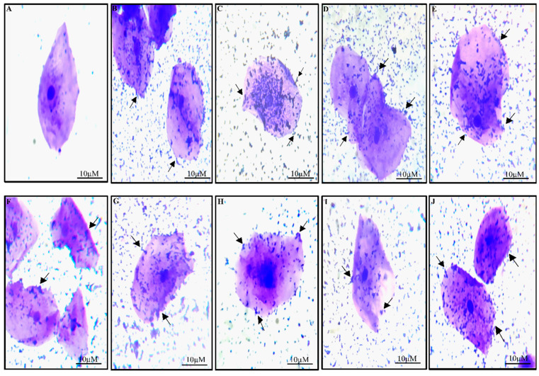
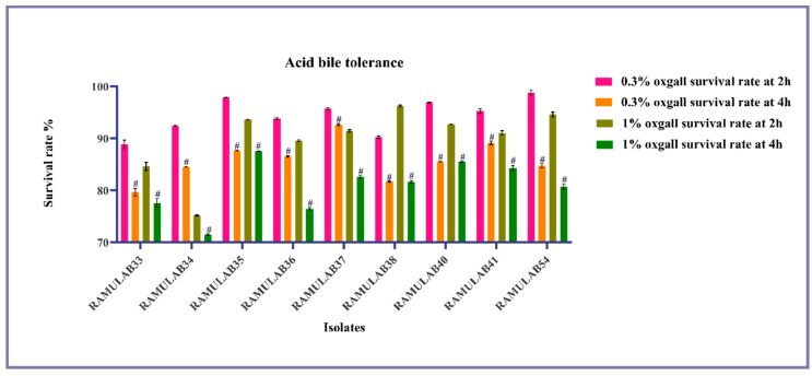
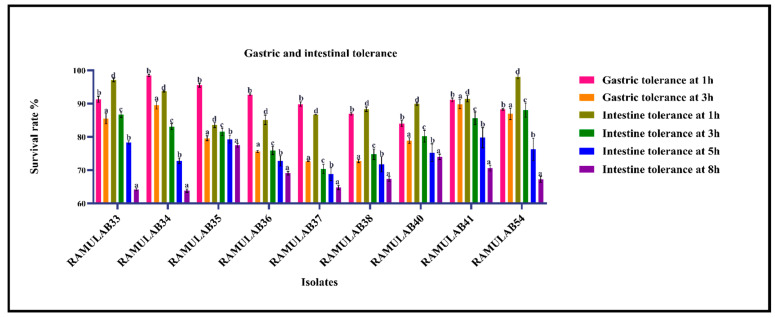
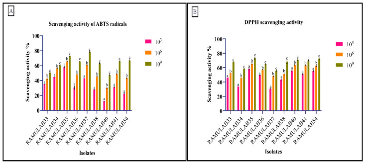
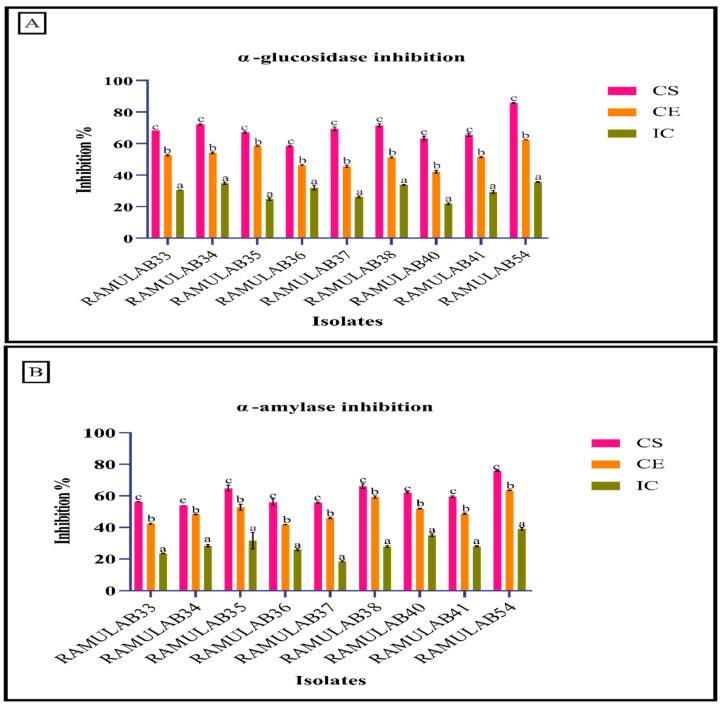
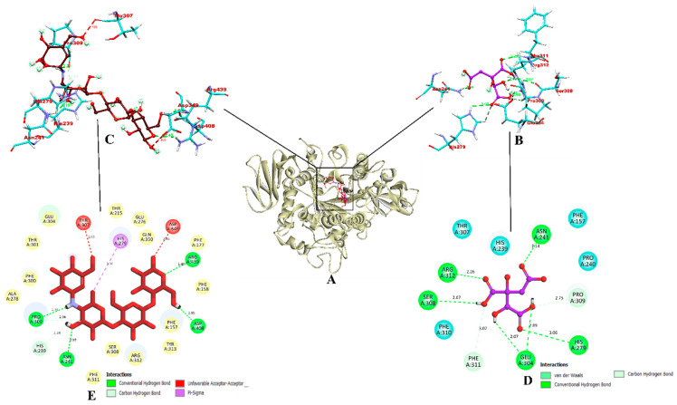
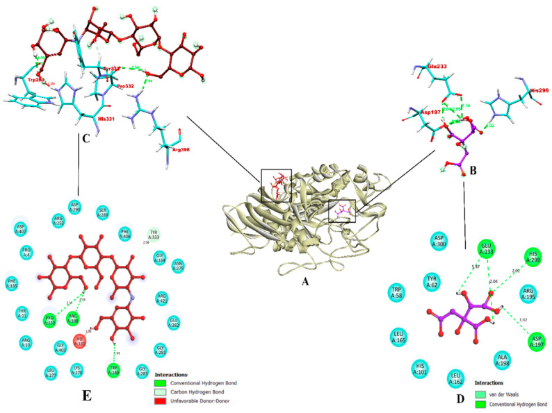
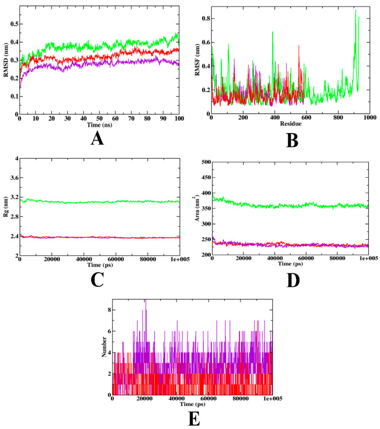
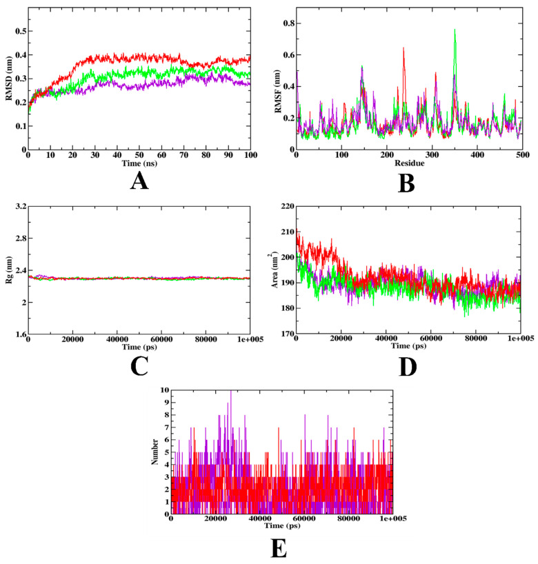
Probiotics are regarded as a potential source of functional foods for improving the microbiota in human gut. When consumed, these bacteria can control the metabolism of biomolecules, which has numerous positive effects on health. Our objective was to identify a probiotic putative Lactobacillus spp. from fermented sugarcane juice that can prevent α-glucosidase and α-amylase from hydrolyzing carbohydrates. Isolates from fermented sugarcane juice were subjected to biochemical, molecular characterization (16S rRNA) and assessed for probiotic traits. Cell-free supernatant (CS) and extract (CE) and also intact cells (IC) were examined for the inhibitory effect on α-glucosidase and α-amylase. CS of the strain showed the highest inhibition and was subjected to a liquid chromatography-mass spectrometry (LCMS) analysis to determine the organic acid profile. The in silico approach was employed to assess organic acid stability and comprehend enzyme inhibitors' impact. Nine isolates were retained for further investigation based on the preliminary biochemical evaluation. Limosilactobacillus spp., Levilactobacillus spp., and Lacticaseibacillus spp. were identified based on similarity > 95% in homology search (NCBI database). The strains had a higher survival rate (>98%) than gastric and intestinal fluids, also a high capacity for adhesion (hydrophobicity > 56%; aggregation > 80%; HT-29 cells > 54%; buccal epithelial cells > 54%). The hemolytic assay indicated that the isolates could be considered safe. The isolates' derivatives inhibited enzymes to varying degrees, with α-glucosidase inhibition ranging from 21 to 85% and α-amylase inhibition from 18 to 75%, respectively. The CS of RAMULAB54 was profiled for organic acid that showed the abundance of hydroxycitric acid, citric acid, and lactic acid indicating their role in the observed inhibitory effects. The in silico approach has led us to understand that hydroxycitric acid has the ability to inhibit both the enzymes (α-glucosidase and α-amylase) effectively. Inhibiting these enzymes helps moderate postprandial hyperglycemia and regulates blood glucose levels. Due to their promising antidiabetic potential, these isolates can be used to enhance intestinal health.

Keywords: lactic acid bacteria; probiotic; sugarcane; α-amylase; α-glucosidase.


Conflict of interest statement

The authors declare no conflict of interest.

Figures

Figure 1
Figure 1
Phylogenetic tree comparison of strains isolated from fermented sugarcane juice based on maximum likelihood bootstrap analysis of 16S rRNA.
Figure 2
Figure 2
(A) The percentage of strains that autoaggregate over time at room temperature; (B) the percentage of LAB strains that coaggregate after two hours at room temperature. The mean ± SD is used to express data. According to the Duncan multiple range tests, the means in aggregation for 2 h with the superscripts (a–e) are significantly different (p ≤ 0.05).
Figure 3
Figure 3
Under a light microscope, LAB strain adherence to buccal epithelial cells has been observed. The buccal epithelial cells in (A) are the control, and the isolates’ (B) RAMULAB33, (C) RAMULAB34, (D) RAMULAB35, (E) RAMULAB36, (F) RAMULAB37, (G) RAMULAB38, (H) RAMULAB40, (I) RAMULAB41 and (J) RAMULAB54 adhesion to these cells is shown. The arrow points to the isolates that are attached to the epithelial cells.
Figure 4
Figure 4
Shows the survival of LAB strains in acidic pH2 circumstances and various bile salt conditions, obtained from a fermented sugarcane juice sample with bile salt concentration parameters 0.3% and 1% for 2 and 4 h (37 °C) in MRS agar plates. The mean and SD are used to express data. Duncan multiple range tests reveal a significant difference (p ≤ 0.05) between the stated averages of the survival rate with a 2 h time interval and superscripts (#).
Figure 5
Figure 5
Shows the isolate survival rates in gastric and intestine juice. The mean ± SD are used to express data. According to the Duncan multiple range test, the means of the survival rates for the time intervals (1 h, 3 h, 5 h, and 8 h) are indicated with distinctive superscripts (a-e) are significantly different (p ≤ 0.05).
Figure 6
Figure 6
Shows of the isolates’ (A) capacity for scavenging ABTS radicals and (B) capacity for scavenging DPPH free radicals. The mean and SD are used to express data. According to the Duncan multiple range test, the means of the scavenging activity of various CFU/mL with distinct superscripts (a–c) are significantly different (p ≤ 0.05).
Figure 7
Figure 7
Shows the isolates’ ability to hinder the enzymes α-glucosidase (A) and α-amylase (B). The mean and SD are used to express data. The means of the inhibitory activity of the CS, CE, and IC with the various superscripts (a–c) are substantially different (p ≤ 0.05), according to the Duncan multiple range test.
Figure 8
Figure 8
The binding interactions of hydroxyacetic acid and acarbose within the inhibitor binding site of target protein α-glucosidase. (A) Ribbon representation of a protein model bound with the ligands (inside the binding site); (B,C) Three-dimensional binding pattern of hydroxyacetic acid and acarbose, respectively. (D,E) Two-dimensional binding pattern of hydroxyacetic acid and acarbose, respectively. Red: acarbose; purple: hydroxyacetic acid. Blue and yellow: surrounding residues; colored: bound residues.
Figure 9
Figure 9
The binding interactions of hydroxyacetic acid and acarbose within the inhibitor binding site of target protein α-amylase. (A) Ribbon representation of a protein model bound with the ligands (inside the binding site); (B,C) Three-dimensional binding pattern of hydroxyacetic acid and acarbose, respectively. (D,E) Two-dimensional binding pattern of hydroxyacetic acid and acarbose, respectively. Red: acarbose; purple: hydroxyacetic acid. Blue: surrounding residues; colored: bound residues.
Figure 10
Figure 10
MD simulation trajectories plotted to show the comparative stability of ligands inside the inhibitor binding site of α-glucosidase (A) RMSD, (B) RMSF, (C) Rg, and (D) SASA, and (E) Ligand H-Bonds. Red: protein-acarbose; purple: protein-hydroxyacetic acid complex, and green: apo-protein.
Figure 11
Figure 11
MD simulation trajectories plotted to show the comparative stability of ligands inside the inhibitor binding site of α-amylase (A) RMSD, (B) RMSF, (C) Rg, and (D) SASA, and (E) Ligand H-Bonds. Red: protein-acarbose; purple: protein-hydroxyacetic acid complex, and green: apo-protein.

Similar articles

Cited by

References

    1. Ramu R., Shirahatti P.S., Zameer F., Ranganatha L.V., Nagendra Prasad M.N. Inhibitory Effect of Banana (Musa Sp. Var. Nanjangud Rasa Bale) Flower Extract and Its Constituents Umbelliferone and Lupeol on α-Glucosidase, Aldose Reductase and Glycation at Multiple Stages. S. Afr. J. Bot. 2014;95:54–63. doi: 10.1016/j.sajb.2014.08.001. - DOI
    1. Sarwar N., Gao P., Kondapally Seshasai S.R., Gobin R., Kaptoge S., di Angelantonio E., Ingelsson E., Lawlor D.A., Selvin E., Stampfer M., et al. Diabetes Mellitus, Fasting Blood Glucose Concentration, and Risk of Vascular Disease: A Collaborative Meta-Analysis of 102 Prospective Studies. Lancet. 2010;375:2215–2222. doi: 10.1016/S0140-6736(10)60484-9. - DOI - PMC - PubMed
    1. Salis S., Virmani A., Priyambada L., Mohan M., Hansda K., de Beaufort C. ‘Old Is Gold’: How Traditional Indian Dietary Practices Can Support Pediatric Diabetes Management. Nutrients. 2021;13:4427. doi: 10.3390/nu13124427. - DOI - PMC - PubMed
    1. Sanders M.E., Merenstein D.J., Reid G., Gibson G.R., Rastall R.A. Probiotics and Prebiotics in Intestinal Health and Disease: From Biology to the Clinic. Nat. Rev. Gastroenterol. Hepatol. 2019;16:605–616. doi: 10.1038/s41575-019-0173-3. - DOI - PubMed
    1. Namai F., Shigemori S., Ogita T., Sato T., Shimosato T. Microbial Therapeutics for Acute Colitis Based on Genetically Modified Lactococcus Lactis Hypersecreting IL-1Ra in Mice. Exp. Mol. Med. 2020;52:1627–1636. doi: 10.1038/s12276-020-00507-5. - DOI - PMC - PubMed

LinkOut - more resources