. 2023 Apr 13;12(4):593.
doi: 10.3390/pathogens12040593.

Leishmania Infection-Induced Proteolytic Processing of SIRPα in Macrophages

Affiliations

Leishmania Infection-Induced Proteolytic Processing of SIRPα in Macrophages

Hana Hirai et al. Pathogens. .

Abstract

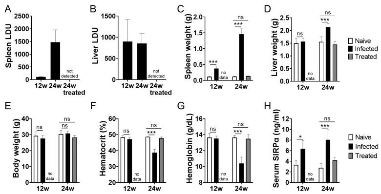
The shedding of cell surface receptors may bring synergistic outcomes through the loss of receptor-mediated cell signaling and competitive binding of the shed soluble receptor to its ligand. Thus, soluble receptors have both biological importance and diagnostic importance as biomarkers in immunological disorders. Signal regulatory protein α (SIRPα), one of the receptors responsible for the 'don't-eat-me' signal, is expressed by myeloid cells where its expression and function are in part regulated by proteolytic cleavage. However, reports on soluble SIRPα as a biomarker are limited. We previously reported that mice with experimental visceral leishmaniasis (VL) manifest anemia and enhanced hemophagocytosis in the spleen accompanied with decreased SIRPα expression. Here, we report increased serum levels of soluble SIRPα in mice infected with Leishmania donovani, a causative agent of VL. Increased soluble SIRPα was also detected in a culture supernatant of macrophages infected with L. donovani in vitro, suggesting the parasite infection promotes ectodomain shedding of SIRPα on macrophages. The release of soluble SIRPα was partially inhibited by an ADAM proteinase inhibitor in both LPS stimulation and L. donovani infection, suggesting a shared mechanism for cleavage of SIRPα in both cases. In addition to the ectodomain shedding of SIRPα, both LPS stimulation and L. donovani infection induced the loss of the cytoplasmic region of SIRPα. Although the effects of these proteolytic processes or changes in SIRPα still remain unclear, these proteolytic regulations on SIRPα during L. donovani infection may explain hemophagocytosis and anemia induced by infection, and serum soluble SIRPα may serve as a biomarker for hemophagocytosis and anemia in VL and the other inflammatory disorders.

Keywords: ADAM; SIRPα; ectodomain shedding; visceral leishmaniasis.


Conflict of interest statement

The authors declare no conflict of interest. The funders had no role in the design of this study; in the collection, analyses, or interpretation of data; in the writing of the manuscript, or in the decision to publish the results.

Figures

Figure 1
Figure 1
Increase serum SIRPα in L. donovani-infected mice. BALB/cA mice were infected with L. donovani. At 12 weeks of infection, some mice received treatment with AmBisome. Representative data of spleen LDU (A), liver LDU (B), spleen weight (C), liver weight (D), body weight (E), hematocrit (F), hemoglobin (G) and serum SIRPα (H) of naïve, L. donovani-infected, or infected/treated BALB/cA mice at 12 or 24 weeks post infection. Mean and SD of each group are shown (n = 5 for each group). * p < 0.05, *** p < 0.001, ns = not significant with two-way ANOVA followed by Tukey’s multiple comparison test.
Figure 2
Figure 2
Release of soluble SIRPα from macrophages during L. donovani infection. (A) Soluble SIRPα in the supernatants was measured with sandwich ELISA. Mean and SD of the representative of two independent experiments with similar results are shown. *** p < 0.001 with one-way ANOVA followed by Dunnett’s multiple comparison test. (B) Both concentrated supernatants and cell lysates were analyzed with Western blotting using anti-SIRPα-ex antibodies. Representatives of two independent experiments with similar results are shown.
Figure 3
Figure 3
Partial inhibition of SIRPα cleavage by an ADAM10 inhibitor. RAW264.7 cells were incubated with GI254023X for 2 h and then LPS or Leishmania promastigotes (MOI = 50) were added to the medium (final concentrations: GI254023X = 0, 1, 10 µM, LPS = 100 ng/mL). After 24 h, supernatants were collected, and soluble SIRPα in the supernatants was measured using sandwich ELISA. Mean and SD of triplicates in each group are shown. Representative of two independent experiments with similar results. ** p < 0.01, *** p < 0.001 with two-way ANOVA followed by Tukey’s multiple comparison test.
Figure 4
Figure 4
Decrease in C-terminal-competent SIRPα during LPS stimulation and Leishmania infection. (A) RAW264.7cells were incubated with medium only, 100 ng/mL LPS, or L. donovani promastigotes (MOI = 50) in complete RPMI1640. After 24 h, cells were harvested and the cell lysates were analyzed with Western blotting using anti-SIRPα N-terminal antibodies or anti-SIRPα C-terminal antibodies. Data shown are triplicate samples representative of two independent experiments with similar results. (B) Densitometric analyses with ImageJ software showing band intensity for SIRPα detected using anti SIRPα-ex or SIRPα-in antibodies. Mean and SD of triplicates in each group are shown. (C) Expression levels of Sirpa mRNA in RAW264.7 of naïve, LPS stimulation, or L. donovani infection were analyzed using qPCR. ** p < 0.01, *** p < 0.001, ns = not significant with one-way ANOVA followed by Dunnett’s multiple comparison test.

References

    1. Goto Y., Cheng J., Omachi S., Morimoto A. Prevalence, Severity, and Pathogeneses of Anemia in Visceral Leishmaniasis. Parasitol. Res. 2017;116:457–464. doi: 10.1007/s00436-016-5313-x. - DOI - PubMed
    1. La Gruta N.L., Kedzierska K., Stambas J., Doherty P.C. A Question of Self-Preservation: Immunopathology in Influenza Virus Infection. Immunol. Cell Biol. 2007;85:85–92. doi: 10.1038/sj.icb.7100026. - DOI - PubMed
    1. Silva-Herzog E., Detweiler C.S. Intracellular Microbes and Haemophagocytosis. Cell. Microbiol. 2008;10:2151–2158. doi: 10.1111/j.1462-5822.2008.01192.x. - DOI - PMC - PubMed
    1. Colomba C., di Carlo P., Scarlata F., Iaria C., Barberi G., Famà F., Cama V., Cascio A. Visceral Leishmaniasis, Hypertriglyceridemia and Secondary Hemophagocytic Lymphohistiocytosis. Infection. 2016;44:391–392. doi: 10.1007/s15010-016-0881-3. - DOI - PubMed
    1. Henter J.I., Horne A., Aricó M., Egeler R.M., Filipovich A.H., Imashuku S., Ladisch S., McClain K., Webb D., Winiarski J., et al. HLH-2004: Diagnostic and Therapeutic Guidelines for Hemophagocytic Lymphohistiocytosis. Pediatr. Blood Cancer. 2007;48:124–131. doi: 10.1002/pbc.21039. - DOI - PubMed

LinkOut - more resources