. 2023 Mar 27;15(4):1072.
doi: 10.3390/pharmaceutics15041072.

Physicochemical, Pre-Clinical, and Biological Evaluation of Viscosity Optimized Sodium Iodide-Incorporated Paste

Affiliations

Physicochemical, Pre-Clinical, and Biological Evaluation of Viscosity Optimized Sodium Iodide-Incorporated Paste

Soo-Jin Chang et al. Pharmaceutics. .

Abstract

This study aimed to investigate the impact of different viscosities of silicone oil on the physicochemical, pre-clinical usability, and biological properties of a sodium iodide paste. Six different paste groups were created by mixing therapeutic molecules, sodium iodide (D30) and iodoform (I30), with calcium hydroxide and one of the three different viscosities of silicone oil (high (H), medium (M), and low (L)). The study evaluated the performance of these groups, including I30H, I30M, I30L, D30H, D30M, and D30L, using multiple parameters such as flow, film thickness, pH, viscosity, and injectability, with statistical analysis (p < 0.05). Remarkably, the D30L group demonstrated superior outcomes compared to the conventional iodoform counterpart, including a significant reduction in osteoclast formation, as examined through TRAP, c-FOS, NFATc1, and Cathepsin K (p < 0.05). Additionally, mRNA sequencing showed that the I30L group exhibited increased expression of inflammatory genes with upregulated cytokines compared to the D30L group. These findings suggest that the optimized viscosity of the sodium iodide paste (D30L) may lead to clinically favorable outcomes, such as slower root resorption, when used in primary teeth. Overall, the results of this study suggest that the D30L group shows the most satisfactory outcomes, which may be a promising root-filling material that could replace conventional iodoform-based pastes.

Keywords: iodoform; root filling dental material; sodium iodide; therapeutic dental paste; viscosity.


Conflict of interest statement

The authors declare no conflict of interest.

Figures

Figure 1
Figure 1
Physical properties of iodoform-based and NaI-based pastes (I30 and D30). (a) Flowability. All values except I30H and I30M were above 17 mm (ISO standard). (b) Radiopacity. All the aluminum thickness values were above the 3 mm Al (ISO standard). (c) Film Thickness. All groups met the ISO standard, which is under 50 µm. (d) Solubility. The solubility of D30 was higher than I30. (e) Optical microscopy images depicting the D30 and I30 groups, distinguished by their colors. The D30 group appears blue-green, whereas the I30 group appears yellow to white. Arrows indicate ISO 68762012 standards. Different superscript letters mean statistically significant differences among groups while ns mean not significant. Error bars mean standard deviations (p < 0.05).
Figure 2
Figure 2
pH analysis and extraction analysis of iodoform-based and NaI-based paste (I30 and D30). (a) The pH was higher in the D30 groups than in the I30 groups. (b,c) Sodium and calcium ions were extracted using inductively coupled plasma atomic spectrometry (ICP/AES). The sodium and calcium concentrations were higher in the D30 group than in the I30 group. (d) Iodoform extracted by ion chromatography. The concentrations of iodoform were higher in the D30 group than in the I30 group. (e) Overall viscosity in the linear graph and viscosity at 0.5 Hz is shown in the bar graph. The viscosity of the I30 group was higher than that of the D30 group. Silicone oil with the L group showed the lowest viscosity compared to H and M silicone oil groups. (f) The highest compressive load is the site when the material extrudes out of the syringe. This peak load is shown in the bar graph. The injection force of the D30 group was lower than the I30 group. Silicone oil with the L group showed the lowest injection force compared to H and M silicone oil groups. Different superscript letters mean statistically significant differences among groups. Error bars mean standard deviations (p < 0.05).
Figure 3
Figure 3
Usability test of NaI-based paste compared to iodoform-based paste. (a) Postoperative radiograph immediately after root canal treatment. (b) Postoperative radiograph after removing filling material using an ultrasonic scaler.
Figure 4
Figure 4
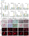
Osteoclast precursors viability test of NaI-based paste using extract compared to iodoform-based paste. (a,b) Cytotoxicity of the extract of NaI-based paste and iodoform-based paste using osteoclast precursors and extracted solutions of different concentrations (control, 12.5, 25, 50, 100%) for 24 h. Arrows indicate ISO standards with the dashed line (70%) represents the cutoff level established by the Cytotoxicity ISO (10993-5), (n = 6, p < 0.05). Both I30L and D30L met ISO recommendations up to 50% concentration in cell viability, but at 100% concentration, the cell viability decreased slightly at D30L. Different superscript letters mean statistically significant differences among groups while ns mean not significant. Error bars mean standard deviations (p < 0.05). (c) Representative images of live/dead staining show live cells stained green and dead cells stained red. The 100% extract from the D30 group displayed a lower count of live cells compared to other groups, as observed in the images.
Figure 5
Figure 5
Osteoclast differentiation test of NaI-based paste using extract compared to iodoform-based paste. (a) RAW 264.7 cells were differentiated into osteoclasts in 12.5% and 25% nontoxic extracts from culture media. After 2, 3, and 4 days of culture, the gene expression of c-FOS, NFATc1, cathepsin K, and TRAP were measured by qPCR. * represents p < 0.05, ** p < 0.01, *** p < 0.001, **** p < 0.0001, n = 3. (b) Representative images of TRAP and phalloidin & DAPI staining of osteoclasts from one of the three experiments are shown. The scale bar is written and the 10×-scale bar is 200 μm and the 20×-scale bar is 100 μm. TRAP staining showed the highest number of differentiated osteoclasts in I30L and the lowest in D30L. The size of the actin ring increased in I30L.
Figure 6
Figure 6
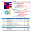
Global gene expression study using quant-sequencing to identify transcriptome features with I30, Ctrl, and D30 under osteoclast differentiation media. (a) Distance-based clustering analysis from global transcriptional changes in 3 groups with 1.2-fold change and over 1 log2 value from differentially expressed gene between I30 and Ctrl. The transcriptome profile with I30 differed from Ctrl and D30L. (b) Gene ontology (GO) analysis using up-regulated genes in I30L compared to Ctrl. Biological process analysis showed that major GO related to active inflammation and apoptosis (negative regulation of proliferation) was enriched in I30L. (c) Venn’s diagram of the comparison of differential gene expression based on Quant-seq data with numbers of co-up or –down and contra-regulated genes. (d) GO analysis using contra-regulated genes (I30L/Ctrl up-regulation & D30L/I30L down-regulation). Biological process analysis showed that major GO related to active inflammation and apoptosis (negative regulation of proliferation) were assigned in down-regulated genes by D30L, meaning a decrease of inflammation and apoptosis could occur in D30L compared to I30L. These transcriptome results support less osteoclast differentiation of precursors in D30L compared to I30L. Collectively, these results indicate that I30L stimulated osteoclast formation, while D30L suppressed the formation of osteoclasts in an in vitro study. (e) Inflammatory gene sets. Among 11 inflammatory up-regulated genes in I30/Ctrl, CD40, ccl22, ccl4, cfh, and lilrb4a were down-regulated by D30, supporting less osteoclastic differentiation potential of D30 than I30.

References

    1. Erausquin J., Muruzábal M. Root canal fillings with zinc oxide-eugenol cement in the rat molar. Oral Surg. Oral Med. Oral Pathol. 1967;24:547–558. doi: 10.1016/0030-4220(67)90436-7. - DOI - PubMed
    1. Cerqueira D.F., Volpi Mello-Moura A.C., Santos E.M., Guedes-Pinto A.C. Cytotoxicity, histopathological, microbiological and clinical aspects of an endodontic iodoform-based paste used in pediatric dentistry: A review. J. Clin. Pediatr. Dent. 2007;32:105–110. doi: 10.17796/jcpd.32.2.k1wx5571h2w85430. - DOI - PubMed
    1. Nurko C., Garcia-Godoy F. Evaluation of a calcium hydroxide/iodoform paste (Vitapex) in root canal therapy for primary teeth. J. Clin. Pediatr. Dent. 1999;23:289–294. - PubMed
    1. Mortazavi M., Mesbahi M. Comparison of zinc oxide and eugenol, and Vitapex for root canal treatment of necrotic primary teeth. Int. J. Paediatr. Dent. 2004;14:417–424. doi: 10.1111/j.1365-263X.2004.00544.x. - DOI - PubMed
    1. Subramaniam P., Gilhotra K. Endoflas, zinc oxide eugenol and metapex as root canal filling materials in primary molars—A comparative clinical study. J. Clin. Pediatr. Dent. 2011;35:365–370. doi: 10.17796/jcpd.35.4.1377v06621143233. - DOI - PubMed

LinkOut - more resources