Review
. 2023 Apr 4;15(4):1143.
doi: 10.3390/pharmaceutics15041143.

Cancer Resistance to Immunotherapy: Comprehensive Insights with Future Perspectives

Affiliations
Review

Cancer Resistance to Immunotherapy: Comprehensive Insights with Future Perspectives

Sawsan Sudqi Said et al. Pharmaceutics. .

Abstract

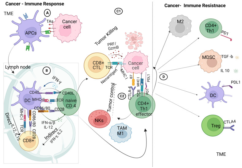
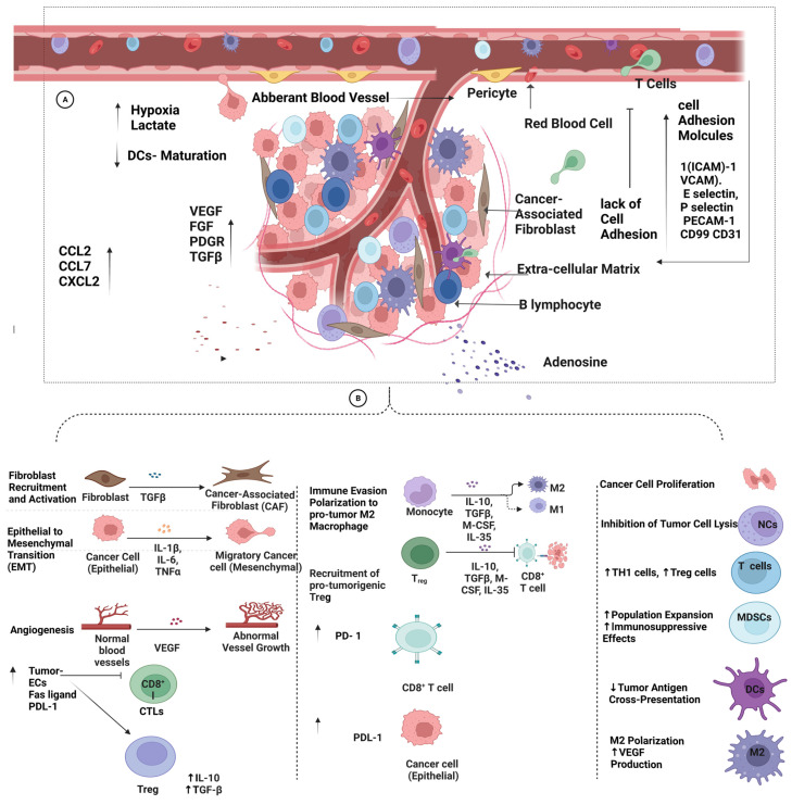
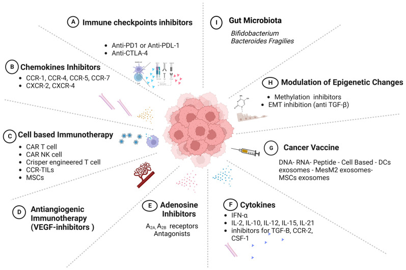
Cancer immunotherapy is a type of treatment that harnesses the power of the immune systems of patients to target cancer cells with better precision compared to traditional chemotherapy. Several lines of treatment have been approved by the US Food and Drug Administration (FDA) and have led to remarkable success in the treatment of solid tumors, such as melanoma and small-cell lung cancer. These immunotherapies include checkpoint inhibitors, cytokines, and vaccines, while the chimeric antigen receptor (CAR) T-cell treatment has shown better responses in hematological malignancies. Despite these breakthrough achievements, the response to treatment has been variable among patients, and only a small percentage of cancer patients gained from this treatment, depending on the histological type of tumor and other host factors. Cancer cells develop mechanisms to avoid interacting with immune cells in these circumstances, which has an adverse effect on how effectively they react to therapy. These mechanisms arise either due to intrinsic factors within cancer cells or due other cells within the tumor microenvironment (TME). When this scenario is used in a therapeutic setting, the term "resistance to immunotherapy" is applied; "primary resistance" denotes a failure to respond to treatment from the start, and "secondary resistance" denotes a relapse following the initial response to immunotherapy. Here, we provide a thorough summary of the internal and external mechanisms underlying tumor resistance to immunotherapy. Furthermore, a variety of immunotherapies are briefly discussed, along with recent developments that have been employed to prevent relapses following treatment, with a focus on upcoming initiatives to improve the efficacy of immunotherapy for cancer patients.

Keywords: cancer immunotherapy; immune checkpoint inhibitors; resistance; tumor microenvironment.


Conflict of interest statement

The authors certify that they have no affiliations with or involvement in any organization or entity with any financial interest or nonfinancial interest in the subject matter or materials discussed in this manuscript.

Figures

Figure 1
Figure 1
The cancer-immunity cycle. The cancer-immunity cycle is a series of steps through which the immune system passes in order to recognize and attack cancer cells. The cycle includes seven stages, starting with the release of cancer-cell antigens and ending with the destruction of cancer cells by T cells. The first stage involves the release of antigens from dying cancer cells. These antigens are then taken up by dendritic cells, which present them to T cells in the lymph nodes. In the second stage, activated T cells migrate to the tumor site and infiltrate the tumor. The third stage involves recognition of cancer cells by T cells, and the fourth stage involves the activation of T cells to recognize and kill cancer cells. In the fifth stage, T cells work to penetrate the tumor and release cytokines that recruit immune cells to the tumor site. In the sixth stage, immune cells, including macrophages and dendritic cells, infiltrate the tumor and destroy cancer cells. Finally, in the seventh stage, the immune system remembers the cancer cells, creating a lasting immune response that can protect against future cancer development.
Figure 2
Figure 2
Crosstalk between cancer and immune cells: (A) innate adaptive immunity, in which the antigen-presenting cells within the tumor microenvironment take up the tumor-associated antigens and process them for antigen-presenting, priming, and activation immune response; (B) adaptive immune response, involving the crosstalk between the dendritic cells and T-cell receptors on naive CD4+ T cells and CD8+ T cells via the major histocompatibility complex class I/II in the presence of co-stimulatory molecules of CD86/80 on DCs and CD28 on CD4+/ CD8+ T cells, ending with the activation of effector T cells; (C1) interaction of effector cytotoxic T cells with tumor cells within the TME; (C2) interaction of effector cytotoxic T cells with tumor cells within the TME and the interaction of effector TH1 cells with tumor cells that trigger the recruitment of tumor-associated macrophages and natural killer cells; (D) immune-checkpoint interactions: activation of PD-1 tumor cells to interact with PD-L1 on effector TH1 lymphocytes, ending with the upregulation of immune suppressive cells; macrophages 2 (M2), regulatory T cells (Tregs), and myeloid-derived suppressor cells (MDSCs) produce IL10 and TGF-β1 with further immune-suppressive actions. Created with BioRender.com.
Figure 3
Figure 3
Schematic presentation of the intrinsic tumor factors associated with resistance to immunotherapy. Multiple mechanisms of cancer resistance to immunotherapy, including (I) loss of neoantigen due to the dedifferentiation or lack of antigen mutations, or mutations in IFN signaling; (II) abnormalities in antigen processing, such as endoplasmic reticulum stress, proteosomes, and TAP protein disturbances; (III) aberrant signaling due to the fact of genetic mutations, such as MAPK, PI3, Wnt/β catenin, and interferon JAK1,2/STAT pathways; (IV) abnormalities in antigen presentation due to the loss of MHC-1 expression, nonclassical MHC-I expression, or β2 microglobulin mutations; (V) expression or exocytosis of immune checkpoint proteins, such as PD-L1; (VI) secretion of metabolites by cancer cells, such as adenosine, epoxy cholesterol, hydroxy cholesterol, and kynurenine, in addition to hypoxia and acidosis, leading to the modulation of the tumor microenvironment; (VII) epigenetic reprogramming of cancer cells through stemness transformation or EMT changes; (VIII) resistance to apoptosis by the aberrant expression of intrinsic and extrinsic apoptotic proteins; (IX) mitochondrial disturbances due to mutations in cytochrome proteins or enzymes in the tricyclic-acetic-acid cycle. Created with BioRender.com.
Figure 4
Figure 4
Schematic presentation of the extrinsic factors contributing to tumor resistance against immunotherapy. (A) Tumor growth and the hypoxic effect on the TME trigger the expression of immunosuppressive and proangiogenic factors, such as TGF-B, VEGF, EGF, FGF, and PDGF. Various vascular changes are mediated by VEGF and immunosuppressive factors, including dysfunctional blood vessels and a lack of both epidermal cells and cell-adhesion molecules (ICAM, P-selectin, and E-selectin). These changes prevent the infiltration of immune cells into the TME and downregulate the immune response. Furthermore, aberrant blood vessels lead to a more stressful and enriched environment, with acidic and hypoxic factors that increase VEGF and prevent DC maturation. Chemokines, such as CCL2, CCL7, and CXCL2 abate/elevate tumor–immune responses and recruit MDSCs, which support tumor growth via TGF-ß. (B) In the tumor microenvironment (TME), further immunosuppressive changes have been observed, such as elevated PD-L1 levels in tumor cells and myeloid-derived suppressor cells (MDSCs), the activation of oxidative and fatty acid metabolism pathways in inactive and resting T cells, and a shortage of glucose that hinders the function of IL-2-stimulated T cells and interferon-gamma (IFN-γ) production.. The upregulation of the TME’s metabolites, such as adenosine, which is produced by TAMs and Tregs, acts as a mediator of immunosuppression. Created with BioRender.com.
Figure 5
Figure 5
Host factors associated with resistance to immunotherapy in cancer patients. (I) Gender is a factor due to the physiological differences between the innate and adaptive immune systems. (II) Age is indicated in studies as the elderly are more likely to develop resistance than their younger counterparts. (III) Malnutrition may interfere with cell-mediated immune responses, which may allow individuals to develop resistance to immunotherapy. (IV) Pregnancy-associated physiological adjustments, including immunotolerance, could interfere with immune recognition and, subsequently, lead to resistance. (V) Microbiomes, such as Bifidobacterium, A. muciniphila, Faecalibacterium, and Bacteroidales, were associated with the enhancement of the immune response in cancer patients, while imbalances in the microbiota were abundant in cancer patients developing resistance to immunotherapy. (VI) Obesity was shown to increase resistance to immunotherapy using animal models of cancer. (VII) Stress is an environmental factor associated with the modulation of immune responses that weaken adaptive immune responses, leading to the failure of the response to immunotherapy. (VIII) Hormones, such as testosterone, estrogen, and progesterone, also modulate the innate and adaptive immune responses, leading to variable responses to immunotherapy in both genders. Created with BioRender.com.
Figure 6
Figure 6
Cancer immunotherapies. Different approaches are highlighted. (A) Immune-checkpoint inhibitors: anti-PD-1/anti-PD-L1 and anti-CTLA4. (B) Blockade of chemokines/chemokine receptors within the TME to inhibit cancer growth or the recruitment of immune-suppressor cells. (C) Cell-based immunotherapies, such as the use of genetically modified T-cell receptors to recognize tumor-antigen-bounded MHC molecules and block cancer-escape pathways, in addition to NKs, MSCs, and TIL cells that are genetically modified to trigger the proliferation and development of immune cells. (D) Antiangiogenic inhibitors reprogram aberrant blood vessels and facilitate the infiltration of the TME by immune cells. (E) Metabolite inhibitors, such as adenosine inhibitors (A2a- and A2b-receptor antagonists). (F) Cytokine therapy provoking immunity in melanoma and renal cancer (IL-2). (G) Different forms of cancer vaccine to encounter immune cells. (H) Modulation of epigenetic changes, such as the epithelial–mesenchymal transition and gene methylation. (I) Gut microbiota to improve the response to ICI therapy. Created with BioRender.com.

References

    1. Ferlay J., Colombet M., Soerjomataram I., Parkin D.M., Piñeros M., Znaor A., Bray F. Cancer statistics for the year 2020: An overview. Cancer Epidemiol. 2021;149:778–789. doi: 10.1002/ijc.33588. - DOI - PubMed
    1. DeVita V.T., Jr., Chu E. A history of cancer chemotherapy. Cancer Res. 2008;68:8643–8653. doi: 10.1158/0008-5472.Can-07-6611. - DOI - PubMed
    1. Wagner E., Elliot K., Enders W., Chobanuk J., Tsui A. BOOK REVIEW: Chemotherapy and Biotherapy Guidelines, and Recommendations for Practice 4th Edition. Can. Oncol. Nurs. J. Rev. Can. De Soins Infirm. En Oncol. 2015;25:480.
    1. Almajali B., Al-Jamal H.A.N., Wan Taib W.R., Ismail I., Johan M.F., Doolaanea A.A., Ibrahim W.N., Tajudin S.A. Thymoquinone Suppresses Cell Proliferation and Enhances Apoptosis of HL60 Leukemia Cells through Re-Expression of JAK/STAT Negative Regulators. Asian Pac. J. Cancer Prev. 2021;22:879–885. doi: 10.31557/apjcp.2021.22.3.879. - DOI - PMC - PubMed
    1. Hanahan D., Weinberg R.A. Hallmarks of cancer: The next generation. Cell. 2011;144:646–674. doi: 10.1016/j.cell.2011.02.013. - DOI - PubMed

LinkOut - more resources