Case Reports
. 2023 Apr 17;23(8):4037.
doi: 10.3390/s23084037.

Longitudinal Gait Analysis of a Transfemoral Amputee Patient: Single-Case Report from Socket-Type to Osseointegrated Prosthesis

Affiliations
Case Reports

Longitudinal Gait Analysis of a Transfemoral Amputee Patient: Single-Case Report from Socket-Type to Osseointegrated Prosthesis

Stefano Di Paolo et al. Sensors (Basel). .

Abstract

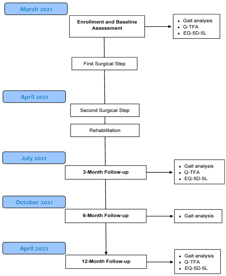
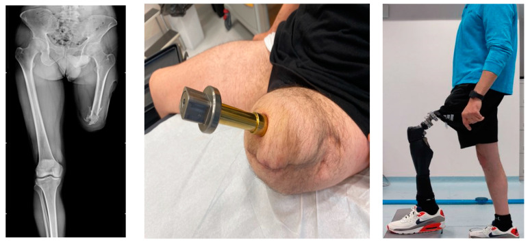
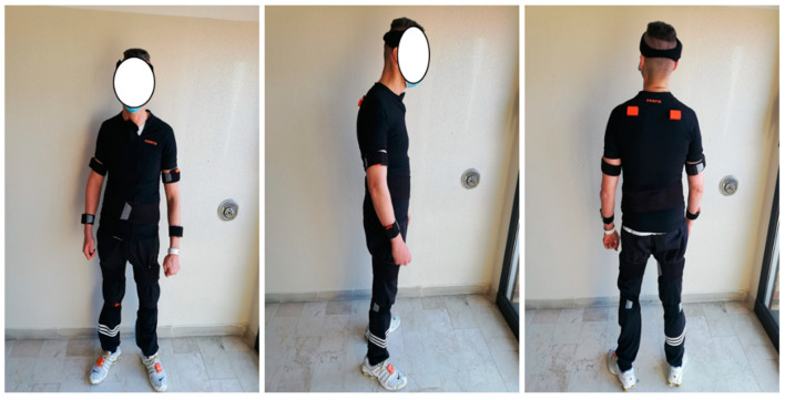
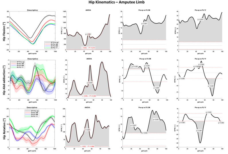
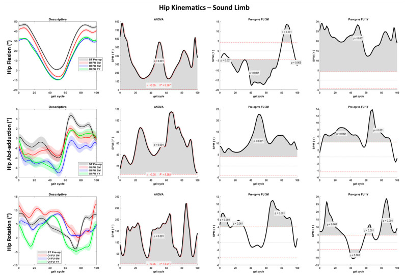
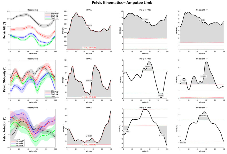
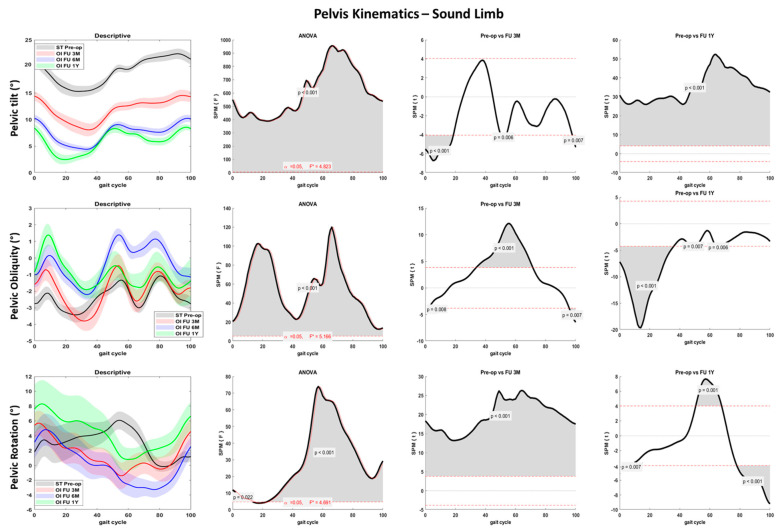
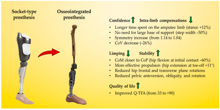
The aim of the present case report was to provide a longitudinal functional assessment of a patient with transfemoral amputation from the preoperative status with socket-type prosthesis to one year after the osseointegration surgery. A 44 years-old male patient was scheduled for osseointegration surgery 17 years after transfemoral amputation. Gait analysis was performed through 15 wearable inertial sensors (MTw Awinda, Xsens) before surgery (patient wearing his standard socket-type prosthesis) and at 3-, 6-, and 12-month follow-ups after osseointegration. ANOVA in Statistical Parametric Mapping was used to assess the changes in amputee and sound limb hip and pelvis kinematics. The gait symmetry index progressively improved from the pre-op with socket-type (1.14) to the last follow-up (1.04). Step width after osseointegration surgery was half of the pre-op. Hip flexion-extension range significantly improved at follow-ups while frontal and transverse plane rotations decreased (p < 0.001). Pelvis anteversion, obliquity, and rotation also decreased over time (p < 0.001). Spatiotemporal and gait kinematics improved after osseointegration surgery. One year after surgery, symmetry indices were close to non-pathological gait and gait compensation was sensibly decreased. From a functional point of view, osseointegration surgery could be a valid solution in patients with transfemoral amputation facing issues with traditional socket-type prosthesis.

Keywords: biomechanics; case report; gait analysis; gait symmetry; osseointegration; socket-type; transfemoral amputation; wearable sensors.


Conflict of interest statement

The authors declare no conflict of interest.

Figures

Figure 1
Figure 1
Flow diagram presenting the longitudinal assessment of the transfemoral amputee patient. The surgical procedure is presented in detail in Section 2.2.
Figure 2
Figure 2
X-ray front view (left), bone-prosthesis connection after the second surgical step (middle), patient wearing the prosthesis at one-year follow-up (right).
Figure 3
Figure 3
Sensors placement. Full-body placement of inertial sensor units for gait analysis in front view (left), side view (middle), and back view (right). Note: the sensors on the thigh were placed on the residual stump for the amputee leg and symmetrically on the sound limb, according to the manufacturer’s instruction.
Figure 4
Figure 4
Hip joint kinematics for the amputee limb over the percentage of the gait cycle for the fast gait in sagittal (first row), frontal (second row), and transverse (third row) planes. Descriptive data (first column) are expressed as mean (solid line) and standard deviation (dashed line). The gray line represents pre-operative gait analysis with socket-type prosthesis (ST pre-op), the red line represents gait analysis at 3 months follow-up with osseointegrated prosthesis (OI FU 3M), the blue line represents gait analysis at 6 months follow-up with osseointegrated prosthesis (OI FU 6M), the green line represents gait analysis at 1-year follow-up with osseointegrated prosthesis (OI FU 1Y). The results of the spm1D repeated-measure ANOVA are presented in the second column. Gray areas represent statistically significant differences (p < 0.05) among the groups. The differences between pre-operative phase and either 3 months follow-up (third column) or 1-year follow-up (fourth column) were assessed through t-test with Bonferroni correction.
Figure 5
Figure 5
Hip joint kinematics for the sound limb over the percentage of the gait cycle for the fast gait in sagittal (first row), frontal (second row), and transverse (third row) planes. Descriptive data (first column) are expressed as mean (solid line) and standard deviation (dashed line). The gray line represents pre-operative gait analysis with socket-type prosthesis (ST pre-op), the red line represents gait analysis at 3 months follow-up with osseointegrated prosthesis (OI FU 3M), the blue line represents gait analysis at 6 months follow-up with osseointegrated prosthesis (OI FU 6M), the green line represents gait analysis at 1-year follow-up with osseointegrated prosthesis (OI FU 1Y). The results of the spm1D repeated-measure ANOVA are presented in the second column. Gray areas represent statistically significant differences (p < 0.05) among the groups. The differences between pre-operative phase and either 3 months follow-up (third column) or 1-year follow-up (fourth column) were assessed through t-test with Bonferroni correction.
Figure 6
Figure 6
Pelvis joint kinematics over the percentage of gait cycle performed with the amputee limb for the fast gait in sagittal (first row), frontal (second row), and transverse (third row) planes. Descriptive data (first column) are expressed as mean (solid line) and standard deviation (dashed line). The gray line represents pre-operative gait analysis with socket-type prosthesis (ST pre-op), the red line represents gait analysis at 3 months follow-up with osseointegrated prosthesis (OI FU 3M), the blue line represents gait analysis at 6 months follow-up with osseointegrated prosthesis (OI FU 6M), the green line represents gait analysis at 1-year follow-up with osseointegrated prosthesis (OI FU 1Y). The results of the spm1D repeated-measure ANOVA are presented in the second column. Gray areas represent statistically significant differences (p < 0.05) among the groups. The differences between pre-operative phase and either 3 months follow-up (third column) or 1-year follow-up (fourth column) were assessed through t-test with Bonferroni correction.
Figure 7
Figure 7
Pelvis joint kinematics over the percentage of gait cycle performed with the sound limb for the fast gait in sagittal (first row), frontal (second row), and transverse (third row) planes. Descriptive data (first column) are expressed as mean (solid line) and standard deviation (dashed line). The gray line represents pre-operative gait analysis with socket-type prosthesis (ST pre-op), the red line represents gait analysis at 3 months follow-up with osseointegrated prosthesis (OI FU 3M), the blue line represents gait analysis at 6 months follow-up with osseointegrated prosthesis (OI FU 6M), the green line represents gait analysis at 1-year follow-up with osseointegrated prosthesis (OI FU 1Y). The results of the spm1D repeated-measure ANOVA are presented in the second column. Gray areas represent statistically significant differences (p < 0.05) among the groups. The differences between pre-operative phase and either 3 months follow-up (third column) or 1-year follow-up (fourth column) were assessed through t-test with Bonferroni correction.
Figure 8
Figure 8
Summary of the functional and quality of life improvements in the patient from socket-type prosthesis to one-year after osseointegrated prosthesis surgery.

Similar articles

Cited by

References

    1. Marks L.J., Michael J.W. Science, medicine, and the future: Artificial limbs. Br. Med. J. 2001;323:732–735. doi: 10.1136/bmj.323.7315.732. - DOI - PMC - PubMed
    1. Kalbaugh C.A., Strassle P.D., Paul N.J., McGinigle K.L., Kibbe M.R., Marston W.A. Trends in Surgical Indications for Major Lower Limb Amputation in the USA from 2000 to 2016. Eur. J. Vasc. Endovasc. Surg. 2020;60:88–96. doi: 10.1016/j.ejvs.2020.03.018. - DOI - PubMed
    1. Van De Meent H., Hopman M.T., Frölke J.P. Walking Ability and Quality of Life in Subjects with Transfemoral Amputation: A Comparison of Osseointegration With Socket Prostheses. Arch. Phys. Med. Rehabil. 2013;94:2174–2178. doi: 10.1016/j.apmr.2013.05.020. - DOI - PubMed
    1. Hagberg K., Brånemark R. Consequences of non-vascular trans-femoral amputation: A survey of quality of life, prosthetic use and problems. Prosthet. Orthot. Int. 2001;25:186–194. doi: 10.1080/03093640108726601. - DOI - PubMed
    1. Buikema K.E.S., Meyerle J.H. Amputation stump: Privileged harbor for infections, tumors, and immune disorders. Clin. Dermatol. 2014;32:670–677. doi: 10.1016/j.clindermatol.2014.04.015. - DOI - PubMed

Publication types