. 2023 Mar 31;11(4):780.
doi: 10.3390/vaccines11040780.

Intradermal Immunization of Soluble Influenza HA Derived from a Lethal Virus Induces High Magnitude and Breadth of Antibody Responses and Provides Complete Protection In Vivo

Affiliations

Intradermal Immunization of Soluble Influenza HA Derived from a Lethal Virus Induces High Magnitude and Breadth of Antibody Responses and Provides Complete Protection In Vivo

Sneha Raj et al. Vaccines (Basel). .

Abstract

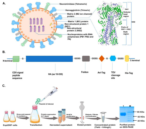
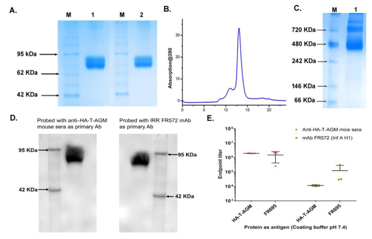
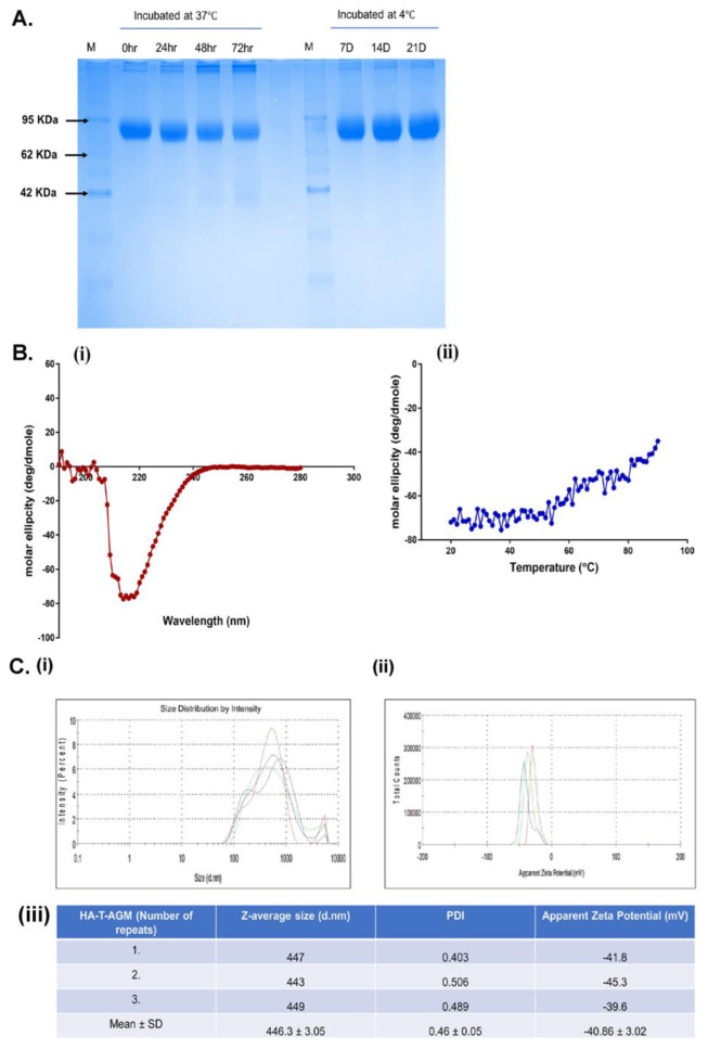
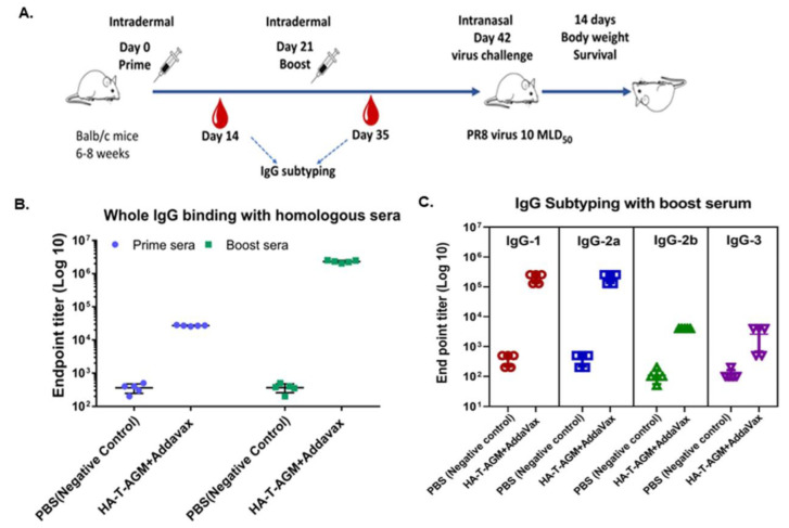
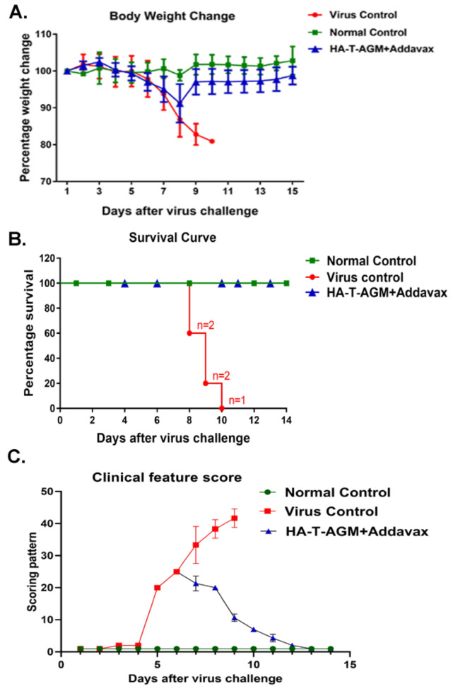
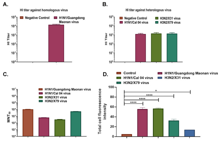
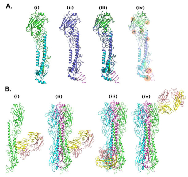
Immunogens mimicking the native-like structure of surface-exposed viral antigens are considered promising vaccine candidates. Influenza viruses are important zoonotic respiratory viruses with high pandemic potential. Recombinant soluble hemagglutinin (HA) glycoprotein-based protein subunit vaccines against Influenza have been shown to induce protective efficacy when administered intramuscularly. Here, we have expressed a recombinant soluble trimeric HA protein in Expi 293F cells and purified the protein derived from the Inf A/Guangdong-Maonan/ SWL1536/2019 virus which was found to be highly virulent in the mouse. The trimeric HA protein was found to be in the oligomeric state, highly stable, and the efficacy study in the BALB/c mouse challenge model through intradermal immunization with the prime-boost regimen conferred complete protection against a high lethal dose of homologous and mouse-adapted InfA/PR8 virus challenge. Furthermore, the immunogen induced high hemagglutinin inhibition (HI) titers and showed cross-protection against other Inf A and Inf B subtypes. The results are promising and warrant trimeric HA as a suitable vaccine candidate.

Keywords: hemagglutinin; influenza; intradermal route; vaccine; virus.


Conflict of interest statement

The authors declare that they have no conflict of interest.

Figures

Figure 1
Figure 1
Construct design and protein production scheme in mammalian cells. (A): Typical Influenza virion structure along with a magnified view of HA-T-AGM 3-D structure (homo-trimer) as generated by the Swiss-model ExPASy online tool based on secondary structure features (purple strands show α-helices and green highlight are β-sheets). (B): Schematic representation of expression cassette used in mammalian pcDNA3.1 expression vector (C): Schematic diagram showing expression and purification of HA-T-AGM protein in Expi293F cells i.e plasmid transfection and protein production and purification stages with final eluted fraction, dialyzed and run on 12% resolving gel.
Figure 2
Figure 2
Antigenic characterization of HA-T-AGM protein. (A): SDS-PAGE results showing purified protein under reduced and non-reduced conditions; Lane 1 with reduced HA-T-AGM and Lane 2 with Non-reduced HA-T-AGM; M is a pre-stained marker (B): Elution profile after purification with Superdex 200 Increase 10/300 column showing a peak at elution volume ~11 mL. (C): Native-PAGE showing oligomerization states of HA-T-AGM soluble protein (D): Immunoblotting of the recombinant HA-T-AGM protein onto PVDF membrane, probing with polyclonal mouse sera (1:500) as primary antibody and another blot with monoclonal antibody IRR FR572 (1:1000) followed by secondary IgG-HRP tagged anti-mouse antibody (1:2000) to develop the blot with help of chemiluminescent substrates (E): ELISA results showing the endpoint titers of HA-T-AGM protein and commercial Influenza HA1 from Influenza A/California/07/2009 (H1N1) pdm09 [FR695]. The binding efficiency of both proteins is tested against polyclonal mouse sera and mouse mAb FR572 raised against Influenza Type A (H1) pdm09.
Figure 3
Figure 3
Biochemical and biophysical characterization of HA-T-AGM. (A): Thermostability profile of the protein after incubation at temperature for hours at 37 °C and days at 4 °C. (B) (i,ii): CD spectroscopy of HA-T-AGM protein. (i) Far-UV CD spectra in the wavelength range of 195–280 nm. (ii) Thermal unfolding of HA-T-AGM as monitored from 25 to 90 °C. (C): Biophysical characterization of the synthesized HA-T-AGM protein. (i) Size distribution profiling, and (ii) Apparent zeta potential distributions of the HA-T-AGM protein. (iii) Statistical analysis and quantifications of the size diameters and zeta potentials. Here, different colors in the graph represent three independent experiments.
Figure 4
Figure 4
Immune response assessment after prime-boost of HA-T-AGM in mice. (A): Schematic representation of antigen administration in BALB/c mice (n = 5) for each test group and n = 5 for naïve control group). The black needle shows the dosage time point, while the red blood drop indicates the time of blood collection. (B): IgG whole (H+L) binding with boost sera using homologous protein and mice group sera. (C): IgG subclass IgG1, IgG 2a, IgG 2b, and IgG3 identification from mouse sera as indicated by ELISA using anti-mouse IgG subtype secondary antibody tagged with HRP. Values plotted are the geometric mean titers mean ± S.E. of triplicate wells.
Figure 5
Figure 5
Protection by HA-T-AGM antigen against virus challenge. (A): Weight change in immunized mice followed for 14 days post virus challenge (Inf A/Puerto Rico/8/34 (PR8)), and comparison made to the immunized vs. non-immunized (virus control) group along with normal naïve control mice group. (B): Survival curve showing the number of dead mice after virus exposure with days passing. (C): The animals were scored from day 1 to day 14 after the virus challenge. The scoring scheme followed from 1–10 where 1 shows NAD (No abnormality), 2 was D (Dull), 3 was MP (Mild piloerection), 4 was PE (Piloerection), 5 was RFD (Reduction in food intake), 6 was reduced movement, huddling (RMH) 7 was H (Hunched), 8 was S (Shivering), 9 was SS (Severe shivering) and 10 represents HLI (Hind limb injury).
Figure 6
Figure 6
A broad range of cross-protection by HA-T-AGM boost sera against virus subtypes. (A): HI titer of Anti-HA-T-AGM vaccinated sera against the homologous strain of the virus (Inf A/H1N1/Guangdong Maonan) (B): HI titer of Anti-HA-T-AGM vaccinated sera against heterologous strains of the virus (Inf A/H1N1/Cal 04; Inf A/H3N2/X-31; Inf A/H3N2/X-79) (C): Microneutralization Assay showing MNT50 titer of HA-T-AGM boost sera against different virus subtypes showing cross-protection. (D): Influenza A/Guangdong HA-T-AGM anti-sera cross-reactivity with other Influenza viruses as shown with Immunofluorescence assay. The data represented here is mean with SE. The normality of the samples was assessed with the Shapiro-Wilk normality test; statistical analysis was then performed using Student’s t-test. * p < 0.05 and **** p < 0.0001.
Figure 7
Figure 7
In-silico analysis of HA-T-AGM. (A). Molecular modeling of HA-T-AGM protein and structural comparison with reported structures: (i) Crystal structure of pdb-id 4LVX, including chain A (green) and chain b (cyan) (ii). Model of HA-T-AGM (blue) (iii) The overlay structure between crystal and model and (iv) The overlay structure (in transparent), to show the changes in amino acids (in licorice). (B). Molecular Docking of HA-T-AGM with reported mAb: (i) monomer chain (in green) with the most likely pose of mAb (pdb-id 3SDY) (yellow and magenta) (ii). Trimer Model of HA in protomer 1 (green), protomer 2 (cyan), and protomer3 (purple) highlighted the most likely pose is consistent in trimer as well (iii) 2nd pose of mAb in trimer and (iv) 3rd pose of mAb in the trimer.

Similar articles

Cited by

References

    1. Kalil A.C., Thomas P.G. Influenza virus-related critical illness: Pathophysiology and epidemiology. Crit. Care. 2019;23:258. doi: 10.1186/s13054-019-2539-x. - DOI - PMC - PubMed
    1. de Francisco Shapovalova N., Donadel M., Jit M., Hutubessy R. A systematic review of the social and economic burden of influenza in low- and middle-income countries. Vaccine. 2015;33:6537–6544. doi: 10.1016/j.vaccine.2015.10.066. - DOI - PubMed
    1. Havers F.P., Campbell A.J.P. Nelson Textbook of Pediatrics. Elsevier; Amsterdam, The Netherlands: 2020. Influenza Viruses; pp. 1727–1732.e1. Chapter 285.
    1. Parrish C.R., Murcia P.R., Holmes E.C. Influenza virus reservoirs and intermediate hosts: Dogs, horses, and new possibilities for influenza virus exposure of humans. J. Virol. 2015;89:2990–2994. doi: 10.1128/JVI.03146-14. - DOI - PMC - PubMed
    1. Petrova V.N., Russell C.A. The evolution of seasonal influenza viruses. Nat. Rev. Microbiol. 2018;16:47–60. doi: 10.1038/nrmicro.2017.118. - DOI - PubMed

LinkOut - more resources