

Shoulder Pseudo-Tumor from COVID-19 Vaccine
- PMID: 37112705
- PMCID: PMC10145291
- DOI: 10.3390/vaccines11040793
Shoulder Pseudo-Tumor from COVID-19 Vaccine
Abstract
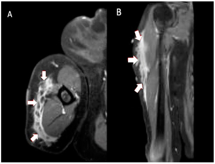
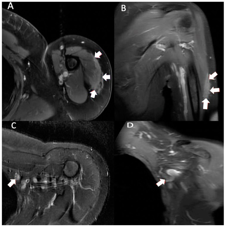
Hypersensitivity reactions to the COVID-19 mRNA vaccines were identified in the initial 2020 trials. Appearance of a soft tissue mass is a rare manifestation of this hypersensitivity reaction. In this patient, bilateral injections resulted in the appearance of shoulder masses. Magnetic resonance imaging showed localized pseudo-tumorous edema in both shoulders, one subcutaneous and the other intramuscular. This is only the second case of a mass-like reaction to the COVID-19 vaccine mimicking a possible soft tissue neoplasm. Improper vaccination administration technique may have contributed to this complication. The case is presented to increase awareness of this potential pseudotumor.
Keywords: COVID-19; SARS-CoV-2; SIRVA; edema; education; orthopedics; shoulder; tumor; vaccine.
Conflict of interest statement
The authors declare no conflict of interest.
Figures
Similar articles
-
SIRVA (Shoulder Injury Related to Vaccine Administration) following mRNA COVID-19 Vaccination: Case discussion and literature review.Vaccine. 2022 Apr 20;40(18):2546-2550. doi: 10.1016/j.vaccine.2022.03.037. Epub 2022 Mar 21. Vaccine. 2022. PMID: 35339304 Free PMC article. Review.
-
Subacromial-subdeltoid bursitis following COVID-19 vaccination: a case of shoulder injury related to vaccine administration (SIRVA).Skeletal Radiol. 2021 Nov;50(11):2293-2297. doi: 10.1007/s00256-021-03803-x. Epub 2021 May 4. Skeletal Radiol. 2021. PMID: 33944967 Free PMC article.
-
Shoulder Injury Related to Vaccine Administration Following Misplaced SARS-CoV-2 Vaccination: A Case Report and Review of Literature.J Orthop Case Rep. 2022 Mar;12(3):100-103. doi: 10.13107/jocr.2022.v12.i03.2736. J Orthop Case Rep. 2022. PMID: 36199926 Free PMC article.
-
Shoulder injury following COVID-19 vaccine administration: a case series and proposed diagnostic algorithm.Expert Rev Vaccines. 2023 Jan-Dec;22(1):299-306. doi: 10.1080/14760584.2023.2189463. Expert Rev Vaccines. 2023. PMID: 36894495 Review.
-
Shoulder Injury Related to Vaccine Administration.Ochsner J. 2022 Fall;22(3):261-264. doi: 10.31486/toj.21.0114. Ochsner J. 2022. PMID: 36189092 Free PMC article.
Cited by
-
Right Biceps Pseudo-Tumor from COVID-19 Vaccination.Vaccines (Basel). 2024 Feb 3;12(2):160. doi: 10.3390/vaccines12020160. Vaccines (Basel). 2024. PMID: 38400143 Free PMC article.
References
Publication types
Grants and funding
LinkOut - more resources
Full Text Sources
Miscellaneous
