. 2023 Apr 14;11(4):845.
doi: 10.3390/vaccines11040845.

Long-Term Follow-Up after Adoptive Transfer of BK-Virus-Specific T Cells in Hematopoietic Stem Cell Transplant Recipients

Affiliations

Long-Term Follow-Up after Adoptive Transfer of BK-Virus-Specific T Cells in Hematopoietic Stem Cell Transplant Recipients

Michael Koldehoff et al. Vaccines (Basel). .

Abstract

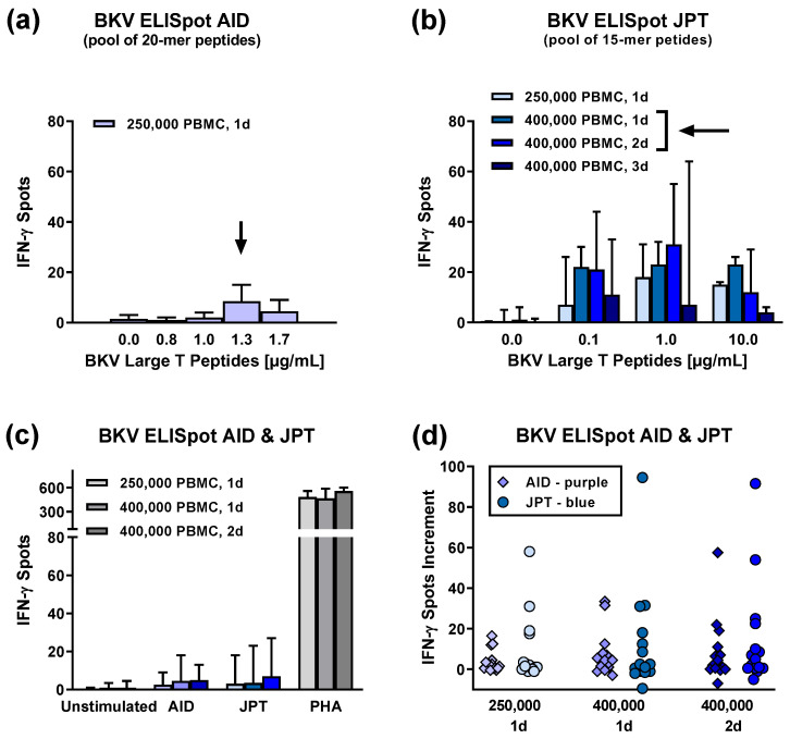
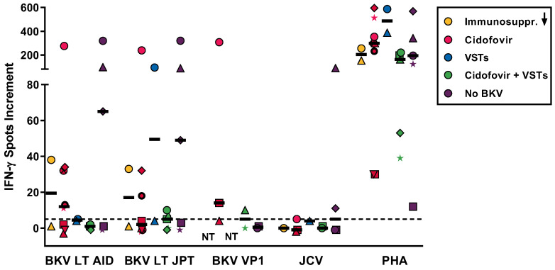
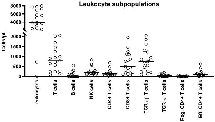
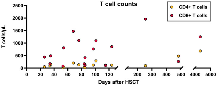
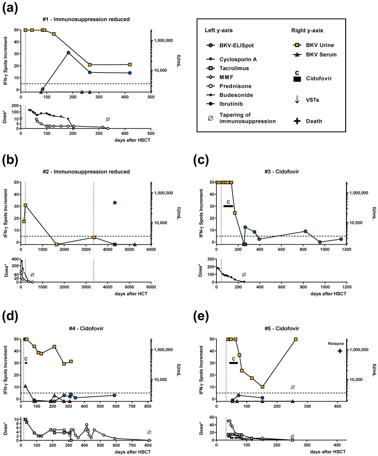
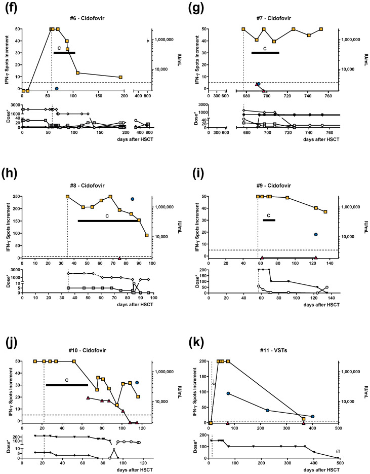
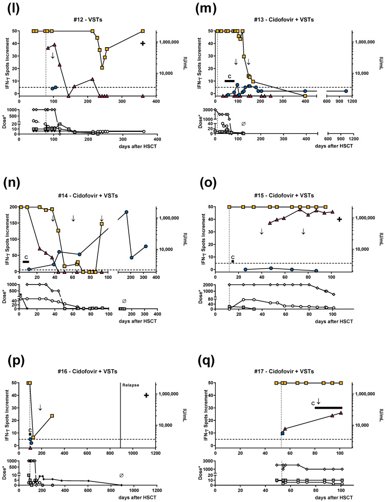
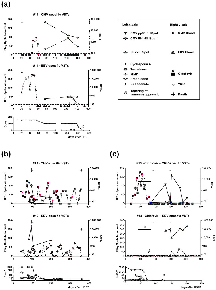
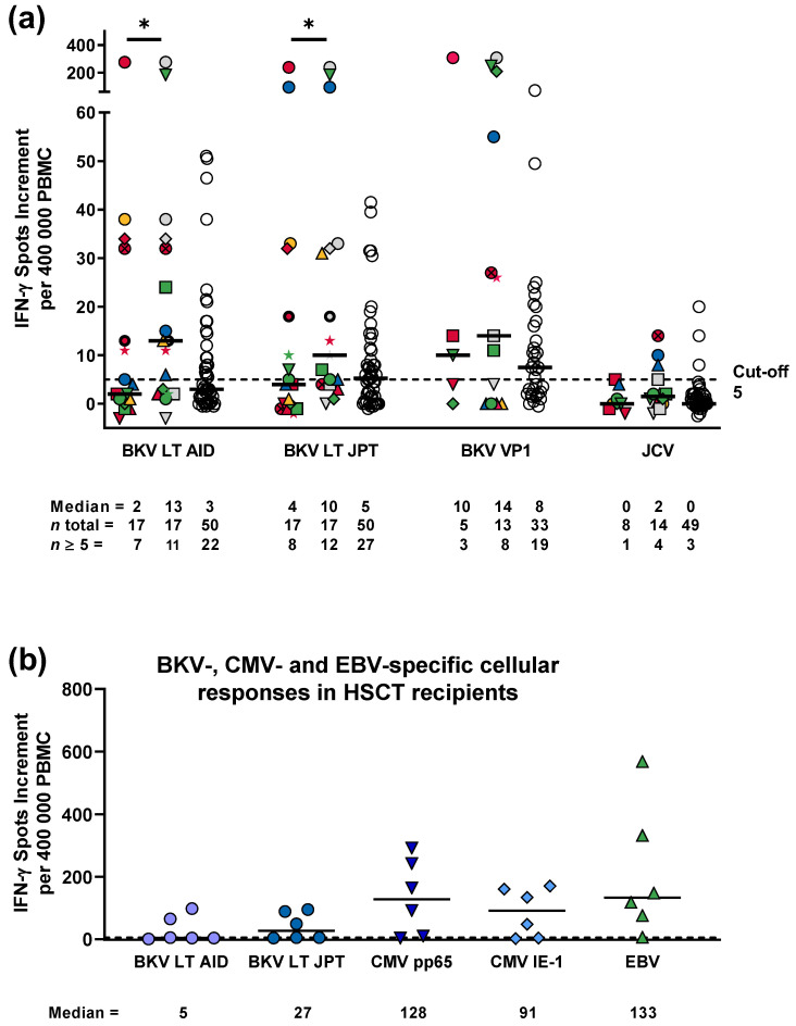
The BK virus (BKV) causes severe hemorrhagic cystitis in hematopoietic stem cell transplant (HSCT) recipients. To eliminate reactivated BKV, symptomatic patients can be treated with a reduction of the immunosuppressive therapy, with the antiviral drug cidofovir, or with virus-specific T cells (VSTs). In the current study, we compared the effect of VSTs to other treatment options, following up specific T cells using interferon-gamma ELISpot assay. We observed BKV large T-specific cellular responses in 12 out of 17 HSCT recipients with BKV-related cystitis (71%). In recipients treated with VSTs, 6 out of 7 showed specific T-cell responses, and that number in those without VSTs was 6 out of 10. In comparison, 27 out of 50 healthy controls (54%) responded. In HSCT recipients treated for BKV-related cystitis, absolute CD4+ T-cell numbers and renal function correlated with BKV-specific cellular responses (p = 0.03 and 0.01, respectively). In one patient, BKV-specific cellular immunity could already be detected at baseline, on day 35 after HSCT and prior to VSTs, and remained increased until day 226 after VSTs (78 vs. 7 spots increment). In conclusion, the ELISpot appears to be suitable to sensitively monitor BKV-specific cellular immunity in HSCT recipients, even early after transplantation or in the long term after VSTs.

Keywords: BK virus; CMV; EBV; ELISpot; JC virus; cidofovir; hematopoietic stem cell transplantation; immunosuppression; monitoring of T-cell immunity; treatment with virus-specific T cells.


Conflict of interest statement

The authors declare no conflict of interest.

Figures

Figure A1
Figure A1
Optimization of ELISpot conditions to determine specific cellular immunity against BK virus (BKV) in patients after hematopoietic stem cell transplantation. Panel (a) shows the titration of 20 mer peptides of BKV large T (AID), using 250,000 peripheral blood mononuclear cells (PBMCs) in 1-day cell cultures (n = 2); panel (b) shows the titration of a 15 mer peptide of BKV large T (JPT) with 250,000 to 400,000 PBMCs in 1–3-day cell cultures (n = 3); and panel (c) shows data from 250,000 and 400,000 PBMCs and various culture durations with both BKV peptides used at optimal concentrations (1.3 and 1 µg/mL per peptide, respectively, indicated by an arrow) (n = 15). Each left column indicates data on 250,000 PBMCs and 1-day cell cultures, each middle column on 400,000 PBMCs and 1-day cell cultures, and each right column on 400,000 PBMCs and 2-day cell cultures. The graphs (ac) show the median and 75% percentile. Panel (d) is based on the data as displayed in panel (c). It indicates individual values, given as spots increment, i.e., response towards BK virus (BKV) large T peptides (from AID or JPT) minus unstimulated controls (n = 15). PHA, phytohemagglutinin (positive control). In panel (c) and (d) the brightest color (either grey, blue or purple) indicates 250,000 PBMC and 1d culture, the intermediate color 400,000 PBMC and 1d culture and the darkest color 400,000 PBMC and 2d culture.
Figure A2
Figure A2
Cellular responses towards BK virus (BKV) large T (LT) peptides (from AID or JPT), as well as towards BKV viral protein 1 (VP1) and JC virus (JCV) peptides. Cell cultures stimulated with phytohemagglutinin (PHA) served as a positive control. The graph shows data on the first timepoint in hematopoietic stem cell transplant recipients with BKV infection, in whom only immunosuppression was reduced (n = 2), who was treated by cidofovir (n = 8), who received virus-specific T cells (VSTs) (n = 2), and who received cidofovir plus VSTs (n = 5). For comparison, we present the results of transplant recipients who suffered from cystitis that was not related to BKV (control group, n = 5). Each patient is depicted by an individual symbol. The values correspond to the first timepoint, as shown in Figure 1, which is a median of 28 days after the onset of cystitis. Data are indicated as spots increment, i.e., BKV-specific spots minus negative control. Solid horizontal lines indicate median values. The dotted horizontal line indicates the cut-off for positive responses (five spots increment). NT, not tested. BKV LT AID—BKV large T peptides (from AID), BKV LT JPT—BKV large T peptides (from JPT), BKV VP1—BKV viral protein 1 peptides.
Figure A3
Figure A3
Distribution of leukocyte subpopulations in the patient cohort shown in Table A1. This figure contains the first dataset of each HSCT recipient with BKV-related cystitis. The median interval between HSCT and study inclusion (first dataset) was 85 days (range 26 days–12 years). Median values are indicated by horizontal lines. Reg.—regulatory, Eff.—effector.
Figure A4
Figure A4
Correlation of the interval between HSCT and study inclusion (days after HSCT) and CD4+ and CD8+ T-cell counts in the patient cohort shown in Table A1. Spearman analysis indicated that CD4+ T-cell counts showed a stronger correlation with days after HSCT (r = 0.46, p = 0.06) than CD8+ T-cell counts (r = 0.28, p = 0.3).
Figure 1
Figure 1
Follow-up data of hematopoietic stem cell transplant (HSCT) recipients who suffered from BK virus (BKV)-related cystitis. The dotted vertical line indicates the onset of BKV-related cystitis. The patients were either treated only with a reduction of their immunosuppressive drugs (a,b); with cidofovir (cj); with BK virus-specific T cells (VSTs), as indicated by an arrow (↓) (k,l); or with a combination of cidofovir and VSTs (mq). Cellular responses towards BKV large T (LT) peptides (from JPT) are indicated on the upper left y-axis as spots increment, i.e., BKV-specific spots minus negative control. The dotted horizontal line indicates the cut-off for positive responses (five spots increment). The upper right y-axis shows the viral load in urine and serum. With one exception, the lower left y-axis indicates the dose of immunosuppressive drugs as mg per day (*). To be visible, the dose of tacrolimus is given as mg * 10 per day. MMF, mycophenolate mofetil.
Figure 1
Figure 1
Follow-up data of hematopoietic stem cell transplant (HSCT) recipients who suffered from BK virus (BKV)-related cystitis. The dotted vertical line indicates the onset of BKV-related cystitis. The patients were either treated only with a reduction of their immunosuppressive drugs (a,b); with cidofovir (cj); with BK virus-specific T cells (VSTs), as indicated by an arrow (↓) (k,l); or with a combination of cidofovir and VSTs (mq). Cellular responses towards BKV large T (LT) peptides (from JPT) are indicated on the upper left y-axis as spots increment, i.e., BKV-specific spots minus negative control. The dotted horizontal line indicates the cut-off for positive responses (five spots increment). The upper right y-axis shows the viral load in urine and serum. With one exception, the lower left y-axis indicates the dose of immunosuppressive drugs as mg per day (*). To be visible, the dose of tacrolimus is given as mg * 10 per day. MMF, mycophenolate mofetil.
Figure 2
Figure 2
Follow-up data of hematopoietic stem cell transplant (HSCT) recipients who received tri-specific virus-specific T cells (VSTs), directed against BK virus, cytomegalovirus (CMV), and Epstein–Barr virus (EBV). The graphs show cellular in vitro responses against CMV phosphoprotein 65 (pp65) and immediate early-1 (IE-1) and EBV and viral load of CMV and EBV. Panel (a) corresponds to the patient shown in Figure 1k, panel (b) corresponds to Figure 1l, and panel (c) corresponds to Figure 1m, where immunity against BKV is displayed. Infusion of VSTs is indicated by an arrow (↓). The dotted horizontal line indicates the cut-off for positive responses (five spots increment). Cellular responses are shown on the upper left y-axis as spots increment, i.e., CMV- or EBV-specific spots minus negative control. The upper right y-axis shows the viral load in whole blood. With one exception, the lower left y-axis indicates the dose of immunosuppressive drugs as mg per day (*). To be visible, the dose of tacrolimus is given as mg * 10 per day. MMF, mycophenolate mofetil.
Figure 3
Figure 3
Specific cellular immunity against peptides of the BK virus (BKV). (a) Responses against large T (LT) and viral protein (VP) 1 peptides of BKV and against a structurally related polyomavirus, the JC virus (JCV), in 17 hematopoietic stem cell transplant (HSCT) recipients with BK virus-related cystitis (coloured symbols) and in 50 healthy controls (HC, white circles). The first dataset in each patient is shown as the left panel, with the maximum response as the middle panel. Each patient is depicted by an individual symbol. Treatment groups are colour-coded (yellow: with reduction of immunosuppression only, red: with cidofovir only, blue: with VSTs only, and green: with cidofovir and VSTs). For patients tested only once, the first set of data is equal to the maximum response, which is shown as a grey symbol (and which is not considered for comparison of the first and maximum response). Please note that only part of the datasets contained VP1 and JCV. One-day cell cultures were performed with 400,000 peripheral blood mononuclear cells and the antigen concentrations defined as optimal. (b) Specific cellular immunity against LT peptides of BKV, cytomegalovirus (CMV) phosphoprotein 65 (pp65), CMV immediate early-1 (IE-1), and Epstein–Barr virus (EBV), analyzed in parallel in six HSCT recipients with BKV-related cystitis. Each viral antigen is indicated by a specific symbol and color. Median values are indicated with solid horizontal lines. The dotted horizontal line indicates the cut-off for positive responses (five spots increment). * p < 0.05 (Wilcoxon matched pairs test). BKV LT AID—BKV large T peptides (from AID), BKV LT JPT—BKV large T peptides (from JPT), BKV VP1—BKV viral protein 1 peptides.

Similar articles

Cited by

References

    1. Saade A., Gras J., Darmon M., Michonneau D., Dhedin N., Feghoul L., Le Goff J., Xhaard A., De Latour R.P., Socie G., et al. Incidence, risk factors and outcome of BK virus hemorrhagic cystitis following allogenic hematopoietic cell transplantation: A retrospective cohort study. Bone Marrow Transpl. 2022;57:1287–1294. doi: 10.1038/s41409-022-01665-y. - DOI - PubMed
    1. Ambalathingal G.R., Francis R.S., Smyth M.J., Smith C., Khanna R. BK Polyomavirus: Clinical Aspects, Immune Regulation, and Emerging Therapies. Clin. Microbiol. Rev. 2017;30:503–528. doi: 10.1128/CMR.00074-16. - DOI - PMC - PubMed
    1. Wong A.S., Cheng V.C., Yuen K.Y., Kwong Y.L., Leung A.Y. High frequency of polyoma BK virus shedding in the gastrointestinal tract after hematopoietic stem cell transplantation: A prospective and quantitative analysis. Bone Marrow Transpl. 2009;43:43–47. doi: 10.1038/bmt.2008.266. - DOI - PMC - PubMed
    1. Egli A., Infanti L., Dumoulin A., Buser A., Samaridis J., Stebler C., Gosert R., Hirsch H.H. Prevalence of polyomavirus BK and JC infection and replication in 400 healthy blood donors. J. Infect. Dis. 2009;199:837–846. doi: 10.1086/597126. - DOI - PubMed
    1. Kaur A., Wilhelm M., Wilk S., Hirsch H.H. BK polyomavirus-specific antibody and T-cell responses in kidney transplantation: Update. Curr. Opin. Infect. Dis. 2019;32:575–583. doi: 10.1097/QCO.0000000000000602. - DOI - PubMed