Meta-Analysis
. 2023 Mar 27;15(4):856.
doi: 10.3390/v15040856.

Understanding Mutations in Human SARS-CoV-2 Spike Glycoprotein: A Systematic Review & Meta-Analysis

Affiliations
Meta-Analysis

Understanding Mutations in Human SARS-CoV-2 Spike Glycoprotein: A Systematic Review & Meta-Analysis

Reetesh Kumar et al. Viruses. .

Abstract

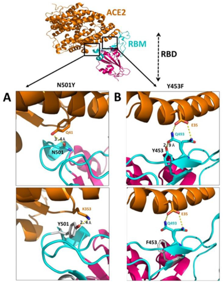
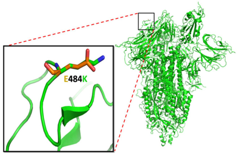
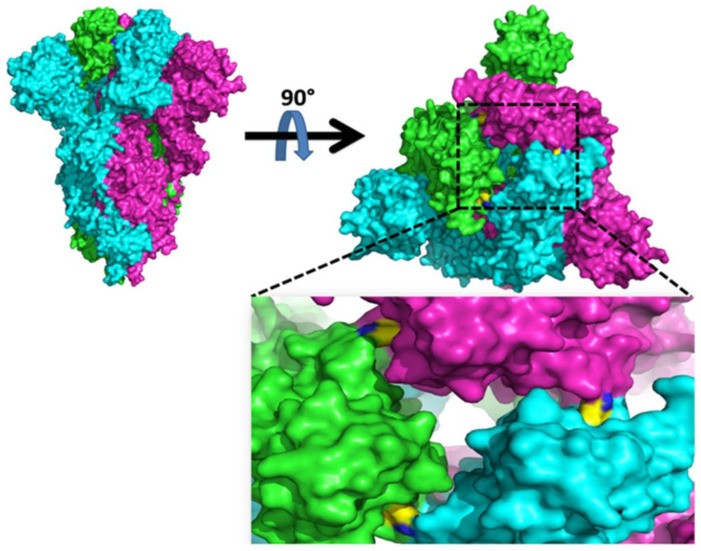
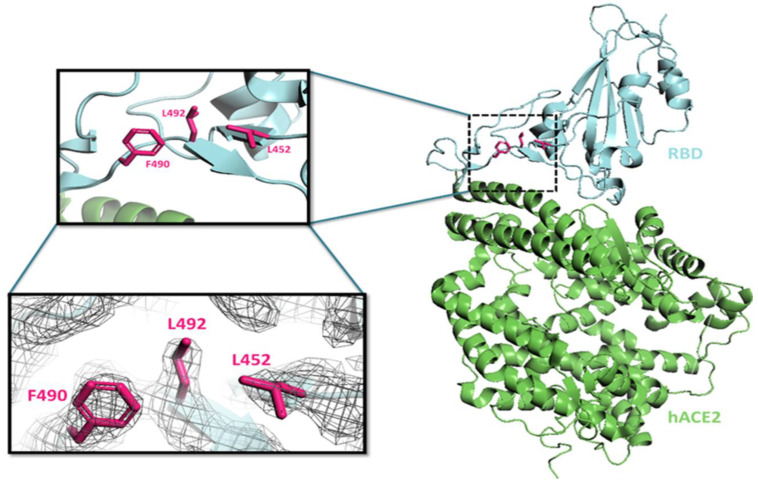
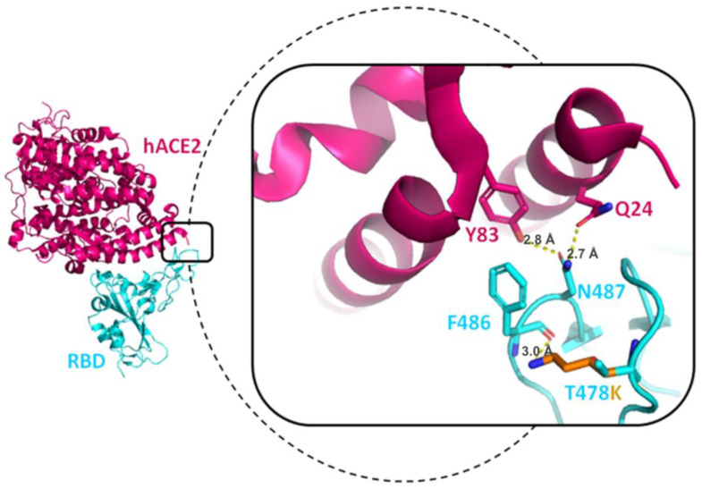
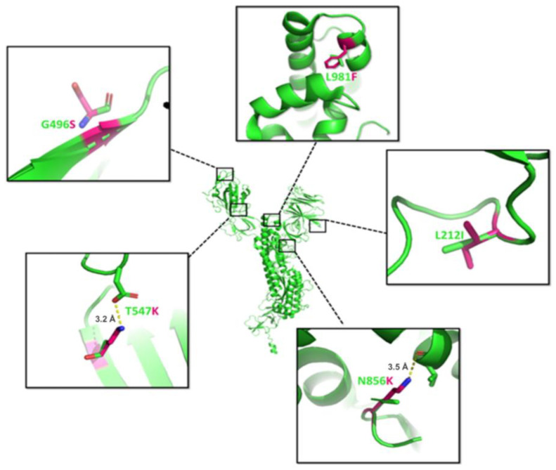
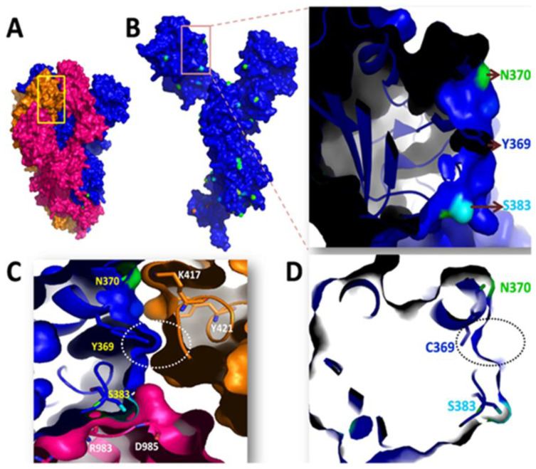
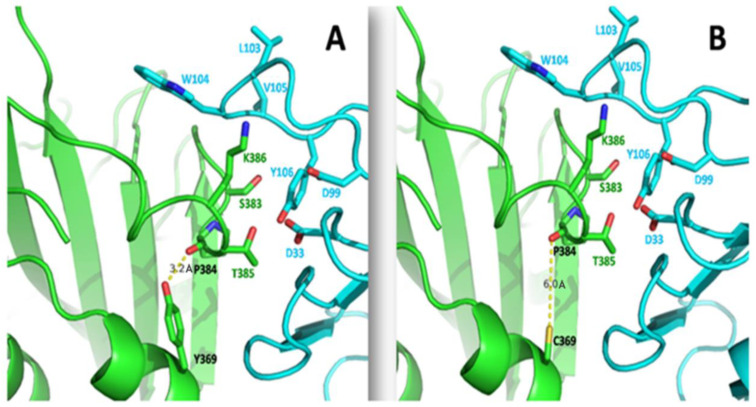
Genetic variant(s) of concern (VoC) of SARS-CoV-2 have been emerging worldwide due to mutations in the gene encoding spike glycoprotein. We performed comprehensive analyses of spike protein mutations in the significant variant clade of SARS-CoV-2, using the data available on the Nextstrain server. We selected various mutations, namely, A222V, N439K, N501Y, L452R, Y453F, E484K, K417N, T478K, L981F, L212I, N856K, T547K, G496S, and Y369C for this study. These mutations were chosen based on their global entropic score, emergence, spread, transmission, and their location in the spike receptor binding domain (RBD). The relative abundance of these mutations was mapped with global mutation D614G as a reference. Our analyses suggest the rapid emergence of newer global mutations alongside D614G, as reported during the recent waves of COVID-19 in various parts of the world. These mutations could be instrumentally imperative for the transmission, infectivity, virulence, and host immune system's evasion of SARS-CoV-2. The probable impact of these mutations on vaccine effectiveness, antigenic diversity, antibody interactions, protein stability, RBD flexibility, and accessibility to human cell receptor ACE2 was studied in silico. Overall, the present study can help researchers to design the next generation of vaccines and biotherapeutics to combat COVID-19 infection.

Keywords: COVID-19; SARS-CoV-2; VoC; evolution; mutations; spike protein.


Conflict of interest statement

The authors declare no conflict of interest. The funders had no role in the design of the study; in the collection, analyses, or interpretation of data; in the writing of the manuscript; or in the decision to publish the results.

Figures

Figure 1
Figure 1
Schematic presentation illustrating the different components of SARS-CoV-2. The three-dimensional SARS-CoV-2 structure (left panel) shows the viral surface proteins (spikes, envelopes, and membranes) embedded in a lipid bilayer envelope. The internal structure (right panel), shows the corresponding protein in addition to the interior nucleocapsid protein which is associated with the single-stranded positive sense viral RNA. The interaction between the S-protein trimer of SARS-CoV-2 and the ACE2 receptor is shown in lower right. Upon binding to the receptor binding domain (RBD) of the S1 subunits, the S protein is primed with TMPRSS2 facilitating the release of the viral genome through S2-assisted fusion.
Figure 2
Figure 2
The D614G mutation in the SARS-CoV-2 spike protein. (A) D614 in the wild-type spike glycoprotein, and (B) G614 the mutated form of D614. The three-dimensional structure of the spike glycoprotein is represented colored by the chain. The yellow dotted line represents the hydrogen bond formation between residues 614 and 647, which shortens when aspartic acid mutates to glycine. The length between residues is shown in units of Angstrom (Å).
Figure 3
Figure 3
The wild-type A222 in the spike glycoprotein mutates to V222, shown in the A222V mutation. The green illustration represents the NTD, with the residues residing close to the RBD in magenta. (A) Neighboring hydrophobic residues surround the wild-type A222 in the spike glycoprotein: Y38, I285, and F220. The representation is shown in the blue circle; (B) The mutated V222 in the S protein is surrounded by hydrophobic residues; Y38, I285, and F220 make a comparatively strong hydrophobic core (shown in the red circle). The dotted/straight (double-headed arrow) illustrates the hydrophobic interaction. The length between residues is shown in units of Angstrom (Å).
Figure 4
Figure 4
The RBD and hACE2 interactions were taken from the PDB structure (PDB: 6M0J). The hACE2 is shown in gray and RBD as a β factor diagram (shown in rainbow). The S477 (wild type) is magnified in the upper right box. The N477 (mutated) is magnified in the lower right box. The side chains of N477 are in different rotamers, increasing its propensity of binding with hACE2 in comparison with the wild type.
Figure 5
Figure 5
The interaction between RBD and hACE2 (adapted from the PDB structure (PDB: 6M0J)). The hACE2 is in orange, RBM is in cyan, and the RBD (except RBM) is in magenta. (A) The N501Y mutation: the wild-type N501 of RBM interacts with Q41 of hACE2 (3.4 Å), which after mutation (Y501) binds more robustly with K353 of hACE2 (2.4 Å); (B) The Y453F mutation: the wild type Y453 interacts with Q493 of RBM (2.9 Å) and subsequently restrains Q493 (reside in two rotamers) interaction with E35 of hACE2. The mutated F453 loses the binding with Q493, subsequently increasing the propensity of Q493 (reside in two rotamers) interaction with E35 of hACE2. The upper panel represents the wild type, whereas the lower panel denotes the mutated interaction. The wild types N501 and Y453 are in cyan, and mutated Y501 and F453 are in gray. The length between residues is shown in units of Angstrom (Å).
Figure 6
Figure 6
The E484K mutation: the structure in green represents the spike glycoprotein. The magnified black box represents the surface-exposed E484K mutation. The glutamic acid and mutated lysine residue were superimposed on the same region (shown in the box). The orange color denotes the negative charge of glutamic acid, and the green color depicts the positive charge of lysine. The mutated lysine changes the surface property because of its positive charge, and length of side chain compared with glutamic acid.
Figure 7
Figure 7
The spike glycoprotein in the trimeric state is shown as surface representation in the (upper left). The protomer is shown in green, cyan, and magenta. The top view shows K417 interaction with N370 (between nearby protomers) shown as a yellow surface (upper right). The black dotted box zooms out to show the K417 and N370 interaction (below right).
Figure 8
Figure 8
The RBD and hACE2 interactions were taken from the PDB structure (PDB: 6M0J), shown on the right. The structure of hACE2 is shown in green and RBD in cyan. The black dotted box is magnified to represent the L452, L492, and F490 residue in the RBD and is shown in magenta in the upper left box. The electron-density map for these residues (L452, L492, and F490) as shown in the below left box.
Figure 9
Figure 9
The RBD and hACE2 interactions were taken from the PDB structure (PDB: 6m0j), shown on the left. The structure of hACE2 is shown in magenta and RBD in green. The dotted line is magnified to represent the result of the T478K mutation. The length between residues is shown in units of Angstrom (Å).
Figure 10
Figure 10
The structure of the spike (PDB: 6VXX) is shown in green (center). The dotted line is magnified to represent the results of L981F, L212I, N856K, T547K, and G496S mutations, where the mutated amino acid is shown in magenta and wild-type amino acids as green sticks. The length between residues is shown in units of Angstrom (Å).
Figure 11
Figure 11
(A) The spike glycoprotein state is illustrated in the trimeric state, with protomers depicted in magenta, blue, and orange; (B) The surface area accessibility of spike protein is represented in one of the protomer states (blue). The inset represents the magnified view of Y369 residue along with nearby exposed amino acids (N370 and S383). The N370 is described in green, and S383 in cyan; (C) The involvement of Y369 residue in cavity filling is illustrated for different protomers (shown in a white circle) for structural integrity. The Y369, N370, and S383 of one protomer (blue) are shown in yellow; K417, Y421, R983, and D985 of nearby protomers (orange and magenta) are shown in white; (D) The mutated C369 lacks the binding cavity as shown in the black circle.
Figure 12
Figure 12
The interactions among the EY6A antibody with spike RBD region were taken from PDB: 6ZDG. The structure in green represents the RBD region of spike glycoprotein. The structure in cyan represents the EY6A Fab. (A) The interaction between Y369 and P384 is indicated, with a bond distance of 3.2 Å; (B) The interaction of mutated Y369C with P384 is shown, where the interaction distance increases to 6.0 Å which subsequently increases the flexibility of the binding region of spike glycoprotein. The yellow dotted line represents the hydrogen bond interaction. The length between residues is shown in units of Angstrom (Å).

References

    1. Lotfi M., Hamblin M.R., Rezaei N. COVID-19: Transmission, prevention, and potential therapeutic opportunities. Clin. Chim. Acta. 2020;508:254–266. doi: 10.1016/j.cca.2020.05.044. - DOI - PMC - PubMed
    1. WHO . Coronavirus Disease 2019 (COVID-19): Situation Report. Volume 51 WHO; Geneva, Switzerland: 2020.
    1. Almehdi A.M., Khoder G., Alchakee A.S., Alsayyid A.T., Sarg N.H., Soliman S.S.M. SARS-CoV-2 spike protein: Pathogenesis, vaccines, and potential therapies. Infection. 2021;49:855–876. doi: 10.1007/s15010-021-01677-8. - DOI - PMC - PubMed
    1. Hu B., Guo H., Zhou P., Shi Z.-L. Characteristics of SARS-CoV-2 and COVID-19. Nat. Rev. Microbiol. 2021;19:141–154. doi: 10.1038/s41579-020-00459-7. - DOI - PMC - PubMed
    1. Jackson C.B., Farzan M., Chen B., Choe H. Mechanisms of SARS-CoV-2 entry into cells. Nat. Rev. Mol. Cell Biol. 2022;23:3–20. doi: 10.1038/s41580-021-00418-x. - DOI - PMC - PubMed

Publication types

Substances