

Identification of B-Cell Linear Epitopes in the Nucleocapsid (N) Protein B-Cell Linear Epitopes Conserved among the Main SARS-CoV-2 Variants
- PMID: 37112903
- PMCID: PMC10145278
- DOI: 10.3390/v15040923
Identification of B-Cell Linear Epitopes in the Nucleocapsid (N) Protein B-Cell Linear Epitopes Conserved among the Main SARS-CoV-2 Variants
Abstract
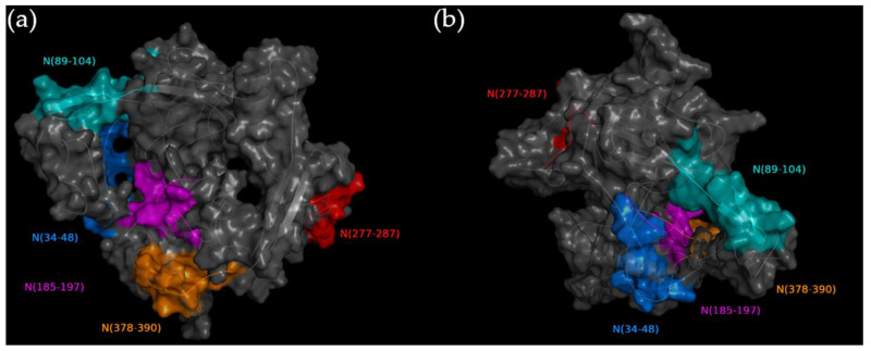
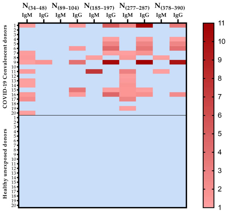
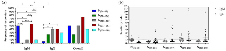
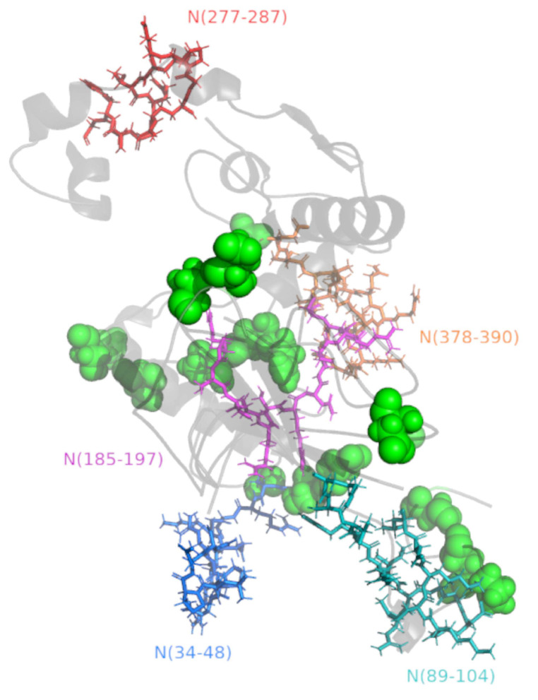
The Nucleocapsid (N) protein is highlighted as the main target for COVID-19 diagnosis by antigen detection due to its abundance in circulation early during infection. However, the effects of the described mutations in the N protein epitopes and the efficacy of antigen testing across SARS-CoV-2 variants remain controversial and poorly understood. Here, we used immunoinformatics to identify five epitopes in the SARS-CoV-2 N protein (N(34-48), N(89-104), N(185-197), N(277-287), and N(378-390)) and validate their reactivity against samples from COVID-19 convalescent patients. All identified epitopes are fully conserved in the main SARS-CoV-2 variants and highly conserved with SARS-CoV. Moreover, the epitopes N(185-197) and N(277-287) are highly conserved with MERS-CoV, while the epitopes N(34-48), N(89-104), N(277-287), and N(378-390) are lowly conserved with common cold coronaviruses (229E, NL63, OC43, HKU1). These data are in accordance with the observed conservation of amino acids recognized by the antibodies 7R98, 7N0R, and 7CR5, which are conserved in the SARS-CoV-2 variants, SARS-CoV and MERS-CoV but lowly conserved in common cold coronaviruses. Therefore, we support the antigen tests as a scalable solution for the population-level diagnosis of SARS-CoV-2, but we highlight the need to verify the cross-reactivity of these tests against the common cold coronaviruses.
Keywords: COVID-19; SARS-CoV-2 variants; antigen test; immunoinformatic.
Conflict of interest statement
The authors declare no conflict of interest and the funders had no role in the design of the study; in the collection, analyses, or interpretation of data; in the writing of the manuscript; or in the decision to publish the results.
Figures
Similar articles
-
Immunoinformatics Identification of the Conserved and Cross-Reactive T-Cell Epitopes of SARS-CoV-2 with Human Common Cold Coronaviruses, SARS-CoV, MERS-CoV and Live Attenuated Vaccines Presented by HLA Alleles of Indonesian Population.Viruses. 2022 Oct 24;14(11):2328. doi: 10.3390/v14112328. Viruses. 2022. PMID: 36366426 Free PMC article.
-
Monoclonal antibodies constructed from COVID-19 convalescent memory B cells exhibit potent binding activity to MERS-CoV spike S2 subunit and other human coronaviruses.Front Immunol. 2022 Dec 22;13:1056272. doi: 10.3389/fimmu.2022.1056272. eCollection 2022. Front Immunol. 2022. PMID: 36618428 Free PMC article.
-
Immune profiling of SARS-CoV-2 epitopes in asymptomatic and symptomatic pediatric and adult patients.J Transl Med. 2023 Feb 14;21(1):123. doi: 10.1186/s12967-023-03963-5. J Transl Med. 2023. PMID: 36788606 Free PMC article.
-
Properties of Coronavirus and SARS-CoV-2.Malays J Pathol. 2020 Apr;42(1):3-11. Malays J Pathol. 2020. PMID: 32342926 Review.
-
Identification of B-Cell Epitopes for Eliciting Neutralizing Antibodies against the SARS-CoV-2 Spike Protein through Bioinformatics and Monoclonal Antibody Targeting.Int J Mol Sci. 2022 Apr 14;23(8):4341. doi: 10.3390/ijms23084341. Int J Mol Sci. 2022. PMID: 35457159 Free PMC article. Review.
Cited by
-
B-Cell Epitopes-Based Chimeric Protein from SARS-CoV-2 N and S Proteins Is Recognized by Specific Antibodies in Serum and Urine Samples from Patients.Viruses. 2023 Sep 5;15(9):1877. doi: 10.3390/v15091877. Viruses. 2023. PMID: 37766284 Free PMC article.
-
The role of cross-reactive immunity to emerging coronaviruses: Implications for novel universal mucosal vaccine design.Saudi Med J. 2023 Oct;44(10):965-972. doi: 10.15537/smj.2023.44.10.20230375. Saudi Med J. 2023. PMID: 37777266 Free PMC article. Review.
-
Epitopes recognition of SARS-CoV-2 nucleocapsid RNA binding domain by human monoclonal antibodies.iScience. 2024 Jan 19;27(2):108976. doi: 10.1016/j.isci.2024.108976. eCollection 2024 Feb 16. iScience. 2024. PMID: 38327783 Free PMC article.
-
Role of SARS‑CoV‑2 nucleocapsid protein in affecting immune cells and insights on its molecular mechanisms.Exp Ther Med. 2023 Sep 12;26(5):504. doi: 10.3892/etm.2023.12203. eCollection 2023 Nov. Exp Ther Med. 2023. PMID: 37822585 Free PMC article.
References
-
- W.H.O . Coronavirus Disease 2019 (COVID-19)-Situation Report 51. W.H.O; Geneva, Switzerland: 2020.
-
- W.H.O WHO Coronavirus (COVID-19) Dashboard. [(accessed on 20 February 2023)]. Available online: https://covid19.who.int/
Publication types
MeSH terms
Substances
Supplementary concepts
LinkOut - more resources
Full Text Sources
Medical
Miscellaneous
