. 2023 Apr 11;15(4):948.
doi: 10.3390/v15040948.

Antiviral Activity of Acetylsalicylic Acid against Bunyamwera Virus in Cell Culture

Affiliations

Antiviral Activity of Acetylsalicylic Acid against Bunyamwera Virus in Cell Culture

Sara Yolanda Fernández-Sánchez et al. Viruses. .

Abstract

The Bunyavirales order is a large group of RNA viruses that includes important pathogens for humans, animals and plants. With high-throughput screening of clinically tested compounds we have looked for potential inhibitors of the endonuclease domain of a bunyavirus RNA polymerase. From a list of fifteen top candidates, five compounds were selected and their antiviral properties studied with Bunyamwera virus (BUNV), a prototypic bunyavirus widely used for studies about the biology of this group of viruses and to test antivirals. Four compounds (silibinin A, myricetin, L-phenylalanine and p-aminohippuric acid) showed no antiviral activity in BUNV-infected Vero cells. On the contrary, acetylsalicylic acid (ASA) efficiently inhibited BUNV infection with a half maximal inhibitory concentration (IC50) of 2.02 mM. In cell culture supernatants, ASA reduced viral titer up to three logarithmic units. A significant dose-dependent reduction of the expression levels of Gc and N viral proteins was also measured. Immunofluorescence and confocal microscopy showed that ASA protects the Golgi complex from the characteristic BUNV-induced fragmentation in Vero cells. Electron microscopy showed that ASA inhibits the assembly of Golgi-associated BUNV spherules that are the replication organelles of bunyaviruses. As a consequence, the assembly of new viral particles is also significantly reduced. Considering its availability and low cost, the potential usability of ASA to treat bunyavirus infections deserves further investigation.

Keywords: Bunyamwera virus; acetylsalicylic acid (ASA); antiviral; bunyavirus; drug repurposing; electron microscopy; high-throughput screening; molecular modeling; viral RNA polymerase; viral replication organelle.


Conflict of interest statement

The authors declare no conflict of interest.

Figures

Figure 1
Figure 1
Molecular modeling and summary of the numeric outcomes. (Left panel): poses adopted by the selected compounds at the endonuclease catalytic site (drugs are represented as colored wireframes and target is sketched in green cartoons). Main residues are indicated by arrows. (Right panel): Computed free energies for the drug–endonuclease complexes (in kcal/mol).
Figure 2
Figure 2
Antiviral activity of ASA against BUNV. Vero cells were absorbed 1 h with BUNV at an MOI of 1 PFU/cell, exposed to increasing concentrations of ASA for 8 h and processed by immunofluorescence. (AD) Fluorescence microscopy of infected cells untreated (A) and treated with 5, 10 and 15 mM ASA (BD). Nuclei are labeled with DAPI (blue) and viral infection with an antibody specific for BUNV nucleoprotein (N) (green). (E) Percentage of infected cells measured by immunofluorescence in the absence of ASA treatment and with three different concentrations of ASA. Data are shown as mean ± s.e.m. of three independent experiments. (F) Dose–response curve (red line) of ASA was determined by nonlinear regression. Cytotoxic effect on Vero cells exposed to increasing concentrations of the drug in the absence of virus is also shown (black line). IC50 and CC50 were determined from dose–response curves based on treatment with seven different concentrations. IC50 was calculated from the percentage of infected cells, and CC50 was determined by the MTT (3-(4,5-dimethylthiazol-2-yl)-2,5-diphenyltetrazolium bromide) cell viability assay. ND, no drug. Scale bars: 50 μm.
Figure 3
Figure 3
Study of ASA antiviral effect by plaque reduction assay. BUNV was propagated in Vero cells in the absence or presence of different concentrations of ASA (5, 10 and 15 mM) for 8 h, the supernatants were collected and the virus titer was tested by plaque assay. (A) A representative picture showing the plaque reduction assay with the drug. (B) BUNV titers from cell supernatants without ASA and with three different concentrations of ASA. Each bar shows the mean value ± s.e.m. Significance was determined by unpaired two-tailed student’s t-test; ns indicates “not significant”, ** p < 0.01 and *** p < 0.001.
Figure 4
Figure 4
Effect of ASA on viral protein expression. Non-infected and BUNV-infected Vero cells at an MOI of 1 PFU/cell for 1 h and incubated in the presence or absence of ASA (5, 10 and 15 mM) for 8 h, were processed for Western blot analysis. An amount of 10 micrograms of the cell lysates were loaded and samples were probed with an antiserum against BUNV. The tubulin signal was used as loading control. NI, non-infected; ND, no drug. One experiment representative of 3 is shown. The tables show the average relative quantity of the Gc protein and N protein ± standard deviation (SD) for all the experiments. The p-values (one- and two-tailed, respectively) were calculated with the student’s t-test.
Figure 5
Figure 5
ASA treatment induces the accumulation of BUNV Gc glycoprotein in the cell perinuclear region. Vero cells were absorbed 1 h with BUNV at an MOI of 1 PFU/cell, incubated in the presence or absence of ASA (5, 10 and 15 mM) for 8 h and processed by immunofluorescence. (AF) Confocal fluorescence microscopy of infected cells untreated and treated with ASA. The nucleus is labeled with DAPI (blue), the BUNV Gc glycoprotein (green) with a specific antibody, and the Golgi complex with the anti-Giantin antibody (red). In untreated cells (A), Gc signal is distributed throughout the cytoplasm, while Gc signal in ASA-treated cells (BD) is retained in a perinuclear region associated with the Golgi complex. (E,F) Three-dimensional reconstructions from a series of confocal images. In the absence of the drug (E), Gc signal is dispersed, while in cells treated with 15 mM ASA (F), Gc signal accumulates near the nucleus in the Golgi complex (white arrows). (G) Average of the number of cells with both N and Gc viral proteins-associated fluorescent signals in the absence and presence of ASA measured by immunofluorescence. This average was determined by counting the number of cells with both N and Gc protein signals. (H) Quantification of the distribution of Gc IF signal shows that ASA induces a retention of Gc signal in the perinuclear region. Quantification was performed by counting the number of cells with Gc protein signal in the cytosol or in the perinuclear region. A total of 50 cells per condition were studied. The results in panels G and H are presented as the mean of the number of cells with viral proteins-associated fluorescence signals ± s.e.m. of three independent experiments. Significance was determined by unpaired two-tailed student’s t-test; ns indicates “not significant”, ** p < 0.01 and *** p < 0.001. ND, no drug. Scale bars: 25 μm in (AD); 10 μm in (E,F).
Figure 6
Figure 6
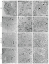
Transmission electron microscopy of BUNV-infected cells untreated or treated with ASA. Vero cells were absorbed 1 h with BUNV at an MOI of 1 PFU/cell, incubated in the presence or absence of ASA (5, 10 and 15 mM) for 8 h and processed by transmission electron microscopy. (AC) BUNV-infected Vero cells not treated with ASA (no drug or ND). (A) Recruitment of cellular organelles to a discrete area of the cytoplasm forming the viral factory (VF). The inset shows extracellular virions. (B) Higher magnification view of the VF shows the Golgi complex (G) surrounded by mitochondria (mi) and endoplasmic reticulum (ER) membranes. Mitochondria have swollen cristae. (C) Bunyavirus replication organelles (spherules and tubules, arrowheads) in Golgi membranes. The inset shows an intracellular virus in the Golgi complex. (DF) BUNV-infected Vero cells treated with ASA 5 mM. The cellular organelles are not recruited to a specific area into the cytoplasm (D). Golgi complex stacks surrounded by endoplasmic reticulum membranes and modified mitochondria (E). The arrowhead in (F) points to a viral spherule in Golgi membranes. (GL) BUNV-infected Vero cells treated with ASA 10 or 15 mM. Low magnification images (G,J) show cellular organelles distributed through the cytoplasm not forming a characteristic VF. Golgi membranes are swollen without bunyavirus spherules and mitochondria have normal morphology and shape (H,K). Details of modified Golgi complex (asterisks) are shown in (I,L). Neither BUNV particles nor spherules were detected. N, nucleus. Scale bars: 2 μm in (A,D,G,J); 500 nm in (B,E,H,K); 200 nm in the inset of (A,C,F,I,L); 100 nm in the inset of (C).
Figure 7
Figure 7
Quantification of spherules, intracellular and extracellular viruses in BUNV-infected cells, with or without ASA using transmission electron microscopy. Vero cells were absorbed 1 h with BUNV at an MOI of 1 PFU/cell, incubated in the presence or absence of ASA (5, 10 and 15 mM) for 8 h and processed by embedding in epoxy resin, ultramicrotomy and electron microscopy. The table shows the absolute and relative frequencies of spherules, intracellular and extracellular viruses counted in BUNV-infected cells in each condition: BUNV ND (infected with no ASA), BUNV + ASA 5 mM, BUNV + ASA 10 mM and BUNV + ASA 15 mM. ASA treatment produces a significant reduction of the number of viral structures. The graphs showing the means of spherules, intracellular and extracellular viruses, counted in a total of 20 cells per condition, are shown near each table, on the right.
Figure 8
Figure 8
Recovery of a productive BUNV infection after elimination of ASA from cell cultures. Vero cells were absorbed 1 h with BUNV at an MOI of 1 PFU/cell and treated with ASA 10 mM for 8 h. The medium was removed and replaced by new DMEM with 2% FBS. After incubation at different time points (2, 4, 6 and 8 h), cells were processed for immunofluorescence, titration by plaque assay and electron microscopy. (A) The percentage of infected cells per condition was determined by fluorescence microscopy using an antibody specific for BUNV N protein. After removing the drug from the cell culture medium, the number of infected cells increased over time. (B) Graph showing the viral titer at different time points. Each bar shows the mean value ± s.e.m. Significance was determined by unpaired two-tailed student’s t-test, where ** indicates p < 0.01. (CH) Electron microscopy study. After 8 h of ASA treatment (C), the Golgi membranes were swollen. Neither BUNV spherules nor viral particles were detected in these infected and ASA-treated cells. (DG) After ASA elimination, BUNV particles (arrows) and spherules (arrowheads) are seen associated with Golgi membranes. (H) High magnification image of a Golgi complex with a viral spherule (arrowhead). N, nucleus; G, Golgi complex. Scale bars: 500 nm in (CG); 200 nm in (H).

Similar articles

Cited by

References

    1. WHO Prioritizing Diseases for Research and Development in Emergency Contexts. [(accessed on 5 April 2023)]. Available online: https://www.who.int/activities/prioritizing-diseases-for-research-and-de....
    1. Buxeraud J., Faure S., Fougere E. Nirmatrelvir/ritonavir (Paxlovid(R)), a treatment for COVID-19. Actual. Pharm. 2022;61:10–12. - PMC - PubMed
    1. Jayk Bernal A., Gomes da Silva M.M., Musungaie D.B., Kovalchuk E., Gonzalez A., Delos Reyes V., Martin-Quiros A., Caraco Y., Williams-Diaz A., Brown M.L., et al. Molnupiravir for Oral Treatment of COVID-19 in Nonhospitalized Patients. N. Engl. J. Med. 2022;386:509–520. doi: 10.1056/NEJMoa2116044. - DOI - PMC - PubMed
    1. Lamontagne F., Agarwal A., Rochwerg B., Siemieniuk R.A., Agoritsas T., Askie L., Lytvyn L., Leo Y.S., Macdonald H., Zeng L., et al. A living WHO guideline on drugs for COVID-19. BMJ. 2020;370:m3379. doi: 10.1136/bmj.m3379. - DOI - PubMed
    1. Schafer A., Martinez D.R., Won J.J., Meganck R.M., Moreira F.R., Brown A.J., Gully K.L., Zweigart M.R., Conrad W.S., May S.R., et al. Therapeutic treatment with an oral prodrug of the remdesivir parental nucleoside is protective against SARS-CoV-2 pathogenesis in mice. Sci. Transl. Med. 2022;14:eabm3410. doi: 10.1126/scitranslmed.abm3410. - DOI - PMC - PubMed

Publication types