. 2023 Apr 14;15(4):968.
doi: 10.3390/v15040968.

Nationwide Laboratory Surveillance of Progressive Multifocal Leukoencephalopathy in Japan: Fiscal Years 2011-2020

Affiliations

Nationwide Laboratory Surveillance of Progressive Multifocal Leukoencephalopathy in Japan: Fiscal Years 2011-2020

Kazuo Nakamichi et al. Viruses. .

Abstract

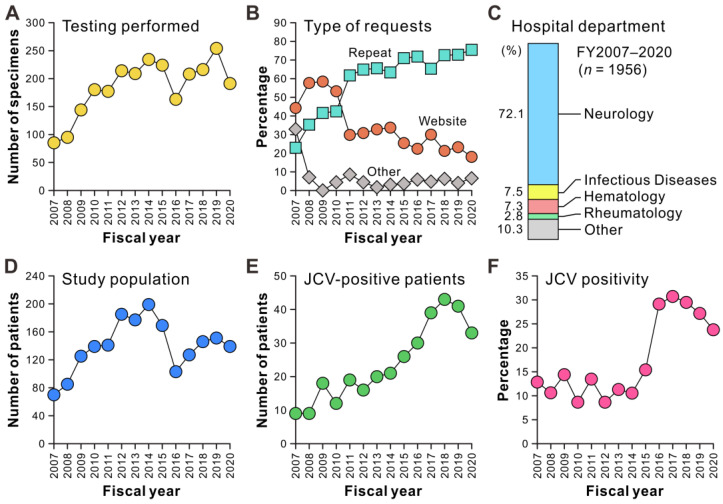
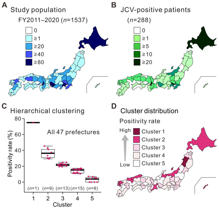
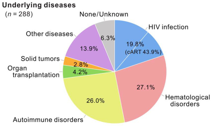
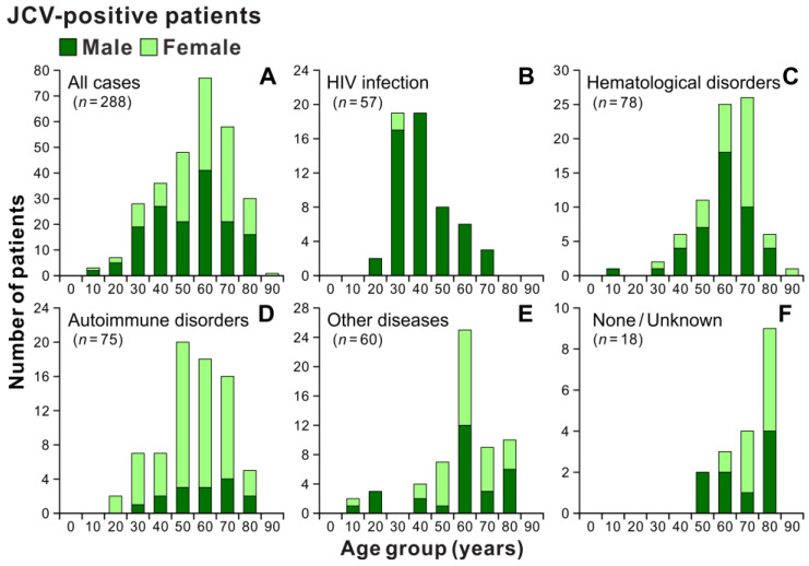
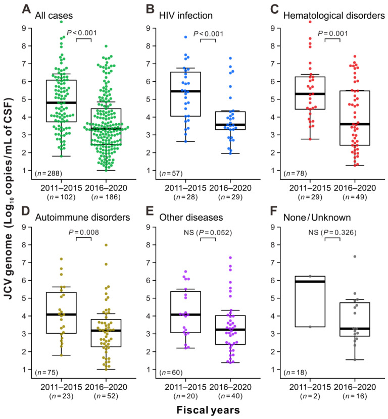
Progressive multifocal leukoencephalopathy (PML) is a devastating demyelinating disease caused by JC virus (JCV), predominantly affecting patients with impaired cellular immunity. PML is a non-reportable disease with a few exceptions, making national surveillance difficult. In Japan, polymerase chain reaction (PCR) testing for JCV in the cerebrospinal fluid (CSF) is performed at the National Institute of Infectious Diseases to support PML diagnosis. To clarify the overall profile of PML in Japan, patient data provided at the time of CSF-JCV testing over 10 years (FY2011-2020) were analyzed. PCR testing for 1537 new suspected PML cases was conducted, and 288 (18.7%) patients tested positive for CSF-JCV. An analysis of the clinical information on all individuals tested revealed characteristics of PML cases, including the geographic distribution, age and sex patterns, and CSF-JCV-positivity rates among the study subjects for each type of underlying condition. During the last five years of the study period, a surveillance system utilizing ultrasensitive PCR testing and widespread clinical attention to PML led to the detection of CSF-JCV in the earlier stages of the disease. The results of this study will provide valuable information not only for PML diagnosis, but also for the treatment of PML-predisposing conditions.

Keywords: JC virus; cerebrospinal fluid; laboratory surveillance; progressive multifocal leukoencephalopathy; real-time PCR testing.


Conflict of interest statement

The authors declare no conflict of interest.

Figures

Figure 1
Figure 1
Track record of laboratory surveillance of PML based on PCR testing for JCV in CSF samples. (A) Number of CSF-JCV testing performed per year during the 4-year start-up period (FY2007–2010) and the study period (FY2011–2020). The result includes follow-up JCV testing after PML diagnosis. (B) Types of requests for CSF-JCV testing from medical departments to the NIID. The requests for CSF-JCV testing for new suspected PML cases in each fiscal year, excluding follow-up testing after PML diagnosis, were classified into three categories: direct contact through the websites, repeat requests from departments that had previously performed the test, and others. (C) Hospital departments of physicians who submitted requests for CSF-JCV testing to the NIID. (D) Number of new patients with suspected PML tested for CSF-JCV in each fiscal year. (E) Number of new patients with JCV-positive CSF in each fiscal year. (F) Proportion of patients with JCV-positive CSF among newly tested individuals in each fiscal year.
Figure 2
Figure 2
Geographic distribution patterns of individuals tested for JCV in the CSF. All individuals tested (A) and patients with JCV-positive CSF (B) during the study period (FY2011–2020) are shown. Each prefecture in Japan is color-coded according to the number of individuals. (C) The hierarchical cluster analysis of the proportion of patients with JCV-positive CSF among individuals in each prefecture. Japanese prefectures were divided into five clusters according to the CSF-JCV-positivity rates. The dots indicate the positivity rates in each prefecture. The thick horizontal line within each box is the median, and the lower and upper boundaries are the 25th and 75th percentiles, respectively. Vertical whiskers extend to minimum and maximum values. (D) Distribution of hierarchical clusters based on CSF-JCV-positivity rates in Japan. Each prefecture is color-coded according to cluster type.
Figure 3
Figure 3
Underlying disease categories of patients with JCV-positive CSF during the study period. The underlying conditions of patients with JCV-positive CSF identified in 10 years (FY2011–2020) are classified into seven categories: HIV infection (n = 57), hematological disorders (n = 78), autoimmune disorders (n = 75), organ transplantation (n = 12), solid tumors (n = 8), other diseases (n = 40), and none/unknown (n = 18). Percentages for each category are shown. cART, combined antiretroviral therapy. All percentages are rounded to two decimal places.
Figure 4
Figure 4
Age and sex distribution patterns of patients with JCV-positive CSF. Patients with JCV-positive CSF during the study period (A) were divided into five groups based on underlying conditions: HIV infection (B), hematologic disorders (C), autoimmune disorders (D), other diseases (E), and none/unknown (F). The “other diseases” group includes patients with a history of organ transplantation, solid tumors, or comorbidities of multiple underlying diseases. The vertical axes show the number of patients, and the numbers below the bars indicate the age groups by decade.
Figure 5
Figure 5
Viral DNA levels in patients with JCV-positive CSF. Patients with JCV-positive CSF during the study period (A) were classified into five groups based on underlying conditions: HIV infection (B), hematologic disorders (C), autoimmune disorders (D), other diseases (E), and none/unknown (F). The copy numbers of the JCV genome in the initial CSF testing that presented positive reactions were indicated using the combinations of beeswarm plots and box-and-whisker plots. In each panel, the data are divided into the first period, when the standard PCR testing was used (FY2011–2015, left), and the second period, when the ultrasensitive PCR testing was implemented (FY2016–2020, right). The vertical axes show the logarithm of the JCV loads measured by quantitative PCR assay. The thick horizontal line within each box is the median, and the lower and upper limits are the 25th and 75th percentiles, respectively. Vertical whiskers extend to 1.5 times the interquartile range of the data. “NS” indicates no significant difference between the two groups.

Similar articles

Cited by

References

    1. Major E.O., Yousry T.A., Clifford D.B. Pathogenesis of Progressive Multifocal Leukoencephalopathy and Risks Associated with Treatments for Multiple Sclerosis: A Decade of Lessons Learned. Lancet Neurol. 2018;17:467–480. doi: 10.1016/S1474-4422(18)30040-1. - DOI - PubMed
    1. Cortese I., Reich D.S., Nath A. Progressive Multifocal Leukoencephalopathy and the Spectrum of JC Virus-Related Disease. Nat. Rev. Neurol. 2021;17:37–51. doi: 10.1038/s41582-020-00427-y. - DOI - PMC - PubMed
    1. Pavlovic D., Patel M.A., Patera A.C., Peterson I. Progressive Multifocal Leukoencephalopathy Consortium. T Cell Deficiencies as a Common Risk Factor for Drug Associated Progressive Multifocal Leukoencephalopathy. Immunobiology. 2018;223:508–517. doi: 10.1016/j.imbio.2018.01.002. - DOI - PubMed
    1. Shishido-Hara Y. Progressive Multifocal Leukoencephalopathy and Promyelocytic Leukemia Nuclear Bodies: A Review of Clinical, Neuropathological, and Virological Aspects of JC Virus-Induced Demyelinating Disease. Acta Neuropathol. 2010;120:403–417. doi: 10.1007/s00401-010-0694-x. - DOI - PMC - PubMed
    1. Walker P.J., Siddell S.G., Lefkowitz E.J., Mushegian A.R., Adriaenssens E.M., Alfenas-Zerbini P., Dempsey D.M., Dutilh B.E., García M.L., Curtis Hendrickson R., et al. Recent Changes to Virus Taxonomy Ratified by the International Committee on Taxonomy of Viruses (2022) Arch. Virol. 2022;167:2429–2440. doi: 10.1007/s00705-022-05516-5. - DOI - PMC - PubMed

Publication types

MeSH terms

LinkOut - more resources