. 2023 Sep;43(9):1475-1489.
doi: 10.1177/0271678X231173187. Epub 2023 Apr 27.

Characteristics of activation of monocyte-derived macrophages versus microglia after mouse experimental intracerebral hemorrhage

Affiliations

Characteristics of activation of monocyte-derived macrophages versus microglia after mouse experimental intracerebral hemorrhage

Fenghui Ye et al. J Cereb Blood Flow Metab. 2023 Sep.

Abstract

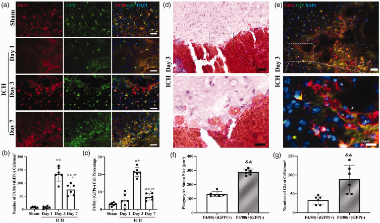
Both monocyte-derived macrophages (MDMs) and brain resident microglia participate in hematoma resolution after intracerebral hemorrhage (ICH). Here, we utilized a transgenic mouse line with enhanced green fluorescent protein (EGFP) labeled microglia (Tmem119-EGFP mice) combined with a F4/80 immunohistochemistry (a pan-macrophage marker) to visualize changes in MDMs and microglia after ICH. A murine model of ICH was used in which autologous blood was stereotactically injected into the right basal ganglia. The autologous blood was co-injected with CD47 blocking antibodies to enhance phagocytosis or clodronate liposomes for phagocyte depletion. In addition, Tmem119-EGFP mice were injected with the blood components peroxiredoxin 2 (Prx2) or thrombin. MDMs entered the brain and formed a peri-hematoma cell layer by day 3 after ICH and giant phagocytes engulfed red blood cells were found. CD47 blocking antibody increased the number of MDMs around and inside the hematoma and extended MDM phagocytic activity to day 7. Both MDMs and microglia could be diminished by clodronate liposomes. Intracerebral injection of Prx2 but not thrombin attracted MDMs into brain parenchyma. In conclusion, MDMs play an important role in phagocytosis after ICH which can be enhanced by CD47 blocking antibody, suggesting the modulation of MDMs after ICH could be a future therapeutic target.

Keywords: Intracerebral hemorrhage; microglia; monocyte-derived macrophages; mouse; peroxiredoxin 2.


Conflict of interest statement

The author(s) declared no potential conflicts of interest with respect to the research, authorship, and/or publication of this article.

Figures

Figure 1.
Figure 1.
Monocyte-derived macrophages (MDMs) infiltrate the brain and form a cell layer after intracerebral hemorrhage (ICH); (a) Representative image of GFP (green fluorescent protein; green) and F4/80 (red) double staining in the perihematomal area in posthemorrhagic Tmem119-EGFP mice from day 1, day 3, day 7 and sham group mice. Sham mice received a needle insertion. Scale bars are 50 µm. (b) Quantification data on the number of perihematomal F4/80(+)GFP(−) cells (MDMs). (c) Quantification data on the percentage of perihematomal F4/80(+)GFP(−) cells. (d) Representative hematoxylin and eosin stained image of giant cells engulfed red blood cells (indicated with *) around hematoma. High magnification images correspond to the hematoma border. Scale bar is 50 µm in the upper panel, 20 µm in the lower panel. (e) Representative image of MDMs gathering around hematoma and forming giant phagocytes. High magnification images correspond to the hematoma border. * indicates F4/80(+)GFP(−) cells. Scale bar is 50 µm in the upper panel, 20 µm in the lower panel. (f) Quantification data on soma size of perihematomal F4/80(+)GFP(+) and F4/80(+)GFP(−) cells and (g) Quantification data on number of giant cells in perihematomal area. **P < 0.001 vs the sham group, ##P < 0.001 vs 3 days. &&P < 0.001 vs the F4/80(+)GFP(+) cells. Values are means ± SD or median (quartile 1, quartile 3); n = 6 per group.
Figure 2.
Figure 2.
CD47 blocking antibody increases the number of monocyte-derived macrophages (MDMs) 3 days after intracerebral hemorrhage (ICH); (a) Representative image of GFP (green fluorescent protein; green) and F4/80 (red) double staining in Tmem119-EGFP mice in ICH+anti-CD47 group and ICH + IgG group 3 days post hemorrhage. Regions of interest were the hematoma core, peri-hematoma, and ipsilateral basal ganglia (BG) areas. (b) Quantification data on the number and percentage of hematoma core F4/80(+)GFP(−) cells. (c) Quantification data on the number and percentage of perihematomal F4/80(+)GFP(−) cells and (d) Quantification data on the number and percentage of ipsilateral BG F4/80(+)GFP(−) cells. *P < 0.05 vs the ICH + IgG group. Scale bars are 50 µm. Values are means ± SD or median (quartile 1, quartile 3); n = 6 per group.
Figure 3.
Figure 3.
CD47 blocking antibody increases the number and extends the duration of phagocytic activity of monocyte-derived macrophages (MDMs) 7 days after intracerebral hemorrhage (ICH); (a) Representative image of GFP (green fluorescent protein; green) and F4/80 (red) double staining in Tmem119-EGFP mice in ICH+anti-CD47 group and ICH + IgG group 7 days post hemorrhage. Regions of interest were the hematoma core, peri-hematoma, and ipsilateral basal ganglia (BG) areas. (b) Quantification data on the number and percentage of hematoma core F4/80(+)GFP(−) cells. (c) Quantification data on the number and percentage of perihematomal F4/80(+)GFP(−) cells and (d) Quantification data on the number and percentage of ipsilateral BG F4/80(+)GFP(−) cells. *P < 0.05 vs the ICH + IgG group. **P < 0.001 vs the ICH + IgG group. Scale bars are 50 µm. Values are means ± SD or median (quartile 1, quartile 3); n = 6 per group.
Figure 4.
Figure 4.
CD47 blocking antibody increases the number of microglia in ipsilateral basal ganglia. (a) Representative native EGFP (enhanced green fluorescent protein; green) fluorescence images of different brain regions from a coronal section of Tmem119-EGFP in ICH+anti-CD47 group and ICH + IgG group 3 days and 7 days post hemorrhage. Regions of interest were hematoma core, peri-hematoma, ipsilateral basal ganglia (BG) areas, and contralateral BG. (b) Quantification data on the number of GFP(+) cells in different brain regions 3 days after hemorrhage and (c) Quantification data on the number of GFP(+) cells in different brain regions 7 days after hemorrhage. *P < 0.05 vs the ICH + IgG group. **P < 0.001 vs the ICH + IgG group. Scale bars are 50 µm. Values are means ± SD or median (quartile 1, quartile 3); n = 6 per group.
Figure 5.
Figure 5.
CD 47 blocking antibody increased the number of giant cells and phagocytosis activity of microglia 3 days after intracerebral hemorrhage (ICH); (a) Representative hematoxylin and eosin stained image of giant cells engulfed red blood cells around the hematoma in ICH + IgG group and ICH+anti-CD47 group. Scale bar is 20 µm. (b) Representative image of GFP (green fluorescent protein; green) and F4/80 (red) double staining. * indicates F4/80(+)GFP(−) giant cells. # indicates F4/80(+)GFP(+) giant cells. Scale bar is 20 µm. (c) Quantification data on number of giant cells in perihematomal area. (d) Quantification data on percentage of F4/80(+)GFP(−) cell in giant cells in perihematomal area and (e) Quantification data on soma size of perihematomal F4/80(+)GFP(+) and F4/80(+)GFP(−) cells. *P < 0.05 vs the ICH + IgG group. Values are means ± SD; n = 6 per group.
Figure 6.
Figure 6.
Clodronate liposomes deplete infiltrating monocyte-derived macrophages (MDMs) and microglia after intracerebral hemorrhage (ICH); (a) Representative image of GFP (green fluorescent protein; green) and F4/80 (red) double staining in Tmem119-EGFP mice in ICH+Clodonate group and ICH+Control group 3 days post hemorrhage. Regions of interest were the hematoma core, peri-hematoma, and ipsilateral basal ganglia (BG) areas. (b) Quantification data on the number of hematoma core F4/80(+)GFP(−) cells and F4/80(+)GFP(+) cells. (c) Quantification data on the number of perihematomal F4/80(+)GFP(−) cells and F4/80(+)GFP(+) cells. (d) Quantification data on the number of ipsilateral BG F4/80(+)GFP(−) cells and F4/80(+)GFP(+) cells. (e) Representative image of DARPP-32 (red) immunoreacitivity in Tmem119-EGFP mice of ICH+Clodonate group and ICH+Control group 3 days post hemorrhage and (f) Quantification data of neuronal loss of ipsilateral BG. *P < 0.05 vs the ICH + IgG group. **P < 0.001 vs the ICH+Control group. Scale bars are 50 μm. Values are means ± SD or median (quartile 1, quartile 3); n = 6 per group.
Figure 7.
Figure 7.
Peroxiredoxin 2 (Prx2) but not thrombin induces the infiltration of monocyte-derived macrophages (MDMs). (a) Representative images of GFP (green fluorescent protein; green) and F4/80 (red) double staining in Tmem119-EGFP mice 1 day after intracerebral Prx2 injection. * indicates F4/80(+)GFP(−) cells. (b) Representative images of GFP (green) and F4/80 (red) double staining in Tmem119-EGFP mice 1 day after fluorescent thrombin injection. (c) Quantification data on the number of F4/80(+)GFP(+) cells and F4/80(+)GFP(−) cells 1 day after Prx2 injection and (d) Quantification data on the number of F4/80(+)GFP(+) cells and F4/80(+)GFP(−) cells 1 day after thrombin injection. **P < 0.001 vs. contralateral side. Dash line indicates the mean value of the sham animal. Values are means ± SD; n = 6 per group. Scale bars are 50 µm in the 2 upper panels, 20 μm in the lower panel.

References

    1. Feigin VL, Lawes CM, Bennett DA, et al.. Worldwide stroke incidence and early case fatality reported in 56 population-based studies: a systematic review. Lancet Neurol 2009; 8: 355–369. - PubMed
    1. Greenberg SM, Ziai WC, Cordonnier C, et al.. 2022 Guideline for the management of patients with spontaneous intracerebral hemorrhage: a guideline from the American Heart Association/American Stroke Association. Stroke 2022; 53: e282–e361. - PubMed
    1. Wilkinson DA, Pandey AS, Thompson BG, et al.. Injury mechanisms in acute intracerebral hemorrhage. Neuropharmacology 2018; 134: 240–248. - PMC - PubMed
    1. Mendelow AD, Gregson BA, Fernandes HM, et al.. Early surgery versus initial conservative treatment in patients with spontaneous supratentorial intracerebral haematomas in the international surgical trial in intracerebral haemorrhage (STICH): a randomised trial. Lancet 2005; 365: 387–397. - PubMed
    1. Mendelow AD, Gregson BA, Rowan EN, et al.. Early surgery versus initial conservative treatment in patients with spontaneous supratentorial lobar intracerebral haematomas (STICH II): a randomised trial. Lancet 2013; 382: 397–408. - PMC - PubMed

Publication types