Case Reports
. 2023 Mar 25;15(3):e36680.
doi: 10.7759/cureus.36680. eCollection 2023 Mar.

Five-Year Sustained Complete Remission With Minimal Adverse Effects Following Radiosurgery for 2-cm Brain Metastasis With Deep Eloquent Location From Lung Adenocarcinoma Despite Low Marginal Dose and High 12 Gy Volume

Affiliations
Case Reports

Five-Year Sustained Complete Remission With Minimal Adverse Effects Following Radiosurgery for 2-cm Brain Metastasis With Deep Eloquent Location From Lung Adenocarcinoma Despite Low Marginal Dose and High 12 Gy Volume

Kazuhiro Ohtakara et al. Cureus. .

Abstract

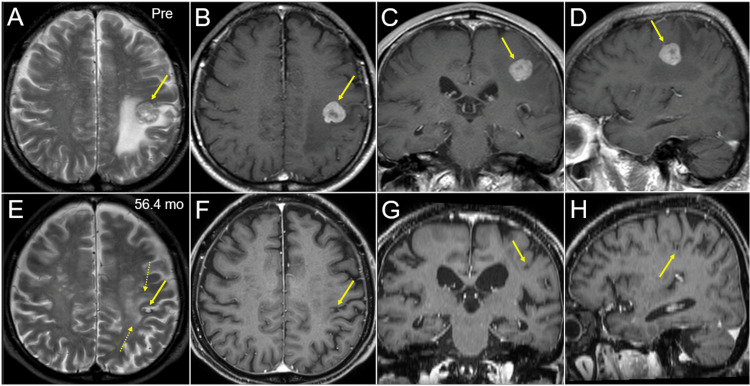
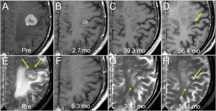
In single-fraction (sf) stereotactic radiosurgery (SRS) for brain metastases (BM) from lung adenocarcinoma (LAC), a marginal dose of ≥22-24 Gy is generally deemed desirable for achieving long-term local tumor control, whereas symptomatic brain radionecrosis significantly increases when the surrounding brain volume receiving ≥12 Gy (V12 Gy) exceeds >5-10 cm3, especially in a deep location. Here, we describe a 75-year-old male with a single LAC-BM of 20 mm in diameter, with a deep eloquent location, which was treated with sfSRS followed by erlotinib, resulting in sustained local complete remission (CR) with minimal adverse radiation effect at nearly five years after sfSRS. The LAC harbored epidermal growth factor receptor (EGFR) mutation. The gross tumor volume (GTV) was defined based on contrast-enhanced computed tomography (CECT) alone. sfSRS was implemented 11 days after planning CECT acquisition. The original GTV had some under- and over-coverage of the enhancing lesion. The D98% values of corrected GTV (cGTV) (3.08 cm3) and 2-mm outside the cGTV were 18.0 Gy with 55% isodose and 14.8 Gy, respectively. The irradiated isodose volumes, including the GTV, receiving ≥22 Gy and ≥12 Gy were 2.18 cm3 and 14.32 cm3, respectively. Erlotinib was administered 13 days after sfSRS with subsequent dose adjustments over 22 months. There was a remarkable tumor response and subsequent nearly CR of the BM were observed at 2.7 and 6.3 months, respectively, with the tumor remnant being visible as a tiny cavitary lesion located in the cortex of the post-central gyrus at 56.4 months. The present case suggests the existence of: (i) extremely radio- and tyrosine kinase inhibitor (TKI)-sensitive LAC-BM for which sfSRS of ≤18 Gy combined with EGFR-TKI is sufficient for attaining long-term CR; and (ii) long-term brain tolerance following sfSRS despite high 12 Gy volume and deep eloquent location in the late 70s The moderate marginal dose of the GTV, the main location of the BM in the cerebral cortex, and the excellent tumor responses with sufficient extrication from the mass effect may render the BM immune to late adverse radiation effect.

Keywords: brain metastasis; brain radionecrosis; dose distribution; dose gradient; lung adenocarcinoma; non-small cell lung cancer; radiotherapy treatment planning; single fraction stereotactic radiosurgery; tyrosine kinase inhibitor; volumetric modulated arc therapy.


Conflict of interest statement

The authors have declared that no competing interests exist.

Figures

Figure 1
Figure 1. Magnetic resonance images before and 4.7 years after stereotactic radiosurgery.
The images show T2-weighted images (WI) (A, E); contrast-enhanced (CE) T1-WI (B-D, F-H); axial images (A, B, E, F); coronal images (C, G); sagittal images (D, H); before stereotactic radiosurgery (SRS) (Pre) (A-D); and at 56.4 months (mo) after SRS (E-H). (A-H) All images were co-registered on MIM MaestroTM software (MIM Software Inc., Beachwood, Ohio, United States) and are shown in the same magnification and coordinates; (A-D) A solid mass lesion (arrows in A-D) in the left frontoparietal lobes is concomitant with surrounding edema almost not extending into the ventral lobe; (E-H) The tumor remnant is observed as a cavitary lesion (arrows in E-H) in the left postcentral gyrus; (E) A slight high-intensity change is seen in the surrounding white matter (dashed arrows in E); (E-H) Progression of brain parenchyma atrophy and relevant ventricular dilatation are seen.
Figure 2
Figure 2. Contrast-enhanced computed tomographic images for stereotactic radiosurgery planning.
The images show CECT images (A-H); axial images (A-D); sagittal images (E-H); original contours of the GTV (B, F); corrected contours of the GTV (C, G); and the superimposed images (D, H). (B-D, F-H) The original GTV contours had some under-coverage (arrows in B, F) and over-coverage (dashed arrows in B, F). CECT: contrast-enhanced computed tomography; GTV: gross tumor volume
Figure 3
Figure 3. Target definition, dose distributions, and dose-volume histograms.
The images show CECT images for SRS planning (A-H); target definition (A, E); dose distributions for the original (B, C, F, G) and alternative plans (D, H); axial images (A-D); sagittal images (arrow in E indicating the ventral side) (E-H); representative isodoses (I); and dose-volume histograms (DVHs) (J-L). (A, E) The original GTV and uniform 2-mm added and reduced objects (GTV + 2 mm, GTV – 2 mm). The contours of margin-added or reduced structures generated on Monaco® (Elekta AB, Stockholm, Sweden) are not smooth but slightly jerky. (B, F) The 20 Gy isodose lines covers 95% of the original GTV (oGTV) with some over-coverage (arrows in B, F). (D, H) The dose gradients just outside and inside the oGTV boundary in the alternative plan (D, H) are steeper and more concentrically laminated than those for the original plan (C, G). (J) The DVHs for the oGTV and oGTV + 2 mm. (K) The DVHs for the corrected GTV (cGTV) and cGTV + 2 mm in addition to the oGTV and oGTV + 2 mm. (L) Comparison of the DVHs for the original and alternative plan. The wall structure is the 8-mm thickness object outside the oGTV + 2 mm boundary. CECT: contrast-enhanced computed tomography; SRS: stereotactic radiosurgery; GTV: gross tumor volume
Figure 4
Figure 4. Magnetic resonance images before and after radiosurgery for brain metastasis.
The images show axial CE-T1-WI (A-D, F); axial T2-WI (E, G, H); 12 days before SRS (Pre) (A, E); at 2.7 months (mo) after SRS (B); at 6.3 months (F); at 39.3 months (C, G); and at 56.4 months (D, H). (B, F) T2-WI at 2.7 and 6.3 months after SRS were not acquired. All images were co-registered and are shown in the same magnification and coordinates. (E) The iso-intensity brain structure (arrows in F), probably the cerebral cortex, is distinguishable from the BM with heterogeneous high intensity. (B) Remarkable tumor shrinkage along with almost disappearance of the peritumoral edema is observed at 2.7 months. (F) Almost complete remission is confirmed at 6.3 months. (C, D, G, H) At 39.3 and 56.4 months, the tumor remnant (arrows in D, H) is visible as a cavitary lesion surrounded by a low-intensity rim on T2-WI with not the whole circumference, just bordering the central sulcus at the ventral side. No abnormal intensity is observed in the surrounding iso-intensity cerebral cortex, although high-intensity changes are observed in the surrounding deep white matter (dashed arrows in G, H). CE: contrast-enhanced; WI: weighted image; SRS: stereotactic radiosurgery; BM: brain metastasis

References

    1. Treatment of brain metastases of non-small cell lung carcinoma. Rybarczyk-Kasiuchnicz A, Ramlau R, Stencel K. Int J Mol Sci. 2021;22:593. - PMC - PubMed
    1. Management of brain metastases in lung cancer: evolving roles for radiation and systemic treatment in the era of targeted and immune therapies. Myall NJ, Yu H, Soltys SG, Wakelee HA, Pollom E. Neurooncol Adv. 2021;3:0–62. - PMC - PubMed
    1. The impact of EGFR-TKI use on clinical outcomes of lung adenocarcinoma patients with brain metastases after Gamma Knife radiosurgery: a propensity score-matched analysis based on extended JLGK0901 dataset (JLGK0901-EGFR-TKI) Yomo S, Serizawa T, Yamamoto M, et al. J Neurooncol. 2019;145:151–157. - PubMed
    1. Estimating survival in patients with lung cancer and brain metastases: an update of the graded prognostic assessment for lung cancer using molecular markers (Lung-molGPA) Sperduto PW, Yang TJ, Beal K, et al. JAMA Oncol. 2017;3:827–831. - PMC - PubMed
    1. Management of brain metastases in tyrosine kinase inhibitor-naïve epidermal growth factor receptor-mutant non-small-cell lung cancer: a retrospective multi-institutional analysis. Magnuson WJ, Lester-Coll NH, Wu AJ, et al. J Clin Oncol. 2017;35:1070–1077. - PubMed

Publication types

LinkOut - more resources