

Concordance in Radiological Parameters of Different Knee Views After Total Knee Arthroplasty
- PMID: 37113460
- PMCID: PMC10129438
- DOI: 10.7759/cureus.38129
Concordance in Radiological Parameters of Different Knee Views After Total Knee Arthroplasty
Abstract
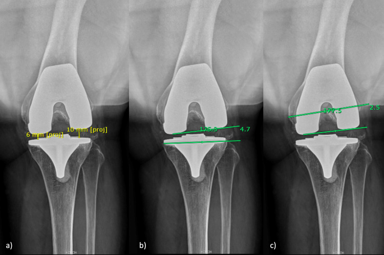
Background Total knee arthroplasty (TKA) is a cost-effective treatment for the end-stage of knee osteoarthritis. Despite the improvements in this surgery, a significant percentage of patients still report dissatisfaction after knee arthroplasty. Radiological results have been used to predict clinical outcomes and satisfaction after knee replacement. This study aims to evaluate the concordance of a set of radiographic views to assess alignment on total knee arthroplasty. Methods A concordance study was designed with 105 patients (130 TKA) that underwent conventional total knee arthroplasty cruciate-retaining design recruited for the study and scheduled for their annual radiograph control. Measurements were performed on the following radiograph after total knee replacement: full-length standing anteroposterior and lateral radiograph, anteroposterior standing, lateral and axial knee view, and the knee "seated view". A musculoskeletal radiologist and a knee surgeon were recruited to perform the radiological measurement and then estimate the interobserver agreement. Results There was an excellent correlation between Limb Length (LL), Hip-knee-ankle angle (HKA), Sagittal mechanical tibial component alignment (smTA), extension lateral and medial joint space (eLJS and eMJS), 90º flexion lateral and medial joint space (fLJS and fMJS) and Sagittal anatomic lateral view tibial component alignment (saLTA); the good correlation between Mechanical lateral femoral component alignment (mLFA), Sagittal anatomic tibial component alignment (saTA), Sagittal anatomic lateral view femoral component alignment 2 (saLFA2), Patella Height (PH); and moderate to poor correlation for the rest of measurements. Conclusion Excellent and good concordance can be achieved for radiographic measurements in different knee views to assess results after TKA. These findings must encourage future studies to address functional and survival outcomes using all knee views and not just one plane.
Keywords: knee replacement; patient; radiograph; satisfaction; total knee arthroplasty.
Copyright © 2023, Barahona et al.
Conflict of interest statement
The authors have declared that no competing interests exist.
Figures


Similar articles
-
Do varus or valgus outliers have higher forces in the medial or lateral compartments than those which are in-range after a kinematically aligned total knee arthroplasty? limb and joint line alignment after kinematically aligned total knee arthroplasty.Bone Joint J. 2017 Oct;99-B(10):1319-1328. doi: 10.1302/0301-620X.99B10.BJJ-2017-0066.R1. Bone Joint J. 2017. PMID: 28963153
-
Using short knee radiographs to predict the coronal alignment after TKA: Is it an accurate proxy for HKA on full-length images?J Orthop Surg Res. 2022 Jul 6;17(1):340. doi: 10.1186/s13018-022-03235-w. J Orthop Surg Res. 2022. PMID: 35794578 Free PMC article.
-
No correlation between coronal alignment of total knee arthroplasty and clinical outcomes: a prospective clinical study using 3D-CT.Knee Surg Sports Traumatol Arthrosc. 2017 Dec;25(12):3892-3900. doi: 10.1007/s00167-016-4400-y. Epub 2016 Dec 22. Knee Surg Sports Traumatol Arthrosc. 2017. PMID: 28005142
-
Comparison of Kinematic Alignment and Mechanical Alignment in Total Knee Arthroplasty: A Meta-analysis of Randomized Controlled Clinical Trials.Orthop Surg. 2020 Dec;12(6):1567-1578. doi: 10.1111/os.12826. Epub 2020 Oct 25. Orthop Surg. 2020. PMID: 33099892 Free PMC article. Review.
-
Component placement accuracy in two generations of handheld robotics-assisted knee arthroplasty.Arch Orthop Trauma Surg. 2021 Dec;141(12):2059-2067. doi: 10.1007/s00402-021-04040-6. Epub 2021 Jul 25. Arch Orthop Trauma Surg. 2021. PMID: 34304279 Review.
References
-
- Knee replacement. Price AJ, Alvand A, Troelsen A, et al. The Lancet. 2018;392:1672–1682. - PubMed
-
- Restoration of pre-operative joint line orientation and alignment does not affect KSS and KOOS 1 year after total knee arthroplasty. D'Amato M, Kosse NM, Wymenga AB. Knee Surg Sports Traumatol Arthrosc. 2021;29:3170–3177. - PubMed
-
- Limb length discrepancy after total knee arthroplasty may contribute to suboptimal functional results. Hinarejos P, Sánchez-Soler J, Leal-Blanquet J, Torres-Claramunt R, Monllau JC. Eur J Orthop Surg Traumatol. 2020;30:1199–1204. - PubMed
LinkOut - more resources
Full Text Sources