Review
. 2023 Apr 10;15(2):199-221.
doi: 10.1007/s12551-023-01059-4. eCollection 2023 Apr.

Raman spectroscopy for viral diagnostics

Affiliations
Review

Raman spectroscopy for viral diagnostics

Jijo Lukose et al. Biophys Rev. .

Abstract

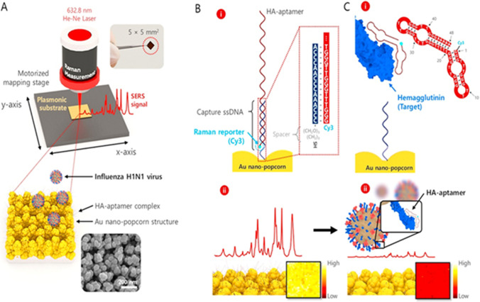
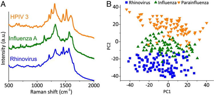
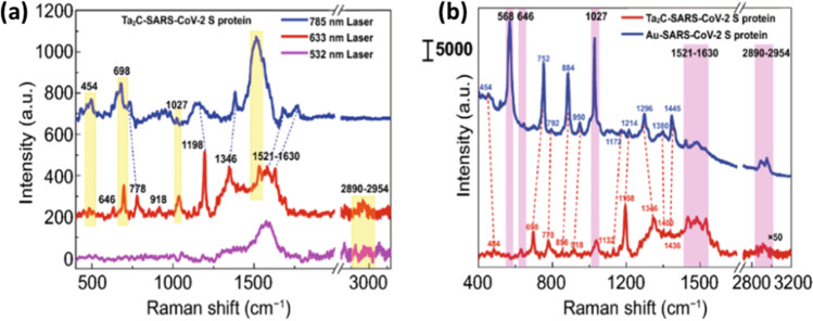
Raman spectroscopy offers the potential for fingerprinting biological molecules at ultra-low concentration and therefore has potential for the detection of viruses. Here we review various Raman techniques employed for the investigation of viruses. Different Raman techniques are discussed including conventional Raman spectroscopy, surface-enhanced Raman spectroscopy, Raman tweezer, tip-enhanced Raman Spectroscopy, and coherent anti-Stokes Raman scattering. Surface-enhanced Raman scattering can play an essential role in viral detection by multiplexing nanotechnology, microfluidics, and machine learning for ensuring spectral reproducibility and efficient workflow in sample processing and detection. The application of these techniques to diagnose the SARS-CoV-2 virus is also reviewed.

Supplementary information: The online version contains supplementary material available at 10.1007/s12551-023-01059-4.

Keywords: Point of care applications; Raman spectroscopy; Raman tweezer; SARS-CoV-2; Surface-enhanced Raman spectroscopy; Tip-enhanced Raman spectroscopy; Virus.


Conflict of interest statement

Conflict of interestThe authors declare no competing interests.

Figures

Fig. 1
Fig. 1
Conceptual representation of scattering process (George 2020)
Fig. 2
Fig. 2
Bacteriophage invasion and corresponding molecular changes probed through Raman tweezer. Reprinted with permission from Pilat et al. (2020)
Fig. 3
Fig. 3
Schematics of identification of individual viruses (H1N1 and coxsackie virus) using TERS multiplexed with CARS. Reprinted from Deckert et al. (2020)
Fig. 4
Fig. 4
Illustration of the signal enhancement process in SERS (adapted from George (2020))
Fig. 5
Fig. 5
Fabrication and working principle ((a)–(h)) of rapid handheld SERS platform for HIV detection (adapted from Yadav et al. (2021))
Fig. 6
Fig. 6
Schematic diagram demonstrating SERS detection of influenza virus (adapted from Chen et al. (2020))
Fig. 7
Fig. 7
SERS spectra of healthy and hepatitis C infected serum samples with different viral load (VL) (adapted from Kashif et al. (2020))
Fig. 8
Fig. 8
A Raman spectra of respiratory virus. B PCA plot of Raman spectral data (adapted from Yeh et al. (2020))
Fig. 9
Fig. 9
IgM/IgG detection using A SERS-LFIA strips and B Au NP-based LFIA strips. C Raman spectra obtained for IgM and IgG lines. D Raman calibration plot obtained for IgM and IgG considering the band at 1328 cm.−1 (adapted from Liu et al. (2021))
Fig. 10
Fig. 10
a Raman spectra of spike protein recorded with the different excitation wavelengths. b S-protein Raman spectra recorded using Ta2C NSs and Au NPS substrates (adapted from Peng et al. (2021))
Fig. 11
Fig. 11
D SERS spectra obtained for various SARS-CoV-2 spike protein concentrations in saliva. E Comparison of Raman spectrum of spike protein in saliva and the blank signal (adapted from Zhang et al. (2021b))
Fig. 12
Fig. 12
Raman spectra of healthy subjects (CTRL), subjects with viral infection (COV +), and subjects with history of viral infection (COV −) (adapted from Carlomagno et al. (2021))
Fig. 13
Fig. 13
Raman detection via collateral cleavage of SERS probe (adapted from Liang et al. (2021))

References

    1. Ali S, Hassan M, Saleem M, Tahir SF. Deep transfer learning based hepatitis B virus diagnosis using spectroscopic images. Int J Imaging Syst Technol. 2021;31:94–105. doi: 10.1002/ima.22462. - DOI
    1. Ambartsumyan O, Gribanyov D, Kukushkin V, Kopylov A, Zavyalova E. SERS-based biosensors for virus determination with oligonucleotides as recognition elements. Int J Mol. 2020;21:3373. doi: 10.3390/ijms21093373. - DOI - PMC - PubMed
    1. Awada C, Abdullah MMB, Traboulsi H, Dab C, Alshoaibi A. SARS-CoV-2 receptor binding domain as a stable-potential target for SARS-CoV-2 detection by surface—enhanced Raman spectroscopy. Sensors. 2021;21:4617. doi: 10.3390/s21134617. - DOI - PMC - PubMed
    1. Bai S, Serien D, Ma Y, Obata K, Sugioka K. Attomolar sensing based on liquid interface-assisted surface-enhanced Raman scattering in microfluidic chip by femtosecond laser processing. ACS Appl Mater Interfaces. 2020;12:42328–42338. doi: 10.1021/acsami.0c11322. - DOI - PubMed
    1. Bankapur A, Barkur S, Chidangil S, Mathur D. A micro-Raman study of live, single red blood cells (RBCs) treated with AgNO3 nanoparticles. PLoS One. 2014;9:e103493. doi: 10.1371/journal.pone.0103493. - DOI - PMC - PubMed

LinkOut - more resources