. 2023 Apr 3:14:1111462.
doi: 10.3389/fphar.2023.1111462. eCollection 2023.

Comprehensive analysis of the cuproptosis-related gene DLD across cancers: A potential prognostic and immunotherapeutic target

Affiliations

Comprehensive analysis of the cuproptosis-related gene DLD across cancers: A potential prognostic and immunotherapeutic target

Weiguang Yang et al. Front Pharmacol. .

Abstract

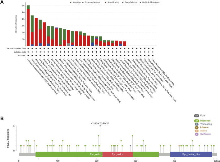
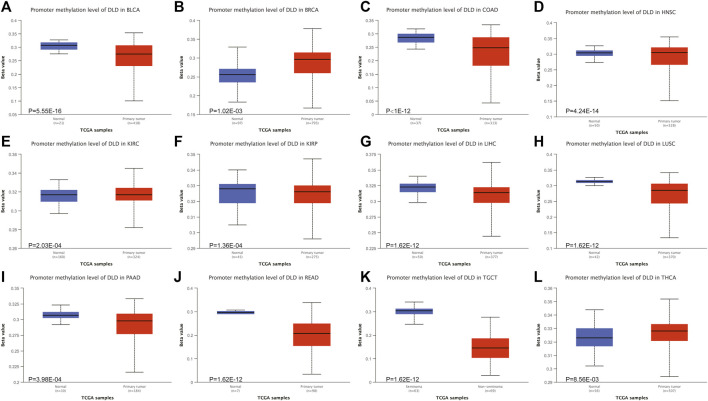
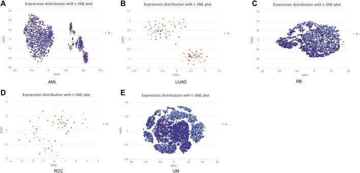
DLD is a key gene involved in "cuproptosis," but its roles in tumor progression and immunity remain unclear. Exploring the potential mechanisms and biological roles of DLD may provide new insights for therapeutic strategies for tumors. In the present study, we analyzed the role of DLD in a variety of tumors by using several bioinformatic tools. The results showed that compared with normal tissues, tumor tissues representing multiple cancers showed significant differential expression of DLD. High DLD expression was associated with a good prognosis in BRCA, KICH, and LUAD. Conversely, high expression levels of DLD were detrimental to patient prognosis in many other tumors, such as COAD, KIRC, and KIRP. In addition, the associations of DLD with infiltrating immune cells, genetic alterations and methylation levels across cancers were assessed. Aberrant expression of DLD was positively correlated with most infiltrating immune cells, especially neutrophils. The DLD methylation level was significantly decreased in COAD, LIHC, and LUSC but significantly increased in BRCA. DLD had the highest mutation rate (6.04%) in ESCA. In LUSC, patients with genetic alterations in DLD showed a poorer prognosis. At the single-cell level, the roles of DLD in regulating cancer-associated biological functions, such as metastasis, inflammation, and differentiation, were explored. Afterward, we further investigated whether several disease-associated genes could be correlated with DLD. GO enrichment analysis indicated that DLD-related genes were mainly associated with mitochondria-related cellular components, aerobic respiration and the tricarboxylic acid cycle. Finally, the correlations between DLD expression and immunomodulatory genes, immune checkpoints, and sensitivity to some antitumor drugs were investigated. It is worth noting that DLD expression was positively correlated with immune checkpoint genes and immunomodulatory genes in most cancers. In conclusion, this study comprehensively analyzed the differential expression, prognostic value and immune cell infiltration-related function of DLD across cancers. Our results suggest that DLD has great potential to serve as a candidate marker for pancancer prognosis and immunotherapy and may provide a new direction for cancer treatment development.

Keywords: bioinformatics analysis; cuproptosis; dld; immune cell infiltration; pan-cancer; prognosis.


Conflict of interest statement

The authors declare that the research was conducted in the absence of any commercial or financial relationships that could be construed as a potential conflict of interest.

Figures

FIGURE 1
FIGURE 1
Flow chart for pancancer analysis of DLD expression.
FIGURE 2
FIGURE 2
Differential expression of DLD across cancers. (A) mRNA level of DLD determined with the TIMER2.0 database. ***p < 0.001; **p < 0.01; *p < 0.05. (B) mRNA level of DLD determined with the GEPIA2.0 database. (C) Protein level of DLD determined with the CPTAC database. (D) Violin plots showing the relationship between the DLD expression level and pathological stage.
FIGURE 3
FIGURE 3
Differential expression of DLD between tumor tissues and corresponding normal tissues from the HPA database. (A) BLAC. (B) LIHC. (C) COAD. (D) BRCA. (E) RCC. (F) LUSC. (G) THCA.
FIGURE 4
FIGURE 4
Survival analysis of DLD in pancancer data from the GEPIA2.0 database. (A) The impact of DLD expression on overall survival. (B) The impact of DLD expression on disease-free survival.
FIGURE 5
FIGURE 5
Genetic alterations of DLD in pancancer data from the cBioPortal tool. (A) The mutational status of DLD in each type of cancer. (B) The mutated site and main mutation types of DLD.
FIGURE 6
FIGURE 6
Survival analysis of DLD alterations in cancers. The correlation between DLD mutation and prognostic value is shown in (A, B) and was determined using cBioPortal (DSS, DFS, PFS, and OS).
FIGURE 7
FIGURE 7
The DNA methylation levels of DLD in tumors. (A–L) The UALCAN database was used to compare the methylation of DLD between primary tumor samples and normal samples.
FIGURE 8
FIGURE 8
Correlations between the expression of DLD and immune cells in data from the TIMER2.0 database. The heatmap depicts the relationships between DLD and the infiltration of B cells (A), CD4+ T cells (B), CD8+ T cells (C), cancer-associated fibroblasts (D), monocytes (E), mast cells (F), myeloid dendritic cells (G) and neutrophils (H).
FIGURE 9
FIGURE 9
The correlations of 14 tumor biological functions with DLD.
FIGURE 10
FIGURE 10
The relationship between DLD and tumor biological function in AML (A), UM (B), RCC (C), RB (D), and LUAD (E). ***p < 0.001; **p < 0.01.
FIGURE 11
FIGURE 11
The distribution of DLD expression at the single-cell level in the above five tumors is demonstrated in t-SNE plots. (A) AML. (B) LUAD. (C) RB. (D) RCC. (E) UM.
FIGURE 12
FIGURE 12
DLD-related gene enrichment analysis. (A) The DLD-interacting molecular network generated with the STRING tool. (B) Correlation scatterplot of the top 6 genes associated with DLD obtained with the GEPIA2.0 tool. (C) GO enrichment analysis of DLD-related genes. (D) KEGG enrichment analysis of DLD-related genes.
FIGURE 13
FIGURE 13
Correlation of DLD expression with immune checkpoint genes or immunoregulatory genes. (A) Immune checkpoint genes. (B) Immunoregulatory genes. *p < 0.05.
FIGURE 14
FIGURE 14
Drug sensitivity analysis. (A) 3D structure of the screened drugs. (B) Relationships between the DLD gene and sensitivity to antitumor drugs.
FIGURE 15
FIGURE 15
(A) Examination of DLD expression in each grade of glioma tissue by IHC analysis. (B) Analysis of DLD expression in each grade of glioma tissue by Student’s t-test. They include the percentage of positive area and The mean optical density values. ***p < 0.001; **p < 0.01; *p < 0.05.

References

    1. Ambrus A., Wang J., Mizsei R., Zambo Z., Torocsik B., Jordan F., et al. (2016). Structural alterations induced by ten disease-causing mutations of human dihydrolipoamide dehydrogenase analyzed by hydrogen/deuterium-exchange mass spectrometry: Implications for the structural basis of E3 deficiency. Biochim. Biophys. Acta 1862 (11), 2098–2109. 10.1016/j.bbadis.2016.08.013 - DOI - PMC - PubMed
    1. Atlante S., Visintin A., Marini E., Savoia M., Dianzani C., Giorgis M., et al. (2018). α-Ketoglutarate dehydrogenase inhibition counteracts breast cancer-associated lung metastasis. Cell Death Dis. 9 (7), 756. 10.1038/s41419-018-0802-8 - DOI - PMC - PubMed
    1. Bray F., Ferlay J., Soerjomataram I., Siegel R. L., Torre L. A., Jemal A. (2018). Global cancer statistics 2018: GLOBOCAN estimates of incidence and mortality worldwide for 36 cancers in 185 countries. CA Cancer J. Clin. 68 (6), 394–424. 10.3322/caac.21492 - DOI - PubMed
    1. Chandrashekar D. S., Karthikeyan S. K., Korla P. K., Patel H., Shovon A. R., Athar M., et al. (2022). Ualcan: An update to the integrated cancer data analysis platform. Neoplasia 25, 18–27. 10.1016/j.neo.2022.01.001 - DOI - PMC - PubMed
    1. Chen F., Chandrashekar D. S., Varambally S., Creighton C. J. (2019). Pan-cancer molecular subtypes revealed by mass-spectrometry-based proteomic characterization of more than 500 human cancers. Nat. Commun. 10 (1), 5679. 10.1038/s41467-019-13528-0 - DOI - PMC - PubMed