

The severity of corneal nerve loss differentiates motor subtypes in patients with Parkinson's disease
- PMID: 37114067
- PMCID: PMC10126700
- DOI: 10.1177/17562864231165561
The severity of corneal nerve loss differentiates motor subtypes in patients with Parkinson's disease
Abstract
Background: Parkinson's disease (PD) is a heterogeneous movement disorder with patients manifesting with either tremor-dominant (TD) or postural instability and gait disturbance (PIGD) motor subtypes. Small nerve fiber damage occurs in patients with PD and may predict motor progression, but it is not known whether it differs between patients with different motor subtypes.
Objective: The aim of this study was to explore whether there was an association between the extent of corneal nerve loss and different motor subtypes.
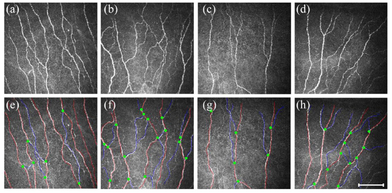
Methods: Patients with PD classified as TD, PIGD, or mixed subtype underwent detailed clinical and neurological evaluation and corneal confocal microscopy (CCM). Corneal nerve fiber density (CNFD), corneal nerve branch density (CNBD), and corneal nerve fiber length (CNFL) were compared between groups, and the association between corneal nerve fiber loss and motor subtypes was investigated.
Results: Of the 73 patients studied, 29 (40%) had TD, 34 (46%) had PIGD, and 10 (14%) had a mixed subtype. CNFD (no./mm2, 24.09 ± 4.58 versus 28.66 ± 4.27; p < 0.001), CNBD (no./mm2, 28.22 ± 11.11 versus 37.37 ± 12.76; p = 0.015), and CNFL (mm/mm2, 13.11 ± 2.79 versus 16.17 ± 2.37; p < 0.001) were significantly lower in the PIGD group compared with the TD group. Multivariate logistic regression showed that higher CNFD (OR = 1.265, p = 0.019) and CNFL (OR = 1.7060, p = 0.003) were significantly associated with the TD motor subtype. The receiver operating characteristic (ROC) analysis demonstrated that combined corneal nerve metrics showed excellent discrimination between TD and PIGD, with an area under the curve (AUC) of 0.832.
Conclusion: Greater corneal nerve loss occurs in patients with PIGD compared with TD, and patients with a higher CNFD or CNFL were more likely to have the TD subtype. CCM may have clinical utility in differentiating different motor subtypes in PD.
Keywords: Parkinson’s disease; corneal nerve fiber; motor subtypes; postural instability and gait disturbance; small nerve fiber; tremor-dominant.
© The Author(s), 2023.
Conflict of interest statement
The authors declared no potential conflicts of interest with respect to the research, authorship, and/or publication of this article.
Figures


References
-
- Nutt JG.Motor subtype in Parkinson’s disease: different disorders or different stages of disease? Mov Disord 2016; 31: 957–961. - PubMed
-
- Zhang YH, Tang BS, Song CY, et al.. The relationship between the phenotype of Parkinson’s disease and levodopa-induced dyskinesia. Neurosci Lett 2013; 556: 109–112. - PubMed
-
- van der Heeden JF, Marinus J, Martinez-Martin P, et al.. Postural instability and gait are associated with severity and prognosis of Parkinson disease. Neurology 2016; 86: 2243–2250. - PubMed
LinkOut - more resources
Full Text Sources
