. 2023 Jun;49(6):120.
doi: 10.3892/or.2023.8557. Epub 2023 Apr 28.

VNN1 overexpression in pancreatic cancer cells inhibits paraneoplastic islet function by increasing oxidative stress and inducing β‑cell dedifferentiation

Affiliations

VNN1 overexpression in pancreatic cancer cells inhibits paraneoplastic islet function by increasing oxidative stress and inducing β‑cell dedifferentiation

Wenjie Qin et al. Oncol Rep. 2023 Jun.

Abstract

Vanin‑1 (VNN1) may be a potential biomarker for the early screening of pancreatic cancer (PC)‑associated diabetes (PCAD). A previous study by the authors reported that cysteamine secreted by VNN1‑overexpressing PC cells induced the dysfunction of paraneoplastic insulinoma cell lines by increasing oxidative stress. In the present study, it was observed that both cysteamine and exosomes (Exos) secreted by VNN1‑overexpressing PC cells aggravated the dysfunction of mouse primary islets. PC‑derived VNN1 could be transported into islets through PC cell‑derived Exos (PC‑Exos). However, β‑cell dedifferentiation, and not cysteamine‑mediated oxidative stress, was responsible for the islet dysfunction induced by VNN1‑containing Exos. VNN1 inhibited the phosphorylation of AMPK and GAPDH, and prevented Sirt1 activation and FoxO1 deacetylation in islets, which may be responsible for the induction of β‑cell dedifferentiation induced by VNN1‑overexpressing PC‑Exos. Furthermore, it was demonstrated that VNN1‑overexpressing PC cells further impaired the functions of paraneoplastic islets in vivo using diabetic mice with islets transplanted under the kidney capsule. On the whole, the present study demonstrates that PC cells overexpressing VNN1 exacerbate the dysfunction of paraneoplastic islets by inducing oxidative stress and β‑cell dedifferentiation.

Keywords: VNN1; exosomes; oxidative stress; pancreatic cancer‑associated diabetes; β‑cell dedifferentiation.


Conflict of interest statement

The authors declare that they have no competing interests.

Figures

Figure 1.
Figure 1.
VNN1-overexpressing PC cells inhibit the viability and function of islets. (A) PANC-1 and CFPAC-1 cells were transfected with empty vector or VNN1 vector, and VNN1 expression in cell lysates was examined using western blot analysis. β-actin was used as the loading control. (B) Primary islets from B6 mice were cultured in vitro. (C) Co-culture system of islets with PC cells was constructed using Transwell chambers. (D) Following co-culture with PC cells for 24 h, islets were dissociated into single cells and cell viability was determined using Trypan blue staining. (E) Following co-culture with PC cells, cleaved caspase-3/9 expression levels in islets were examined using western blot analysis. β-actin was used as the loading control. (F) Following co-culture with PC cells, the insulin secretion of islets was determined using radioimmunoassay. Data are presented as the mean ± SD (n=3). Data were analyzed using one-way ANOVA followed by Tukey's post-hoc test. *P<0.05 and **P<0.01 compared with the PANC-1, CFPAC-1 or control group. #P<0.05 compared with the PANC-1, PE, CFPAC-1 or CE groups. VNN1, Vanin-1; PC, pancreatic cancer; PV, PANC-1 cells with the stable overexpression of VNN1; PE, PANC-1 cells transfected with empty vector; CV, CFPAC-1 cells with the stable overexpression of VNN1; CE, CFPAC-1 cells transfected with empty vector.
Figure 2.
Figure 2.
The viability and function of paraneoplastic islets are improved after suppressing VNN1-induced oxidative stress by GSH or TZD. (A and B) The extracellular and intracellular cysteamine concentrations were detected using high-performance liquid chromatography. (C) ROS contents in islets were analyzed using flow cytometry. (D) GSH concentrations in islets were detected using spectrophotometry. (E) PPARγ expression in islets was examined using western blot analysis. β-actin was used as the loading control. (F) Following islet pre-treatment with GSH or TZD, islet viability was determined using Trypan blue staining. (G) Following islet pre-treatment with GSH or TZD, cleaved caspase-3/9 levels in islets were examined using western blot analysis. β-actin was used as the loading control. (H) Following islet pre-treatment with GSH or TZD, the insulin secretion was determined using radioimmunoassay. Data are presented as the mean ± SD (n=3). Data were analyzed using one-way ANOVA followed by Tukey's post-hoc test. *P<0.05 compared with the PANC-1, CFPAC-1, PV or control group. ROS, reactive oxygen species; PPARγ, peroxisome proliferator activated receptor gamma; GSH, glutathione; TZD, thiazolidinedione; PV, PANC-1 cells with the stable overexpression of VNN1; PE, PANC-1 cells transfected with empty vector; CV, CFPAC-1 cells with the stable overexpression of VNN1; CE, CFPAC-1 cells transfected with empty vector; VNN1, Vanin-1.
Figure 3.
Figure 3.
Islet dysfunction was exacerbated by Exos extracted from VNN1-overexpressing PC cells. (A and B) Cysteamine contents in conditioned media were determined using high-performance liquid chromatography. After the islets wereco-cultured with different conditioned media, (C) the cysteamine contents in islets were detected using high-performance liquid chromatography and (D) insulin secretion was determined using radioimmunoassay. (E) Transmission microcopy images of PC- Exos. (F) Size distributions of PC-Exos. (G) Exo markers (TSG101 and Alix) and negative marker (C-myc) in PC-Exos were determined using western blot analysis. TSG101 and Alix were used as the positive loading controls, and C-myc was used as the negative loading control. (H) Following treatment with different PC-Exos, the insulin secretion of islets was determined using radioimmunoassay. (I) Following treatment with different PC-Exos, the insulin content in islets was also determined using radioimmunoassay. Data are presented as the mean ± SD (n=3). Data were analyzed using one-way ANOVA followed by Tukey's post-hoc test. *P<0.05 compared with the PANC-1, PE + CM, PANC1-Exos or control group. #P<0.05 compared with the PE + CM group. $P<0.05 compared with the PE + cysteamine group. &P<0.05 compared with the PANC1-Exos or PE-Exos group. CM, complete medium; Exos, exosomes; VNN1, Vanin-1; PC, pancreatic cancer; PV, PANC-1 cells with the stable overexpression of VNN1; TSG101, tumor susceptibility gene 101; PE, PANC-1 cells transfected with empty vector.
Figure 4.
Figure 4.
Exos derived from VNN1-overexpressing PC cells induce β-cell dedifferentiation. (A) VNN1 in PC-Exos was determined using western blot analysis. As an Exo marker, Alix was used as the loading control. (B) Following treatment with PC-Exos, VNN1 expression in islets was determined using western blot analysis. β-actin was used as the loading control. Following co-culture with PC-Exos, (C) VNN1 expression in islets was analyzed using immunohistochemistry, and (D) cysteamine in islets was determined using high-performance liquid chromatography. (E) Following incubation with various concentrations of PV-Exos, cysteamine in islets was determined using high-performance liquid chromatography. (F) Following co-culture with PC-Exos, β-cell differentiation markers (Pdx1, Mafa and NeuroD1) in islets were examined using western blot analysis. β-actin was used as the loading control. Data are presented as the mean ± SD (n=3). Data were analyzed using one-way ANOVA followed by Tukey's post-hoc test. *P<0.05 compared with the PANC1-Exos group. VNN1, Vanin-1; Exos, exosomes; Pdx1, pancreatic and duodenal homeobox 1; Mafa, MAF BZIP transcription factor A; NeuroD1, neurogenic differentiation 1; PC, pancreatic cancer; PV, PANC-1 cells with the stable overexpression of VNN1; PE, PANC-1 cells transfected with empty vector.
Figure 5.
Figure 5.
VNN1 in PC-Exos inhibits the AMPK/GAPDH/Sirt1/FoxO1 signaling pathway in islets. (A, left panel) p-AMPK levels in PC cells were determined using western blot analysis. β-actin was used as the loading control. (A, right panel) Following treatment with PC-Exos, p-AMPK levels in islets were examined using western blot analysis. β-actin was used as the loading control. (B) Following treatment with PC-Exos, nuclear-localized GAPDH in islets were examined using western blot analysis. Tubulin and Lamin B were used as the loading controls. (C) Following co-culture with PC-Exos, the interaction of GAPDH or FoxO1 with Sirt1 in islets was detected using co-immunoprecipitation. GAPDH, FoxO1 and Sirt1 were used as the loading controls. (D) Following co-culture with PC-Exos, acetylated-lysine of FoxO1 in islets was detected using co-immunoprecipitation. FoxO1 was used as the loading control. Data are presented as the mean ± SD (n=3). Data were analyzed using one-way ANOVA followed by Tukey's post-hoc test. *P<0.05 compared with the PANC-1 or PANC1-Exos group. p-, phosphorylated; Exos, exosomes; VNN1, Vanin-1; AMPK, AMP-activated protein kinase; PC, pancreatic cancer; PV, PANC-1 cells with the stable overexpression of VNN1; PE, PANC-1 cells transfected with empty vector.
Figure 6.
Figure 6.
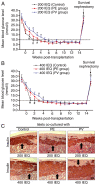
Secretions of VNN1-overexpressing PC cells aggravate islet dysfunction in vivo. (A and B) Average blood glucose levels of B6 diabetic mice before and after transplantation with 200 or 400 IEQ islets co-cultured or not with PC cells under the kidney capsule (5 mice in each group). (C) Immunohistochemical analysis of insulin in islets under the kidney capsule (indicated with black arrows). Data are presented as the mean ± SD (n=5). Data were analyzed using one-way ANOVA followed by Tukey's post-hoc test. *P<0.05, PV group compared with the PE group; #P<0.05, untreated group compared with the PE or PV group; $P<0.05 compared with untreated, PE or PV group at 14th week. IEQ, islet equivalent; VNN1, Vanin-1; PV, PANC-1 cells with the stable overexpression of VNN1; PE, PANC-1 cells transfected with empty vector.
Figure 7.
Figure 7.
Schematic diagram illustrating the mechanisms through which VNN1 in PC cells induces β-cell dedifferentiation by inhibiting the AMPK/GAPDH/Sirt1/FoxO1 signaling pathway. (A) PC cell membrane acquires VNN1 by endocytosis and forms a small vesicle. (B) The multivesicular body fuses with the PC cell membrane and releases VNN1-containing Exos. (C) VNN1 is transferred into β-cells by PC-Exos and inhibits the phosphorylation of AMPK. (D) The phosphorylation of cytoplasmic GAPDH is inhibited by inactivated AMPK. (E) GAPDH is prevented from redistributing into the nucleus and interacting with Sirt1; hence, Sirt1 activity is inhibited. (F) Sirt1 is prevented from binding with FoxO1. (G) FoxO1 cannot be deacetylated. (H) β-cell dedifferentiation is initiated thereof. PC, pancreatic cancer; VNN1, Vanin-1; AMPK, AMP-activated protein kinase; FoxO1, Forkhead box protein O1; Sirt1, sirtuin 1.

Similar articles

Cited by

References

    1. Cai J, Chen H, Lu M, Zhang Y, Lu B, You L, Zhang T, Dai M, Zhao Y. Advances in the epidemiology of pancreatic cancer: Trends, risk factors, screening, and prognosis. Cancer Lett. 2021;520:1–11. doi: 10.1016/j.canlet.2021.06.027. - DOI - PubMed
    1. Sinha V, Shinde S, Saxena S, Thakur S, Walia T, Dixit V, Tiwari AK, Vishvakarma NK, Dwivedi M, Shukla D. A comprehensive review of diagnostic and therapeutic strategies for the management of pancreatic cancer. Crit Rev Oncog. 2020;25:381–404. doi: 10.1615/CritRevOncog.2020035971. - DOI - PubMed
    1. Pannala R, Basu A, Petersen GM, Chari ST. New-onset diabetes: A potential clue to the early diagnosis of pancreatic cancer. Lancet Oncol. 2009;10:88–95. doi: 10.1016/S1470-2045(08)70337-1. - DOI - PMC - PubMed
    1. Pelaez-Luna M, Takahashi N, Fletcher JG, Chari ST. Resectability of presymptomatic pancreatic cancer and its relationship to onset of diabetes: A retrospective review of CT scans and fasting glucose values prior to diagnosis. Am J Gastroenterol. 2007;102:2157–2163. doi: 10.1111/j.1572-0241.2007.01480.x. - DOI - PubMed
    1. Boursi B, Finkelman B, Giantonio BJ, Haynes K, Rustgi AK, Rhim AD, Mamtani R, Yang YX. A clinical prediction model to assess risk for pancreatic cancer among patients with new-onset diabetes. Gastroenterology. 2017;152:840–850.e3. doi: 10.1016/S0016-5085(17)31215-5. - DOI - PMC - PubMed