. 2023 Jul;601(13):2621-2634.
doi: 10.1113/JP284195. Epub 2023 May 11.

Basal NAD(H) redox state permits hydrogen peroxide-induced mesenteric artery dilatation

Affiliations

Basal NAD(H) redox state permits hydrogen peroxide-induced mesenteric artery dilatation

Sean M Raph et al. J Physiol. 2023 Jul.

Abstract

Smooth muscle voltage-gated K+ (Kv) channels in resistance arteries control vascular tone and contribute to the coupling of blood flow with local metabolic activity. Members of the Kv1 family are expressed in vascular smooth muscle and are modulated upon physiological elevation of local metabolites, including the glycolytic end-product l-lactate and superoxide-derived hydrogen peroxide (H2 O2 ). Here, we show that l-lactate elicits vasodilatation of small-diameter mesenteric arteries in a mechanism that requires lactate dehydrogenase (LDH). Using the inside-out configuration of the patch clamp technique, we show that increases in NADH that reflect LDH-mediated conversion of l-lactate to pyruvate directly stimulate the activity of single Kv1 channels and significantly enhance the sensitivity of Kv1 activity to H2 O2 . Consistent with these findings, H2 O2 -evoked vasodilatation was significantly greater in the presence of 10 mM l-lactate relative to lactate-free conditions, yet was abolished in the presence of 10 mM pyruvate, which shifts the LDH reaction towards the generation of NAD+ . Moreover, the enhancement of H2 O2 -induced vasodilatation was abolished in arteries from double transgenic mice with selective overexpression of the intracellular Kvβ1.1 subunit in smooth muscle cells. Together, our results indicate that the Kvβ complex of native vascular Kv1 channels serves as a nodal effector for multiple redox signals to precisely control channel activity and vascular tone in the face of dynamic tissue-derived metabolic cues. KEY POINTS: Vasodilatation of mesenteric arteries by elevated external l-lactate requires its conversion by lactate dehydrogenase. Application of either NADH or H2 O2 potentiates single Kv channel currents in excised membrane patches from mesenteric artery smooth muscle cells. The binding of NADH enhances the stimulatory effects of H2 O2 on single Kv channel activity. The vasodilatory response to H2 O2 is differentially modified upon elevation of external l-lactate or pyruvate. The presence of l-lactate enhances the vasodilatory response to H2 O2 via the Kvβ subunit complex in smooth muscle.

Keywords: calcium signalling; endothelium; metabolism; smooth muscle; voltage-gated potassium channels.


Conflict of interest statement

Competing interests: The authors declare no competing interests.

Figures

Figure 1.
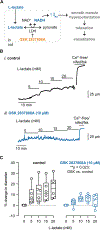
Figure 1.. Vasodilation in response to L-lactate requires LDH activity.
(A) Proposed model of redox-dependent vasoregulation upon elevation of external L-lactate. Internalization and elevation of cytosolic L-lactate results in LDH-mediated interconversion of lactate to pyruvate, elevation of [NADH]i, and Kvβ-dependent smooth muscle hyperpolarization and relaxation. (B) Representative arterial diameter recordings obtained in pressurized (80 mmHg) and preconstricted (100 nM U46619) mesenteric arteries before and after application of L-lactate (5–20 mM) in the absence (i.) and presence (ii.) of the LDH inhibitor GSK 2837808A (10 μM). Maximum passive diameters in Ca2+-free perfusate containing nifedipine (1 μM) and forskolin (0.5 μM) at the end of the experiments were 203 μm (i.) and 148 μm (ii.). (C) Symbol plots showing summarized percent change in diameter from baseline (100 nM U46619) in response to L-lactate (5 – 20 mM) in the absence (left; control) and presence (right) of GSK 2837808A. n = 6 arteries from 6 mice for each, **p = 0.023, normalized L-lactate-induced change in diameter (%) in GSK-treated arteries vs. control (– GSK), two-way ANOVA.
Figure 2.
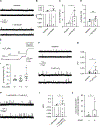
Figure 2.. Smooth muscle Kv1 channel activity potentiation is enhanced in the presence of NADH.
(A) Exemplary inside-out patch clamp recordings of single Kv channel activity obtained in excised membrane patches of isolated mesenteric artery smooth muscle cells before (baseline) and after application of 1 mM NADH in the bath solution. Dashed lines represent open state. (Β-D) Summarized open probabilities (nPo), relative nPo (normalized to baseline nPo), and open state dwell times recorded before and after application of 1 mM NADH. n = 7 cells from 6 mice for each; B, *p = 0.047, Wilcoxon matched-pairs signed rank test; C, *p = 0.031, Wilcoxon signed rank test; D, ns: p = 0.399, Paired t test. (E) Representative diameter recording (i.) obtained in pressurized and preconstricted (80 mmHg, 100 nM U46619) mesenteric artery before and after application of H2O2 in the perfusate (0.1 – 10 μM). The maximum passive diameter (155 μm) was recorded at the end of the experiment in Ca2+-free perfusate containing 1 μΜ nifedipine (nifed) and 0.5 μM forskolin (fsk); and, summary of % change in diameter from baseline in the presence of 0.1, 1, and 10 μM H2O2. n = 8 arteries from 5 mice. *p = 0.011, (%). (F) Exemplary inside-out patch clamp recordings of single Kv channel activity obtained in excised membrane patches of isolated mesenteric artery smooth muscle cells before (baseline) and after application of 10 μM H2O2 in the bath solution. (G) Summarized nPo recorded before and after application of 10 μM H2O2. n = 6 cells from 5 mice; *p = 0.031, Wilcoxon matched-pairs signed rank test. (H) Exemplary inside-out patch clamp recordings of single Kv channel activity obtained in excised membrane patches of isolated mesenteric artery smooth muscle cells after the application of 10 μM H2O2 with 1 mM NADH in the bath solution. (I-J) Summarized nPo recorded in the presence of 1 mM NADH or 1 mM NADH + 10 μM H2O2 in the bath solution (I) and summarized change in nPo upon application of 10 μM H2O2 in the absence (−) and presence (+) of 1 mM NADH. Data are expressed as relative to baseline nPo values. I, n = 7 cells from 6 mice each, *p = 0.016, Wilcoxon matched-pairs signed rank test; J, n = 6–7 cells from 5–6 mice, *p = 0.042, unpaired t test using Log-transformed data.
Figure 3:
Figure 3:. Overexpression of Kvβ1.1 in smooth muscle does not influence mesenteric artery vasodilation in response to H2O2.
(A) Exemplary Western blot showing immunoreactive bands for Kvβ1.1 in mesenteric artery lysates from sm22α-rtTA and sm22α-rtTA:TRE-Kcnab1 transgenic mice. Total protein is shown as a loading control. Representative of 3 independent experiments. (B) Schematic diagram illustrating stoichiometric change in Kvβ subunit composition (i.e., increased Kvβ1:Kvβ2 ratio) upon treatment of sm22α-rtTA:TRE-Kcnab1 mice with doxycycline for 10–14 days, as supported by data published in (Ohanyan et al., 2021). (C) Summary of change in diameter (percent of maximum diameter) elicited by application of H2O2 (0.1 – 10 μM) for U46619-preconstricted mesenteric arteries isolated from doxycycline-treated sm22α-rtTA:TRE-Kcnab1 (n = 6 arteries from 6 mice) and control sm22α-rtTA (n = 6 arteries from 6 mice) transgenic mice. p = 0.103, sm22α-rtTA:TRE-Kcnab1 vs. sm22α-rtTA, Mixed-effects model.
Figure 4:
Figure 4:. Vascular NAD(H) redox status determines the magnitude of H2O2-induced vasodilation.
(A) Representative arterial diameter traces obtained from U46619-preconstricted mesenteric arteries isolated from sm22α-rtTA control mice (dox-treated for 10–14 days) in the presence of either L-lactate or pyruvate (10 mM). Maximum passive diameters were obtained at the end of each recording in Ca2+-free perfusate with nifedipine (1 μΜ; nifed) and forskolin (0.5 μM; fsk). (B) Summarized 10 μM H2O2-induced change in diameter in the absence (ct) or presence of 10 mM L-lactate or pyruvate. Data are expressed as a percent of maximum passive diameter for each experiment. n = 5–7 arteries from 5–6 mice, *p = 0.027 lactate vs. ct, **p = 0.001 pyruvate vs. lactate, Ordinary one-way ANOVA with Šídák’s multiple comparisons test. (C-D) Representative diameter traces and summarized H2O2-induced changes in diameter, as described in A and B, respectively, for mesenteric arteries isolated from sm22α-rtTA:TRE-Kcnab1 mice. n = 5–11 arteries from 5–9 mice, ns: p = 0.938 lactate vs. ct, *p = 0.043 pyruvate vs. lactate, Ordinary one-way ANOVA with Šídák’s multiple comparisons test.
Figure 5:
Figure 5:. Amplified Kv activity in response to hydrogen peroxide via redox sensing Kvβ proteins.
Proposed model of integrated redox sensing capacity of Kvβ proteins to regulate native Kv function and vascular tone. Changes in cellular metabolic activity in surrounding tissue leads to accumulation of metabolites L-lactate and H2O2 in the perivascular space. Internalization and interconversion of L-lactate to pyruvate and associated elevation of cytosolic NADH potentiates Kv1 activity and result in vasodilation. Binding of NADH to Kvβ proteins sensitizes the channel to other redox-dependent vasoactive signals. The magnitude of H2O2-dependent potentiation of Kv1 activity is amplified by elevation of cytosolic NADH, suggesting synergistic actions of multiple redox signals on native vascular Kv channels. Figure created with BioRender.com.

References

    1. Ahmed K, Tunaru S, Tang C, Muller M, Gille A, Sassmann A, Hanson J & Offermanns S. (2010). An autocrine lactate loop mediates insulin-dependent inhibition of lipolysis through GPR81. Cell Metab 11, 311–319. - PubMed
    1. Andersen LW, Mackenhauer J, Roberts JC, Berg KM, Cocchi MN & Donnino MW. (2013). Etiology and therapeutic approach to elevated lactate levels. Mayo Clin Proc 88, 1127–1140. - PMC - PubMed
    1. Barlow RS & White RE. (1998). Hydrogen peroxide relaxes porcine coronary arteries by stimulating BKCa channel activity. The American journal of physiology 275, H1283–1289. - PubMed
    1. Barski OA, Tipparaju SM & Bhatnagar A. (2009). Kinetics of nucleotide binding to the beta-subunit (AKR6A2) of the voltage-gated potassium (Kv) channel. Chem Biol Interact 178, 165–170. - PMC - PubMed
    1. Beyer AM, Zinkevich N, Miller B, Liu Y, Wittenburg AL, Mitchell M, Galdieri R, Sorokin A & Gutterman DD. (2017). Transition in the mechanism of flow-mediated dilation with aging and development of coronary artery disease. Basic Res Cardiol 112, 5. - PMC - PubMed

LinkOut - more resources