. 2023 Jun 9;51(10):5255-5270.
doi: 10.1093/nar/gkad314.

Allosteric regulation and crystallographic fragment screening of SARS-CoV-2 NSP15 endoribonuclease

Affiliations

Allosteric regulation and crystallographic fragment screening of SARS-CoV-2 NSP15 endoribonuclease

Andre Schutzer Godoy et al. Nucleic Acids Res. .

Abstract

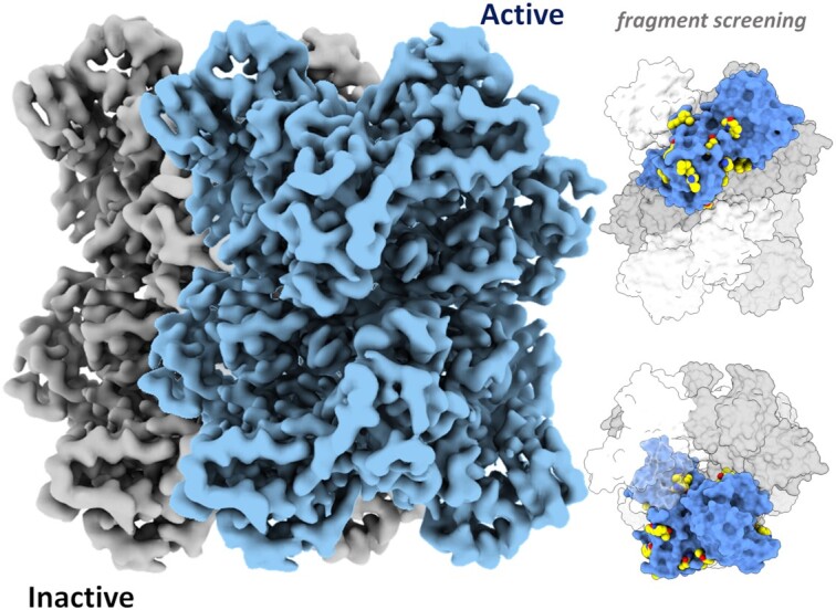
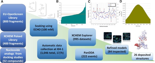
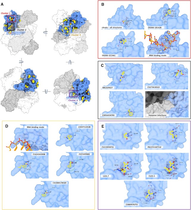
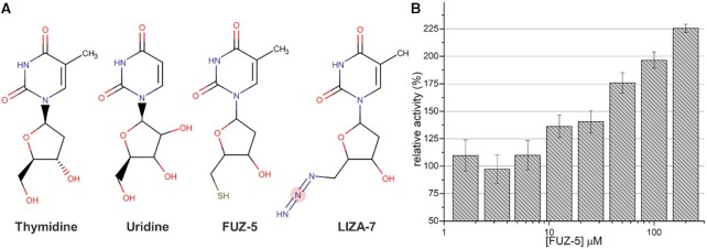
Severe acute respiratory syndrome coronavirus 2 (SARS-CoV-2) is the causative agent of coronavirus disease 2019 (COVID-19). The NSP15 endoribonuclease enzyme, known as NendoU, is highly conserved and plays a critical role in the ability of the virus to evade the immune system. NendoU is a promising target for the development of new antiviral drugs. However, the complexity of the enzyme's structure and kinetics, along with the broad range of recognition sequences and lack of structural complexes, hampers the development of inhibitors. Here, we performed enzymatic characterization of NendoU in its monomeric and hexameric form, showing that hexamers are allosteric enzymes with a positive cooperative index, and with no influence of manganese on enzymatic activity. Through combining cryo-electron microscopy at different pHs, X-ray crystallography and biochemical and structural analysis, we showed that NendoU can shift between open and closed forms, which probably correspond to active and inactive states, respectively. We also explored the possibility of NendoU assembling into larger supramolecular structures and proposed a mechanism for allosteric regulation. In addition, we conducted a large fragment screening campaign against NendoU and identified several new allosteric sites that could be targeted for the development of new inhibitors. Overall, our findings provide insights into the complex structure and function of NendoU and offer new opportunities for the development of inhibitors.


Figures

Graphical Abstract
Graphical Abstract
SARS-CoV-2 NendoU shifts between active and inactive conformation, and fragment screening revealed multiple allosteric sites.
Figure 1.
Figure 1.
Summarized enzymatic profile characterization of NendoU. (A) Time-course reaction of NendoUhex at different temperatures. (B) Calculated Hill constant for NendoUhex, showing a positive cooperative index of 2. (C) Michaelis–Menten plot of NendoUmon, showing a typical first-order enzymatic profile. (D) The relative enzymatic activity of NendoUhex in thepresence of different concentrations of MnCl2 and EDTA. (E) Acrylamide gel showing the activity of NendoUhex against poly(U) RNA in the presence of different concentrations of MnCl2 and EDTA. (F) Calculated initial velocities of mutant T114L and wild-type (WT) NendoUhex.
Figure 2.
Figure 2.
Overview of the NendoU structure. (A) Two distinct views of the NendoU hexamer surface (PDB 7KF4), highlighting regions of interest, including the active site, the S groove and the switch region. Citrate molecules on the active site are depicted as green spheres. (B) NendoU–dsRNA binding mode on the surface of the hexamer, occupying one active site and interacting with the switch region. (C) Detailed dsRNA binding mode on the active site of NendoU. (D) Detailed dsRNA binding mode on the switch region of NendoU. The structure of NendoU is colored according to its electrostatic potential projected on surface charge (–5 to 5 f kJ/mol/e in red-white-blue color model). dsRNA is colored in green/blue and was depicted from PDB 7TJ2.
Figure 3.
Figure 3.
Cryo-EM maps of NendoU in different conditions. (A) Cryo-EM maps of NendoU in HEPES at pH 7.5 (PDB 7RB0) in the closed state, with chains A and B colored in blue and cyan, respectively. The box shows a detailed view of the map and model from the switch region between these two chains. (B) Cryo-EM maps of NendoU in PBS at pH 6.0 (PDB 7ME0) in the open state, with chains A and B colored in blue and light blue, respectively. The box shows a detailed view of the map and model from the switch region between these two chains. (C) Cryo-EM maps of NendoU in complex with dsRNA (PDB 7TJ2) and in the open state, with chains A and B colored in blue and salmon pink, respectively. THe dsRNA map is colored in light blue. The black box shows a detailed view of the three models aligned in the active site region. The green box shows detailed views of the middle domain of the three models as cartoons superposed on adjacent NendoU. 7RB0, 7ME0 and 7TJ2 are colored in dark blue, cyan and salmon pink, respectively.
Figure 4.
Figure 4.
Overview of supramolecular organization of NendoU. (A) 2D classification of selected filaments of NendoU observed from samples collected in Bis-Tris at pH 6.0. (B) Crystal structure of NendoU in the presence of oligo(dT) reveals high resolution details of contacts between the switch region of two NendoU hexamers (PDB 7KF4). The protein is depicted as a cartoon, with chains C, D, E and F colored in blue, green, orange and pink, respectively. Contacting residues are shown as colored sticks. (C) Stacking model based on the surface charge of NendoU. The structure of NendoU is colored according to its electrostatic potential projected on surface charge (–0.5 to 0.5 f kJ/mol/e in a red-white-blue color model).
Figure 5.
Figure 5.
Overall schematic showing steps and data processing of the fragment screening campaign against NendoU. (A) Schematic summarizing steps from library screening to refinement and deposition of models. Panels show event maps of selected fragments (1.0 sigma) and the overlap of all structures colored according to B-factors. (B) Graph showing the obtained resolution per dataset collected and processed. The dotted line marks the average resolution of all 997 usable datasets (2.2 Å). (C) Graph showing resolution and refinement statistics of the final models deposited. Resolution is colored in red triangles, Rwork is colored in blue squares and Rfree is colored in dark blue diamonds. (D) Normal distribution graph of the molecular weitght from identified fragments.
Figure 6.
Figure 6.
Overall visualization of fragments bound to NendoU. (A) Overall view of all NendoU sites bound to fragments in multiple orientations. All chains containing fragments were supperposed into one, colored as blue surface, while adjacent chains of biological units are colored in shades of gray. Colored squares highlight the cluster of fragments identified. (B) Detailed view of cluster 1 (active site) showing citrate, multiple fragment contacts and the RNA binding mode. (C) Detailed view of cluster 2 showing multiple fragment contacts and the oligomerization interface. (D) Detailed view of cluster 3, showing multiple fragment contacts and the RNA binding mode. (E) Detailed view of cluster 4, showing multiple fragment contacts with NendoU. In all, the NendoU surface is colored in blue, fragments are depicted as sticks or spheres with yellow carbons, while interacting NendoU residues are depicted as gray carbon sticks. RNA carbons are colored in orange, and coordinates were obtained from PDB 7TJ2.
Figure 7.
Figure 7.
Fragments bound to cluster 4 of NendoU. (A) Structural comparison between thymidine, uridine, FÜZS-5 and LIZA-7 fragments. (B) Effect of different FÜZS-5 concentrations on NendoU relative enzymatic activity determined using a fluorogenic substrate.

References

    1. Zhou P., Yang X.L., Wang X.G., Hu B., Zhang L., Zhang W., Si H.R., Zhu Y., Li B., Huang C.L.et al. .. A pneumonia outbreak associated with a new coronavirus of probable bat origin. Nature. 2020; 579:270–273. - PMC - PubMed
    1. Zhu N., Zhang D., Wang W., Li X., Yang B., Song J., Zhao X., Huang B., Shi W., Lu R.et al. .. A novel coronavirus from patients with pneumonia in China, 2019. N. Engl. J. Med. 2020; 382:727–733. - PMC - PubMed
    1. Li H., Zhou Y., Zhang M., Wang H., Zhao Q., Liu J.. Updated approaches against SARS-CoV-2. Antimicrob. Agents Chemother. 2020; 64:1–7. - PMC - PubMed
    1. Lu R., Zhao X., Li J., Niu P., Yang B., Wu H., Wang W., Song H., Huang B., Zhu N.et al. .. Genomic characterisation and epidemiology of 2019 novel coronavirus: implications for virus origins and receptor binding. Lancet North Am. Ed. 2020; 395:565–557. - PMC - PubMed
    1. Li H., Liu Z., Ge J.. Scientific research progress of COVID-19/SARS-CoV-2 in the first five months. J. Cell. Mol. Med. 2020; 24:6558. - PMC - PubMed

Publication types

Substances

LinkOut - more resources