

Development of a simple standardized scoring system for assessing large vessel vasculitis by 18F-FDG PET-CT and differentiation from atherosclerosis
- PMID: 37115211
- PMCID: PMC10317865
- DOI: 10.1007/s00259-023-06220-5
Development of a simple standardized scoring system for assessing large vessel vasculitis by 18F-FDG PET-CT and differentiation from atherosclerosis
Abstract
Purpose: This study is to develop a structured approach to distinguishing large-artery vasculitis from atherosclerosis using 18-fluorodeoxyglucose positron emission tomography combined with low-dose computed tomography (FDG PET/CT).
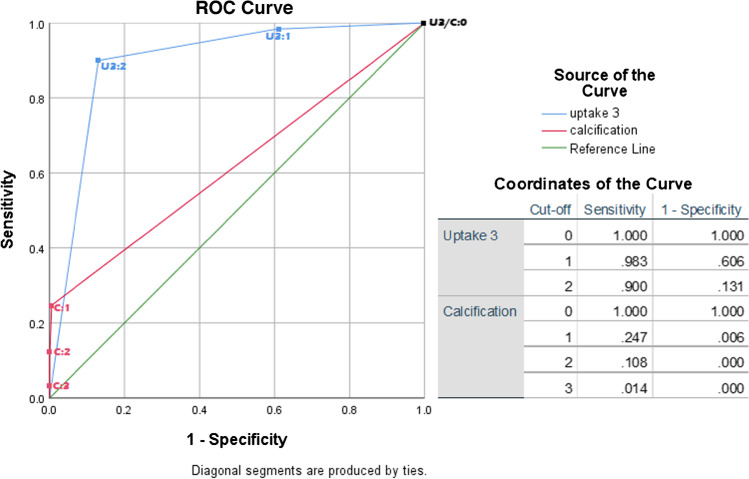
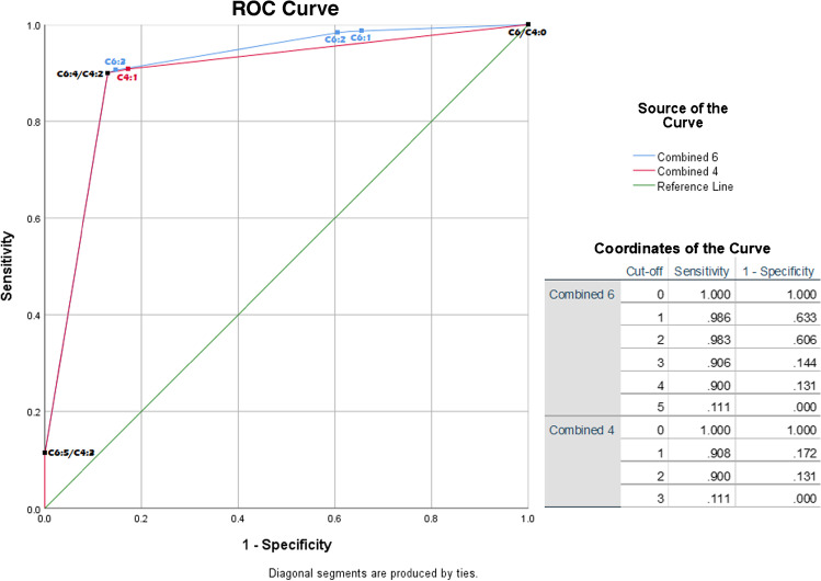
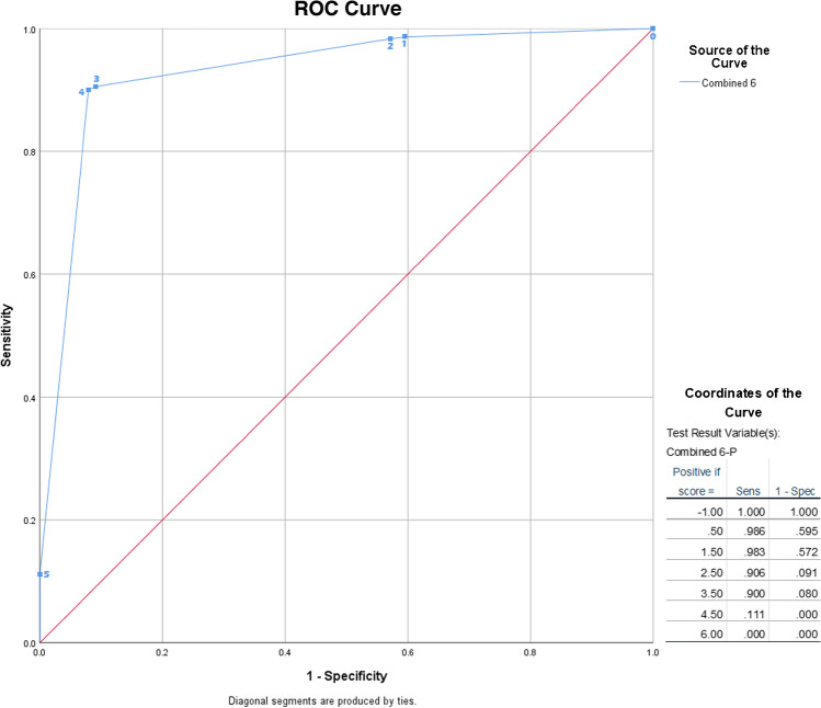
Methods: FDG PET/CT images of 60 patients were evaluated, 30 having biopsy-proven giant cell arteritis (GCA; the most common form of large-artery vasculitis), and 30 with severe atherosclerosis. Images were evaluated by 12 nuclear medicine physicians using 5 criteria: FDG uptake pattern (intensity, distribution, circularity), the degree of calcification, and co-localization of calcifications with FDG-uptake. Criteria that passed agreement, and reliability tests were subsequently analysed for accuracy using receiver operator curve (ROC) analyses. Criteria that showed discriminative ability were then combined in a multi-component scoring system. Both initial and final 'gestalt' conclusion were also reported by observers before and after detailed examination of the images.
Results: Agreement and reliability analyses disqualified 3 of the 5 criteria, leaving only FDG uptake intensity compared to liver uptake and arterial wall calcification for potential use in a scoring system. ROC analysis showed an area under the curve (AUC) of 0.90 (95%CI 0.87-0.92) for FDG uptake intensity. Degree of calcification showed poor discriminative ability on its own (AUC of 0.62; 95%CI 0.58-0.66). When combining presence of calcification with FDG uptake intensity into a 6-tiered scoring system, the AUC remained similar at 0.91 (95%CI 0.88-0.93). After exclusion of cases with arterial prostheses, the AUC increased to 0.93 (95%CI 0.91-0.95). The accuracy of the 'gestalt' conclusion was initially 89% (95%CI 86-91%) and increased to 93% (95%CI 91-95%) after detailed image examination.
Conclusion: Standardised assessment of arterial wall FDG uptake intensity, preferably combined with assessment of arterial calcifications into a scoring method, enables accurate, but not perfect, distinction between large artery vasculitis and atherosclerosis.
Keywords: 18-Fluorodeoxyglucose positron emission tomography; Atherosclerosis; Computed tomography; Diagnosis; Vasculitis.
© 2023. The Author(s).
Conflict of interest statement
The authors declare no competing interests.
Figures
References
-
- Hellmich B, Agueda A, Monti S, Buttgereit F, de Boysson H, Brouwer E, Cassie R, Cid MC, Dasgupta B, Dejaco C, Hatemi G, Hollinger N, Mahr A, Mollan SP, Salvarani C, Sivakumar R, Tian X, Tomasson G, Turesson C, Schmidt W, Viliger PM, Watts R, Toung C, Luqmani RA. 2018 update of the EULAR recommendations for the management of large-vessel vasculitis. Ann Rheum Dis. 2020;79:19–30. doi: 10.1136/annrheumdis-2019-215672. - DOI - PubMed
MeSH terms
Substances
Grants and funding
LinkOut - more resources
Full Text Sources
Medical
