. 2023 May 1;133(9):e161453.
doi: 10.1172/JCI161453.

Pressure overload induces ISG15 to facilitate adverse ventricular remodeling and promote heart failure

Affiliations

Pressure overload induces ISG15 to facilitate adverse ventricular remodeling and promote heart failure

Veera Ganesh Yerra et al. J Clin Invest. .

Abstract

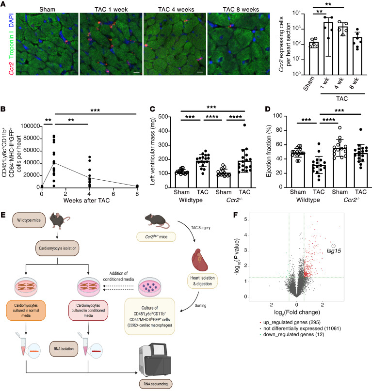
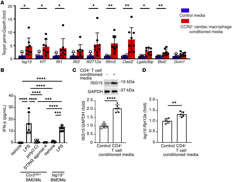
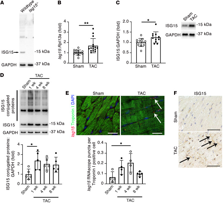
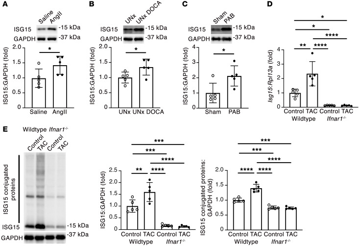
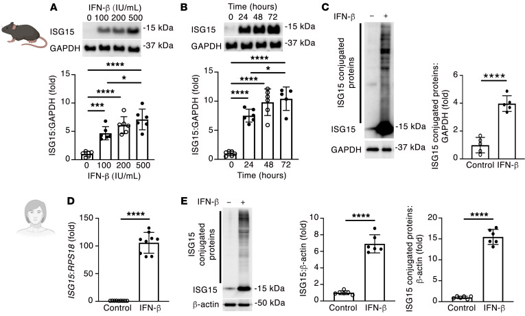
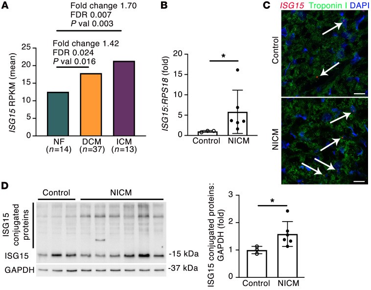
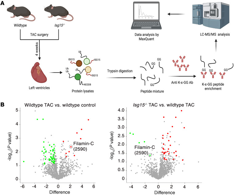
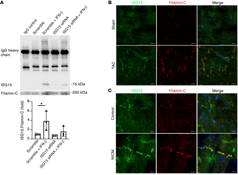
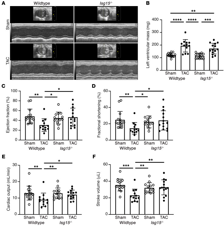
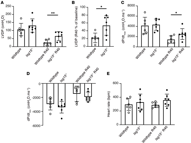
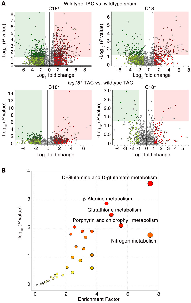
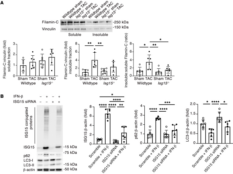
Inflammation promotes adverse ventricular remodeling, a common antecedent of heart failure. Here, we set out to determine how inflammatory cells affect cardiomyocytes in the remodeling heart. Pathogenic cardiac macrophages induced an IFN response in cardiomyocytes, characterized by upregulation of the ubiquitin-like protein IFN-stimulated gene 15 (ISG15), which posttranslationally modifies its targets through a process termed ISGylation. Cardiac ISG15 is controlled by type I IFN signaling, and ISG15 or ISGylation is upregulated in mice with transverse aortic constriction or infused with angiotensin II; rats with uninephrectomy and DOCA-salt, or pulmonary artery banding; cardiomyocytes exposed to IFNs or CD4+ T cell-conditioned medium; and ventricular tissue of humans with nonischemic cardiomyopathy. By nanoscale liquid chromatography-tandem mass spectrometry, we identified the myofibrillar protein filamin-C as an ISGylation target. ISG15 deficiency preserved cardiac function in mice with transverse aortic constriction and led to improved recovery of mouse hearts ex vivo. Metabolomics revealed that ISG15 regulates cardiac amino acid metabolism, whereas ISG15 deficiency prevented misfolded filamin-C accumulation and induced cardiomyocyte autophagy. In sum, ISG15 upregulation is a feature of pathological ventricular remodeling, and protein ISGylation is an inflammation-induced posttranslational modification that may contribute to heart failure development by altering cardiomyocyte protein turnover.

Keywords: Cardiology; Heart failure.


Conflict of interest statement

Conflict of interest: CHB is the chief scientific officer of MRM Proteomics Inc. and the chief technology officer of Molecular You.

Figures

Figure 1
Figure 1. CCR2+ macrophage accumulation impairs cardiac function in pressure overload.
(A) RNAscope in situ hybridization for Ccr2 and immunostaining for troponin I in mouse hearts 8 weeks after sham surgery or 1, 4, or 8 weeks after transverse aortic constriction (TAC). Sham, n = 5; 1 week TAC, n = 5; 4 weeks TAC, n = 5; 8 weeks TAC, n = 7. Scale bars: 10 μm. (B) Enumeration of CCR2+ cardiac monocyte-derived macrophages (CD45+Ly6ChiCD11b+CD64+MHC-IIhiGFP+ cells) in Ccr2gfp/+ mice. Control, n = 5; 1 week TAC, n = 18; 4 weeks TAC, n = 13; 8 weeks TAC, n = 6. (C and D) LV mass (C) and ejection fraction (D) in WT and Ccr2–/– mice 8 weeks after sham or TAC. WT sham, n = 16; WT TAC, n = 17; Ccr2–/– sham, n = 14; Ccr2–/– TAC, n = 17. (E) Design of RNA sequencing experiments. (F) Volcano plot of genes expressed by mouse cardiomyocytes exposed to CCR2+ cardiac macrophage–conditioned medium (n = 4 per condition). Values are mean ± SD. **P < 0.01, ***P < 0.001, ****P < 0.0001 by 1-way ANOVA followed by Dunnett’s post hoc test (A) or Tukey’s post hoc test (BD).
Figure 2
Figure 2. CCR2+ cardiac macrophages induce a cardiomyocyte IFN response.
(A) qRT-PCR for IFN response genes (Isg15, Irf7, Ifit1, Ifit3, Ifi2712a, Ifitm3, Oasl2, Lgals3bp, Bst2, Gvin1) in mouse cardiomyocytes in medium conditioned by CCR2+ cardiac macrophages isolated from Ccr2gfp/+ mouse hearts 1 week after TAC, or under control conditions. Control, n = 4; CCR2+ cardiac macrophage–conditioned medium, n = 8. (B) Culture medium IFN-β concentration in bone marrow–derived macrophages (BMDMs) from Ccr2gfp/+ mice or Isg15–/– mice incubated with LPS (1 μg/mL), poly(I:C) (500 ng/mL), or STING agonist-4 (5 μmol/L) for 24 hours (n = 6 per condition). (C and D) Immunoblotting (C; n = 6 per condition) and qRT-PCR (D; n = 5 per condition) for ISG15 in mouse cardiomyocytes exposed to CD4+ T cell–conditioned medium for 24 hours. Values are mean ± SD. *P < 0.05, **P < 0.01, ***P < 0.001, ****P < 0.0001 by unpaired 2-tailed Mann-Whitney test (A), 1-way ANOVA followed by Tukey’s post hoc test (B), or unpaired 2-tailed Student’s t test (C and D).
Figure 3
Figure 3. Cardiomyocyte ISG15 is upregulated in mouse hearts after TAC.
(A) Immunoblotting of WT and Isg15–/– mouse hearts confirming specificity of the ISG15 antibody (clone 1H9L21). (B) qRT-PCR for Isg15 in mouse hearts 8 weeks after sham or TAC. Sham, n = 13; TAC, n = 15. (C) Immunoblotting for ISG15 in mouse hearts 8 weeks after sham or TAC. Sham, n = 11; TAC, n = 11. (D) Immunoblotting for ISG15-conjugated proteins in mouse hearts 8 weeks after sham surgery or 1, 4, or 8 weeks after TAC (n = 5 per group). (E) RNAscope in situ hybridization for Isg15 and immunofluorescence for troponin I in heart sections from mice 4 weeks after sham or TAC. The arrows mark Isg15 RNAscope puncta in troponin I+ cardiomyocytes. Scale bars: 20 μm. n = 4 per group, except 8 weeks after TAC (n = 5). (F) Immunohistochemistry for ISG15 in mouse hearts 4 weeks after sham or TAC. The arrows mark positive immunostaining at, or close to, intercalated discs. Scale bars: 50 μm. Values are mean ± SD. *P < 0.05, **P < 0.01 by unpaired 2-tailed Student’s t test (B and C), or 1-way ANOVA followed by Dunnett’s post hoc test (D and E).
Figure 4
Figure 4. Pressure overload induces IFNAR-dependent ISG15 upregulation.
(A) Immunoblotting for ISG15 in LV tissue of mice infused with Ang II (2 mg/kg/d) or saline for 14 days (n = 5 per group). (B) Immunoblotting for ISG15 in LV tissue of uninephrectomized rats (UNx) or UNx DOCA-salt rats followed for 4 weeks (UNx DOCA) (n = 5 per group). (C) Immunoblotting for ISG15 in right ventricular tissue of rats 6 weeks after sham or pulmonary artery banding (PAB) (n = 5 per group). (D and E) qRT-PCR (D) and immunoblotting (E) for ISG15 in WT and Ifnar1–/– mouse hearts 1 week after TAC (n = 5 per group). Values are mean ± SD. *P < 0.05, **P < 0.01, ***P < 0.001, ****P < 0.0001 by unpaired 2-tailed Mann-Whitney test (A), unpaired 2-tailed Student’s t test (B and C), or 1-way ANOVA followed by Tukey’s post hoc test (D and E).
Figure 5
Figure 5. ISG15 is inducible in mouse and human cardiomyocytes.
(A) ISG15 induction by recombinant IFN-β for 24 hours in primary mouse cardiomyocytes (n = 6 per condition). (B) ISG15 induction by 500 IU/mL recombinant IFN-β in primary mouse cardiomyocytes (n = 6 per condition, except 72 hours [n = 5]). (C) Immunoblotting for ISG15-conjugated proteins following stimulation with 500 IU/mL recombinant IFN-β for 48 hours (n = 5 per condition). (D) qRT-PCR for ISG15 in human cardiomyocytes incubated with 500 IU/mL IFN-β for 24 hours (n = 9 per condition). (E) Immunoblotting for ISG15 in human cardiomyocytes incubated with 500 IU/mL IFN-β for 48 hours (n = 6 per condition). Values are mean ± SD. *P < 0.05, ***P < 0.001, ****P < 0.0001 by 1-way ANOVA followed by Tukey’s post hoc test (A and B), or unpaired 2-tailed Student’s t test (CE).
Figure 6
Figure 6. ISG15 is upregulated in human NICM.
(A) ISG15 in human LV samples from patients with nonfailing hearts (NF), dilated cardiomyopathy (DCM), or ischemic cardiomyopathy (ICM) (28). Differential expression determined by linear model ANOVA; P value (pval) adjusted for Benjamini-Hochberg FDR ≤ 0.05 (28). RPKM, reads per kilobase of transcript, per Mmllion mapped reads. (B) qRT-PCR in control human heart tissue (n = 3) and tissue from patients with end-stage heart failure due to NICM (n = 6). (C) RNAscope in situ hybridization for ISG15 and immunofluorescence for troponin I in human LV tissue. The arrows mark ISG15 RNAscope puncta in troponin I+ cardiomyocytes. Scale bars: 20 μm. (D) Immunoblotting for ISG15 and quantification of ISG15 protein conjugates in human control (n = 3) and NICM (n = 6) heart tissue. Values are mean ± SD. *P < 0.05 by unpaired 2-tailed Mann-Whitney test.
Figure 7
Figure 7. Identification of ISGylation targets in mouse hearts after TAC.
(A) Design for diGLY proteomics experiments. (B) Volcano plots for the comparison of diGLY-enriched sites in WT TAC versus WT control and Isg15–/– TAC versus WT TAC (n = 3 per group). “Difference” indicates difference in the means of log2-transformed values between groups.
Figure 8
Figure 8. ISG15 associates with filamin-C in mouse and human hearts.
(A) Immunoprecipitation for filamin-C and immunoblotting for ISG15 in human cardiomyocytes following knockdown of ISG15 with siRNA and incubation with 500 IU/mL IFN-β for 48 hours (n = 3 per condition). (B and C) Dual immunofluorescence staining for ISG15 and filamin-C in the hearts of sham-operated mice and mice 1 week after TAC (B) and human control tissue and LV tissue from a human with NICM (C). Scale bars: 10 μm. Values are mean ± SD. *P < 0.05 by 1-way ANOVA followed by Dunnett’s post hoc test.
Figure 9
Figure 9. ISG15 deficiency attenuates LV systolic dysfunction in mice after TAC.
(A and B) Representative M-mode echocardiographs (A) and LV mass (B) in WT and Isg15–/– mice 8 weeks after sham or TAC surgery. (CF) Ejection fraction (C), fractional shortening (D), cardiac output (E), and stroke volume (F) in WT and Isg15–/– mice 8 weeks after sham or TAC. WT sham, n = 15; WT TAC, n = 12; Isg15–/– sham, n = 13; Isg15–/– TAC, n = 14. Values are mean ± SD. *P < 0.05, **P < 0.01, ***P < 0.001, ****P < 0.0001 by 1-way ANOVA followed by Tukey’s post hoc test (skew-distributed data in BF were log-transformed before statistical comparison).
Figure 10
Figure 10. ISG15 deficiency improves contractile recovery of isolated mouse hearts.
(A) Left ventricular developed pressure (LVDP) at baseline and 40 minutes after ischemia/reperfusion (R40) in isolated perfused hearts from WT (n = 6) and Isg15–/– (n = 7) mice. (B) Percentage recovery of LVDP 40 minutes after reperfusion. (C) dP/dtmax. (D) dP/dtmin. (E) Heart rate. Values are mean ± SD. *P < 0.05, **P < 0.01 by unpaired 2-tailed Student’s t test (A and B) or unpaired 2-tailed Mann-Whitney test (C).
Figure 11
Figure 11. ISG15 deficiency alters amino acid metabolism in pressure-overloaded mouse hearts.
(A) Volcano plots of untargeted metabolomic comparison of WT and Isg15–/– mouse hearts 8 weeks after sham or TAC surgery. Top: WT TAC hearts (n = 4) versus WT sham (n = 3). Bottom: Isg15–/– TAC hearts (n = 4) versus WT TAC (n = 4). (B) KEGG pathway analysis of metabolic pathways enriched in Isg15–/– mouse hearts versus WT mouse hearts 8 weeks after TAC. Enrichment factor = ratio of significant pathway hits versus expected pathway hits (n = 4 per group).
Figure 12
Figure 12. ISG15 induction impairs cardiomyocyte protein turnover.
(A) Immunoblotting for filamin-C in the soluble and insoluble fractions of WT and Isg15–/– mouse hearts 8 weeks after sham or TAC surgery (n = 6 per group). (B) Immunoblotting for ISG15, ISG15-conjugated proteins, p62, and LC3 in human cardiomyocytes transfected with siRNA directed against ISG15 for 6 hours before incubation with 500 IU/mL IFN-β for 48 hours. Values are mean ± SD. *P < 0.05, **P < 0.01, ***P < 0.001, ****P < 0.0001 by 1-way ANOVA followed by Tukey’s post hoc test.

References

    1. Cohn JN, et al. Cardiac remodeling—concepts and clinical implications: a consensus paper from an international forum on cardiac remodeling. On behalf of an international forum on cardiac remodeling. J Am Coll Cardiol. 2000;35(3):569–582. doi: 10.1016/S0735-1097(99)00630-0. - DOI - PubMed
    1. King KR, et al. IRF3 and type I interferons fuel a fatal response to myocardial infarction. Nat Med. 2017;23(12):1481–1487. doi: 10.1038/nm.4428. - DOI - PMC - PubMed
    1. Martini E, et al. Single-cell sequencing of mouse heart immune infiltrate in pressure overload-driven heart failure reveals extent of immune activation. Circulation. 2019;140(25):2089–2107. doi: 10.1161/CIRCULATIONAHA.119.041694. - DOI - PubMed
    1. Revelo XS, et al. Cardiac resident macrophages prevent fibrosis and stimulate angiogenesis. Circ Res. 2021;129(12):1086–1101. doi: 10.1161/CIRCRESAHA.121.319737. - DOI - PMC - PubMed
    1. Bajpai G, et al. The human heart contains distinct macrophage subsets with divergent origins and functions. Nat Med. 2018;24(8):1234–1245. doi: 10.1038/s41591-018-0059-x. - DOI - PMC - PubMed

Publication types