. 2023 Apr 28;18(4):e0284012.
doi: 10.1371/journal.pone.0284012. eCollection 2023.

Sex-specific effects of CD248 on metabolism and the adipose tissue lipidome

Affiliations

Sex-specific effects of CD248 on metabolism and the adipose tissue lipidome

Kieran Patrick et al. PLoS One. .

Abstract

Cd248 has recently been associated with adipose tissue physiology, demonstrated by reduced weight gain in high fat diet-fed mice with genetic deletion of Cd248 relative to controls. Here we set out to determine the metabolic consequences of loss of Cd248. Strikingly, we find these to be sex specific; By subjecting Cd248-/- and Cd248+/+ mice to a high fat diet and indirect calorimetry study, we identified that only male Cd248-/- mice show reduced weight gain compared to littermate control wildtype mice. In addition, male (but not female) mice showed a lower respiratory exchange ratio on both chow and high fat diets, indicating a predisposition to metabolise lipid. Lipidomic studies on specific fat depots found reduced triglyceride and diglyceride deposition in male Cd248-/- mice, and this was supported by reduced expression of lipogenic and adipogenic genes. Finally, metabolomic analysis of isolated, differentiated preadipocytes found alterations in metabolic pathways associated with lipid deposition in cells isolated from male, but not female, Cd248-/- mice. Overall, our results highlight the importance of sex controls in animal studies and point to a role for Cd248 in sex- and depot-specific regulation of lipid metabolism.


Conflict of interest statement

I have read the journal’s policy and the authors of this manuscript have the following competing interests: XT, MSG, LC, SH, DB, CC, GD are employees of AstraZeneca and may own stocks and/or restricted stocks. This does not alter our adherence to PLOS ONE policies on sharing data and materials.

Figures

Fig 1
Fig 1
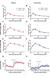
Weight gain ±SD over 16 weeks of high fat diet in (A) males and (B) females. Food intake ±SD at baseline (C) day and (D) night. Food intake over 12 hours after 12 weeks of high fat diet during the (E) day and (F) night. Body weight data over time were analysed for significant differences by t-test. Food intake was analysed by t-test. In all graphs: Cd248+/+ males (red), Cd248-/- males (cyan), Cd248+/+ females (black), Cd248-/- females (blue). Significant * p ≤ 0.05.
Fig 2
Fig 2
Respiratory exchange ratio (RER) as calculated from indirect calorimetry measurements over 48 hours: Chow-fed (A) males and (B) females; HFD-fed (C) males and (D) females. Line graphs indicate hourly averages over time whilst bar graphs show overall averages at night ± SD. Body weight data over time were analysed for significant differences by t-test. RER data were analysed for significance by ANCOVA test to determine if the body weight covariable is responsible for the significant changes in RER. In all graphs: Cd248+/+ males (red), Cd248-/- males (cyan), Cd248+/+ females (black), Cd248-/- females (blue). Significant * p ≤ 0.05.
Fig 3
Fig 3
Glucose tolerance test (GTT) was performed at baseline in (A) males and (B) females (6–8 weeks old mice) and following high fat diet-feeding for 6 weeks (C) males and (D) females, then 15 weeks (E) males and (F) females. Response to CL316,243 injection was measured as average VO2 over time in (G) males and (H) females. In all graphs: Cd248+/+ males (red), Cd248-/- males (cyan), Cd248+/+ females (black), Cd248-/- females (blue). Data were analysed for significant differences by ANOVA. No significant differences were found. All error bars ± SEM.
Fig 4
Fig 4. Gene expression and lipidomic results from fat pads following high fat diet feeding for 16 weeks.
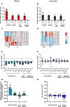
CD248 expression analysis in each fat pad confirmed loss of CD248 in Cd248-/- tissues from (A) males (B) females. Heatmap showing z—scores (scale bar from blue = -3 to red = +3 shown—far right) of significantly altered lipids in (C) male Cd248-/- perirenal fat pad compared to Cd248+/+ with (D) female shown for comparison where no significant differences were detected. Lipid notation follows the nomenclature described in Liebsch et al. 2013 [22]. Detail of the MSI identification level for each of the lipid species is given in S1 Table. Gene expression array showing significantly altered genes from (E) male Cd248-/- perirenal fat pad, with (F) female shown for comparison where no significant differences were detected. Adipoq expression data for all four fat depots in (G) male and (H) female mice are also shown. Results analysed for significant differences by t-test. Considered significant (*) if t-test yielded p ≤ 0.05 and fold change or z score from control ≥ 1.5. All error bars ± SD.
Fig 5
Fig 5. Results from differentiated preadipocytes.
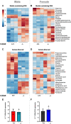
Heat map showing z–scores for metabolites identified as significantly altered in cells isolated from male mice. Cd248+/+ and Cd248-/- differentiated preadipocytes from chow fed (A) males and (B) females were cultured in 10% FBS media (Media containing FBS) or following 2 hours of serum starved conditions: (C) males and (D) females. Scale bar from blue = -2 to red = +2 (shown top left). Analysis for significant differences by t-test. Considered significant if t-test yielded p ≤ 0.05 and z—score from control ≥ 1.5. Detail of the MSI identification level for each of the metabolite species is given in S2 and S3 Tables for culture in media containing FBS and serum starved conditions, respectively. Expression levels of adipocyte differentiation marker, CD36 (normalised to housekeeping gene) are shown for (E) male and (F) female-derived cells. No significant difference was detected.

References

    1. Zwick RK, Guerrero-Juarez CF, Horsley V, Plikus M V. Anatomical, Physiological, and Functional Diversity of Adipose Tissue. Cell Metab. 2018;27: 68–83. doi: 10.1016/j.cmet.2017.12.002 - DOI - PMC - PubMed
    1. Yang C-F, Liu W-W, Wang H-Q, Zhang J-L, Li K, Diao Z-Y, et al.. Gonadal white adipose tissue is important for gametogenesis in mice through maintenance of local metabolic and immune niches. Journal of Biological Chemistry. 2022;298: 101818. doi: 10.1016/j.jbc.2022.101818 - DOI - PMC - PubMed
    1. van Beek L, van Klinken JB, Pronk ACM, van Dam AD, Dirven E, Rensen PCN, et al.. The limited storage capacity of gonadal adipose tissue directs the development of metabolic disorders in male C57Bl/6J mice. Diabetologia. 2015;58: 1601–1609. doi: 10.1007/s00125-015-3594-8 - DOI - PMC - PubMed
    1. Tramunt B, Smati S, Grandgeorge N, Lenfant F, Arnal J-F, Montagner A, et al.. Sex differences in metabolic regulation and diabetes susceptibility. Diabetologia. 2020;63: 453–461. doi: 10.1007/s00125-019-05040-3 - DOI - PMC - PubMed
    1. Bi Y, Jiang M, Guo W, Guan X, Xu M, Ren S, et al.. Sex-Dimorphic and Sex Hormone–Dependent Role of Steroid Sulfatase in Adipose Inflammation and Energy Homeostasis. Endocrinology. 2018;159: 3365–3377. doi: 10.1210/en.2018-00531 - DOI - PMC - PubMed

Publication types