. 2023 May 18;83(10):1743-1760.e11.
doi: 10.1016/j.molcel.2023.03.027. Epub 2023 Apr 27.

Modular antibodies reveal DNA damage-induced mono-ADP-ribosylation as a second wave of PARP1 signaling

Affiliations

Modular antibodies reveal DNA damage-induced mono-ADP-ribosylation as a second wave of PARP1 signaling

Edoardo José Longarini et al. Mol Cell. .

Abstract

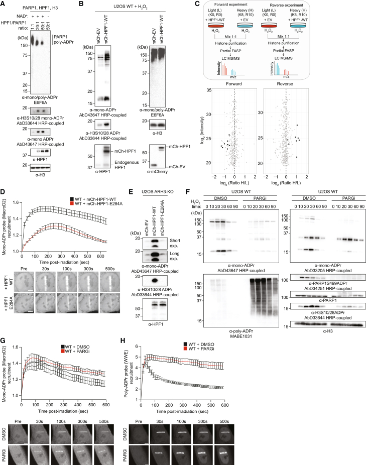
PARP1, an established anti-cancer target that regulates many cellular pathways, including DNA repair signaling, has been intensely studied for decades as a poly(ADP-ribosyl)transferase. Although recent studies have revealed the prevalence of mono-ADP-ribosylation upon DNA damage, it was unknown whether this signal plays an active role in the cell or is just a byproduct of poly-ADP-ribosylation. By engineering SpyTag-based modular antibodies for sensitive and flexible detection of mono-ADP-ribosylation, including fluorescence-based sensors for live-cell imaging, we demonstrate that serine mono-ADP-ribosylation constitutes a second wave of PARP1 signaling shaped by the cellular HPF1/PARP1 ratio. Multilevel chromatin proteomics reveals histone mono-ADP-ribosylation readers, including RNF114, a ubiquitin ligase recruited to DNA lesions through a zinc-finger domain, modulating the DNA damage response and telomere maintenance. Our work provides a technological framework for illuminating ADP-ribosylation in a wide range of applications and biological contexts and establishes mono-ADP-ribosylation by HPF1/PARP1 as an important information carrier for cell signaling.

Keywords: ADP-ribosylation; DNA damage response; HPF1; PARP1; RNF114; SpyTag; antibodies; telomere.


Conflict of interest statement

Declaration of interests E.J.L., H.D., J.J.B., T.C., and I.M. declare the following competing financial interests: Max-Planck-Innovation, the technology transfer center of the Max Planck Society, has licensed the antibodies AbD33204, AbD33205, AbD33644, AbD34251, AbD33641, and AbD43647 to Bio-Rad Laboratories.

Figures

None
Graphical abstract
Figure 1
Figure 1
SpyTag-based modular antibodies enable the sensitive and versatile detection of mono-ADPr (A) Schematic illustration of modular antibody engineering: a monovalent Fab antibody with the SpyTag peptide is covalently conjugated to functionalized SpyCatchers to generate various formats, including but not limited to “IgG-like” antibodies with different Fc chains (i.e., rabbit, mouse, and human), HRP-coupled antibodies, and fluorophore-coupled antibodies (“Fab probe”). (B) Mono-ADPr immunoblotting with AbD33204 or AbD43647. (C) Specificity of AbD43647 by immunoblotting. See also Figure S2A. (D) Immunoprecipitation and immunoblotting of histones with AbD43647 (top panel). Histone levels by Ponceau staining (bottom panel). (E) Left: immunofluorescence (IF) of mono- and poly-ADPr. Scale bars, 10 μm. Right: quantified nuclear signal intensities. (F) Top: IF images of mono-ADPr in telomere-localized DNA damage. Bottom: quantified mono-ADPr positive telomeres (%). Scale bars, 5 μm. (G) IF images showing mitochondria and mono-ADPr co-localization. Scale bars, 10 μm. (H) Dot blot of genomic DNA (gDNA) ADPr by the indicated antibodies. See also Figures S1 and S2 and Methods S1. Graphs indicate mean ± SEM from a representative of 3 independent experiments. ∗∗∗∗p < 0.0001; ns, not significant.
Figure 2
Figure 2
Fluorescence-based sensors reveal DNA damage-induced serine mono-ADPr as second wave of PARP1 signaling (A) Real-time live-cell detection of mono-ADPr by bead-loaded Fab antibodies. (B–E) Recruitment kinetics and representative confocal images of: (B) mono-ADPr Fab probe (fluorophore-coupled AbD33205), scale bars, 10 μm. (C) Genetically encoded poly-ADPr probe (RNF146 WWE domain), scale bars, 10 μm. (D) Genetically encoded mono-ADPr probe (macrodomain of MacroD2), scale bars, 5 μm. (E) Poly- and mono-ADPr probes, scale bars, 10 μm. (F) Left: IF images of WT U2OS cells, treated with H2O2 for the indicated times. Right: quantified mean nuclear intensity from mono- or poly-ADPr antibodies. Scale bar, 10 µm. (G) Immunoblotting of WT U2OS cells treated with H2O2 for the indicated time. See also Figure S2. Data in (B)–(E) are shown as mean ± SEM from a representative of 3 independent experiments.
Figure 3
Figure 3
Cellular HPF1/PARP1 ratios regulate mono-ADPr levels (A) Immunoblotting of in vitro HPF1/PARP1 ADPr reactions with increasing concentrations of recombinant HPF1. (B) Immunoblotting of WT U2OS cells transfected with mCherry-empty vector (mCh-EV) or mCherry-HPF1-WT (mCh-HPF1-WT) and H2O2 treated. (C) Top: schematics of SILAC-based proteomics of histone mono-ADPr marks on HPF1 overexpression and H2O2 treatment. Bottom: scatterplot showing SILAC quantification. Mono-ADPr peptides (black) and other peptides (gray). (D) Top: mono-ADPr probe recruitment kinetics in WT U2OS cells overexpressing mCherry-HPF1-WT (black) or mCherry-HPF1-E284A (red). Bottom: representative confocal images. (E) Immunoblotting of ARH3-KO U2OS cells transfected with mCherry-EV, mCherry-HPF1-WT, or mCherry-HPF1-E284A. (F) Immunoblotting showing mono-ADPr levels on PARGi and H2O2 time-course treatment. (G) Top: mono-ADPr probe recruitment kinetics in WT U2OS cells treated with DMSO (black) or PARGi (red). Bottom: representative confocal images. (H) Top: poly-ADPr probe recruitment kinetics in WT U2OS cells treated with DMSO (black) or 1 μM PARGi (red). Bottom: representative confocal images. Data in (D), (G), and (H) are shown as mean ± SEM from a representative of 3–4 independent experiments. Scale bars, 5 μm.
Figure 4
Figure 4
Identification of mono-ADPr readers by chromatin proteomics (A) Quantitative proteomics workflows to identify interactomes of Ser-mono-ADPr peptides (1) and H3S10ADPr nucleosome (2). (B) Scatterplot showing proteins enriched (red) by H3S10 mono-ADPr peptide compared with unmodified peptide. n = 2 biological replicates. (C) Chemoenzymatic generation of site-specific H3S10ADPr nucleosomes. (D) Scatterplot showing proteins enriched (red) by the H3S10ADPr nucleosome compared with unmodified nucleosome. n = 2 biological replicates. (E) Subcellular fractionation proteomics workflows for analysis of the mono-ADPr-dependent chromatin-associated proteome. (F) WT U2OS cells were H2O2-treated, and the chromatin fraction (as in E) was subjected to LC-MS/MS analysis. n = 3 biological replicates. (G) Top: immunoblotting of HPF1-KO U2OS cells transfected with mCherry-HPF1 WT or mCherry-HPF1-E284A, treated with H2O2 for 20 min. Immunoblotting (top) or LC-MS/MS of chromatin fractions. Bottom: volcano plot showing the log2-fold change of identified proteins. n = 4 biological replicates. (H–J) ARH3-KO (H) and WT (I) U2OS cells were treated with DMSO or 1 μM olaparib for 48 h, and the chromatin fraction was subjected to LC-MS/MS. Volcano plot showing the log2-fold change of identified proteins. (J) Heatmap showing log2-fold change of chromatin-associated proteins in the indicated condition. Data from (H)–(J) come from the same experiment. n = 3 biological replicates. (K) Immunoblotting of WT U2OS cells transfected with GFP-EV or GFP-RNF114, olaparib- and H2O2-treated then subjected to anti-GFP immunoprecipitation. For (B), (D), and (F)–(I), the red dotted line represents significance with p value = 0.05 (−log10(adj. p value) > 1.3) cutoff. Significant proteins are indicated in red or blue. See also Figures S3–S5 and Table S1.
Figure 5
Figure 5
Chromatin mono-ADPr functions as a recruitment signal for RNF114 (A) Chromatin fraction analysis of H2O2-treated WT U2OS cells. Volcano plots showing the log2-fold change of identified proteins. Red dotted lines represent significance with p value = 0.05 (−log10(adj. p value) > 1.3) cutoff. Significant proteins are indicated in red. n = 4 biological replicates. (B–D) Recruitment kinetics and representative confocal images for GFP-RNF114-WT in: (B) WT U2OS cell untreated (black) or 30 μm olaparib treated (red); (C) HPF1-KO U2OS cells expressing mCherry-HPF1-WT (black) or mCherry-HPF1-E284A (red); (D) WT (black) or ARH3-KO (red) U2OS cells. Scale bars, 5 μm. (E–H) Recruitment kinetics of: poly-ADPr probe (E), APLF (F), mono-ADPr probe (G), and RNF114 (H) in ARH3-KO U2OS cells. Cells were treated (red) or not (black) with 30 μM olaparib 210 s after laser microirradiation. (I–K) Recruitment kinetics of: poly-ADPr probe (I), mono-ADPr probe (J), and RNF114 (K) in ARH3-KO U2OS cells expressing mCherry-ARH3-WT (red) or mCherry-ARH3-D77/78N (black). (L) Recruitment kinetics of GFP-RNF114 (red) and mCherry-ALC1 (black). (M) Effective diffusion coefficient measured by FCS for GFP-RNF114 (left) and mono-ADPr probe (right). ∗∗∗∗p value < 0.0001, ∗∗∗p value < 0.001 (unpaired Student’s t test assuming unequal variances). Data in (B)–(L) are shown as mean ± SEM from a representative of 3 independent experiments. See also Figure S6 and Table S1.
Figure 6
Figure 6
RNF114 recruitment to DNA lesions is mediated by its zinc-finger domains (A) Dot blots of recombinant full-length RNF114 with indicated peptides or poly-ADP-ribose. Bovine serum albumin (BSA) and anti-mono/poly-ADPr (E6F6A) were used as negative and positive controls of ADPr binding, respectively. (B) Dot blots of equal moles of recombinant APLF, ALC1, and RNF114. (C) Domain architectures of RNF114 and deletion mutants. RING (RING-finger domain), Zn1 (zinc finger 1), Zn2 (zinc finger 2), Zn3 (zinc finger 3), and UIM (ubiquitin-interacting motif). Numbers indicate the motifs amino-acid positions. (D) Top: recruitment kinetics of GFP-RNF114-WT or individual GFP-RNF114 deletion constructs (as in C). Bottom: representative confocal images. Scale bars, 5 μm. (E) Top: recruitment kinetics of GFP-RNF114-WT or GFP-RNF114-C176A (as in C). Bottom: representative confocal images. Scale bars, 5 μm. (F) Dot blot of recombinant RNF114 and deletion constructs. (G) Immunoblotting images of WT U2OS cells transfected with indicated plasmids, H2O2 treated and subjected to anti-GFP immunoprecipitation. Bound proteins were immunoblotted and stained with the indicated antibodies. Data in (D) and (E) are shown as mean ± SEM from a representative of 5 independent experiments.
Figure 7
Figure 7
RNF114 modulates the alternative lengthening of telomeres pathway and the DNA damage response (A) IF images (left) and quantified ABPs (right) in WT and HPF1-KO U2OS cells transfected with indicated plasmids. (B) Left: representative images of WT, ARH3-KO, and HPF1-KO U2OS cells co-transfected with indicated plasmids. Right: quantification of RNF114 positive telomeres (%). See also Figure S7. (C) Quantification of APBs in WT U2OS cells transfected with siRNA for control (siControl), HPF1 (siHPF1), RNF114 (siRNF114), or HPF1 + siRNF114. See also Figure S7E. (D) Quantified APBs in WT and RNF114-KO U2OS cells complemented with GFP-RNF114-WT or GFP-RNF114-C176A. (E) Quantified relative amounts of DNA synthesis occurring at damaged telomeres (%Edu + telomeres). (F) Clonogenic cell survival assay of WT and RNF114-KO U2OS cells. (G) Representative IF images (left) and quantified 53BP1 foci (right) in IR-treated WT or RNF114-KO U2OS cells. (H) IF images (left) and quantified γH2Ax foci (right) in IR-treated WT HeLa cells, transfected with siControl or siRNF114. (I) Representative IF images (left) and quantified 53BP1 foci (right) in IR-treated WT, or RNF114-KO U2OS cells stably complemented with GFP-EV, GFP-RNF114-WT, or GFP-RNF114-C176A. (J) Top: recruitment kinetics of GFP-tagged H2AK13/15Ub probe in WT (black) or ARH3-KO (red) U2OS cells. Bottom: representative confocal images. (K) Representative IF images (left) and quantified RIF1 foci (right) in IR-treated WT or RNF114-KO U2OS cells. See also Figure S7. (L) Representative IF images (left) and quantification (right) of RNF168 foci in WT, or RNF114-KO U2OS cells stably complemented with GFP-EV, GFP-RNF114-WT, or GFP-RNF114-C176A treated with 5 Gy IR for 1 h, fixed with PFA and stained with the indicated antibody. The mean ± SEM from 100 cells from a representative of 3 independent experiments is shown. (M) Quantified relative NHEJ efficiency. Quantification of GFP-positive U2OS-EJ5 cells (relative NHEJ efficiency) after transfection with I-SceI and the indicated siRNAs. Data were normalized to siControl set to 100%. Data in (A)–(M) are shown as mean ± SEM from 3 to 4 independent experiments. ∗∗∗∗p value < 0.0001; ∗∗∗p value < 0.001; ∗∗p value < 0.01; p value < 0.05; ns, not significant (two-tailed Student’s t test). (G–L) Scale bars, 5 μm.

Comment in

References

    1. Gupte R., Liu Z., Kraus W.L. PARPs and ADP-ribosylation: recent advances linking molecular functions to biological outcomes. Genes Dev. 2017;31:101–126. doi: 10.1101/gad.291518.116. - DOI - PMC - PubMed
    1. Cohen M.S., Chang P. Insights into the biogenesis, function, and regulation of ADP-ribosylation. Nat. Chem. Biol. 2018;14:236–243. doi: 10.1038/Nchembio.2568. - DOI - PMC - PubMed
    1. Gibson B.A., Zhang Y., Jiang H., Hussey K.M., Shrimp J.H., Lin H., Schwede F., Yu Y., Kraus W.L. Chemical genetic discovery of PARP targets reveals a role for PARP-1 in transcription elongation. Science. 2016;353:45–50. doi: 10.1126/science.aaf7865. - DOI - PMC - PubMed
    1. Huang D., Kraus W.L. The expanding universe of PARP1-mediated molecular and therapeutic mechanisms. Mol. Cell. 2022;82:2315–2334. doi: 10.1016/j.molcel.2022.02.021. - DOI - PMC - PubMed
    1. Leidecker O., Bonfiglio J.J., Colby T., Zhang Q., Atanassov I., Zaja R., Palazzo L., Stockum A., Ahel I., Matic I. Serine is a new target residue for endogenous ADP-ribosylation on histones. Nat. Chem. Biol. 2016;12:998–1000. doi: 10.1038/nchembio.2180. - DOI - PMC - PubMed

Publication types