. 2023 May 9;56(5):998-1012.e8.
doi: 10.1016/j.immuni.2023.04.001. Epub 2023 Apr 27.

Methotrexate suppresses psoriatic skin inflammation by inhibiting muropeptide transporter SLC46A2 activity

Affiliations

Methotrexate suppresses psoriatic skin inflammation by inhibiting muropeptide transporter SLC46A2 activity

Ravi Bharadwaj et al. Immunity. .

Abstract

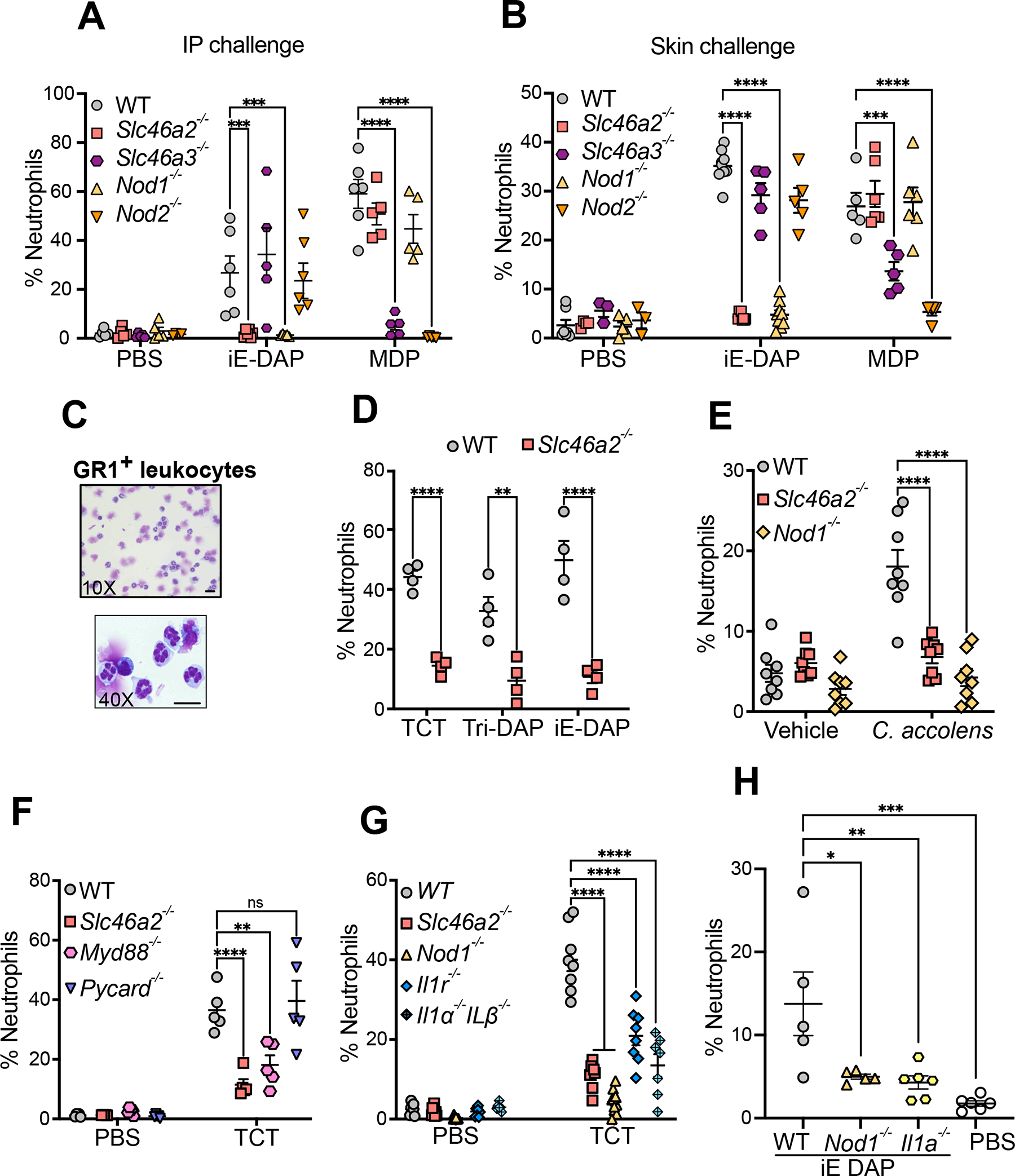
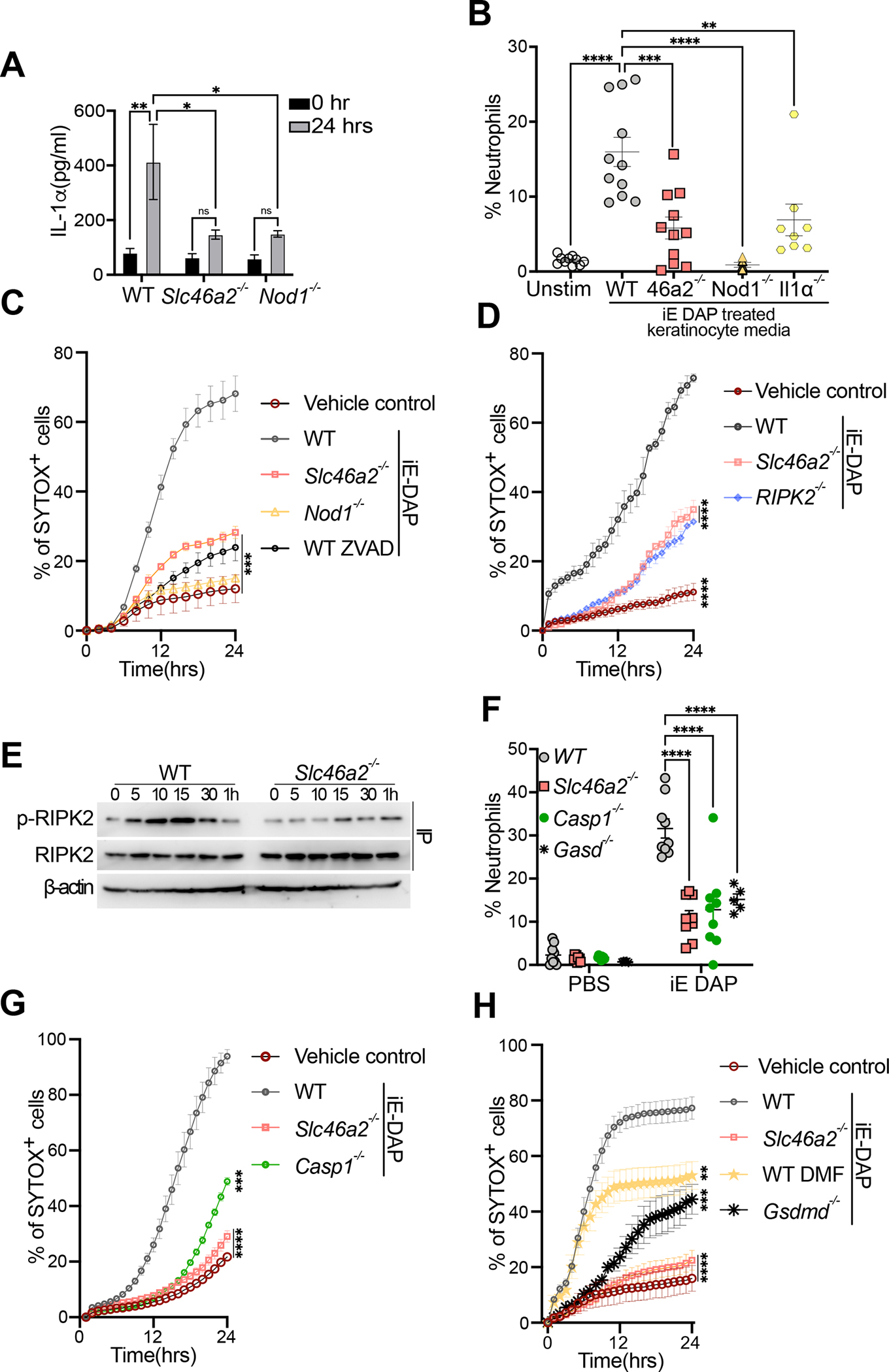
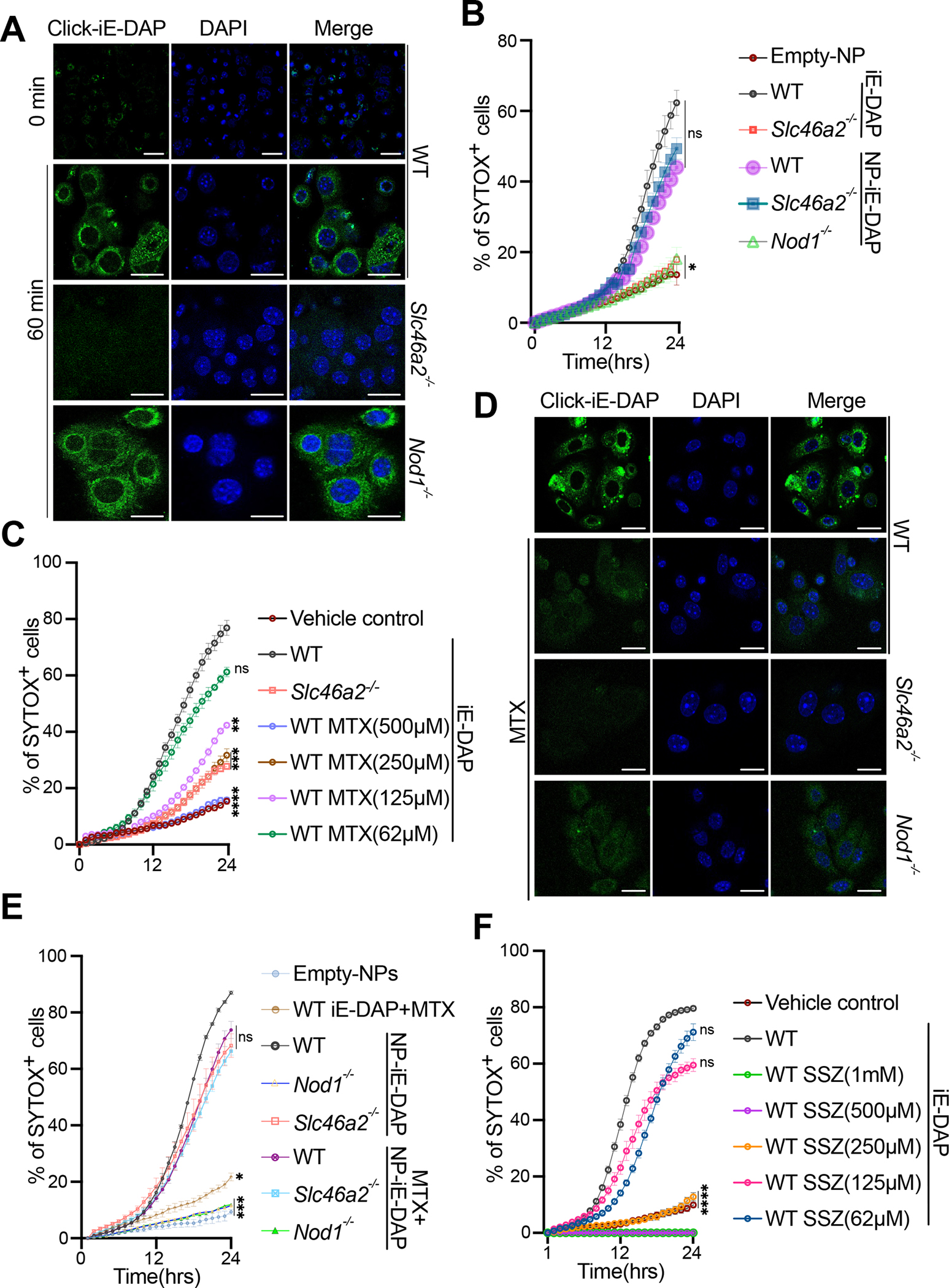
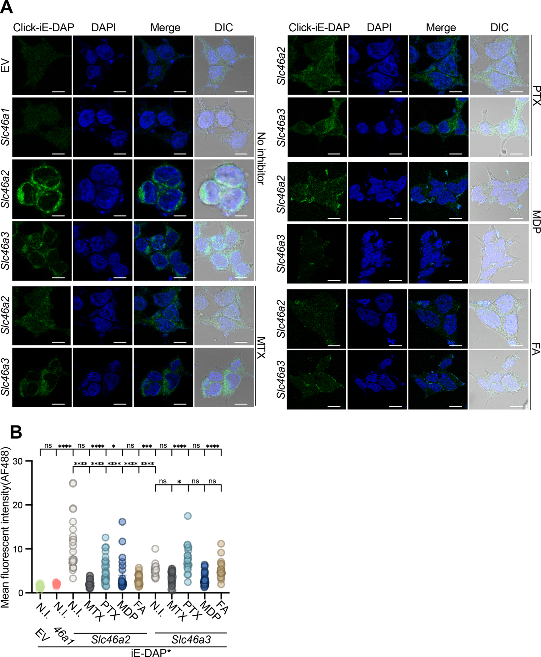
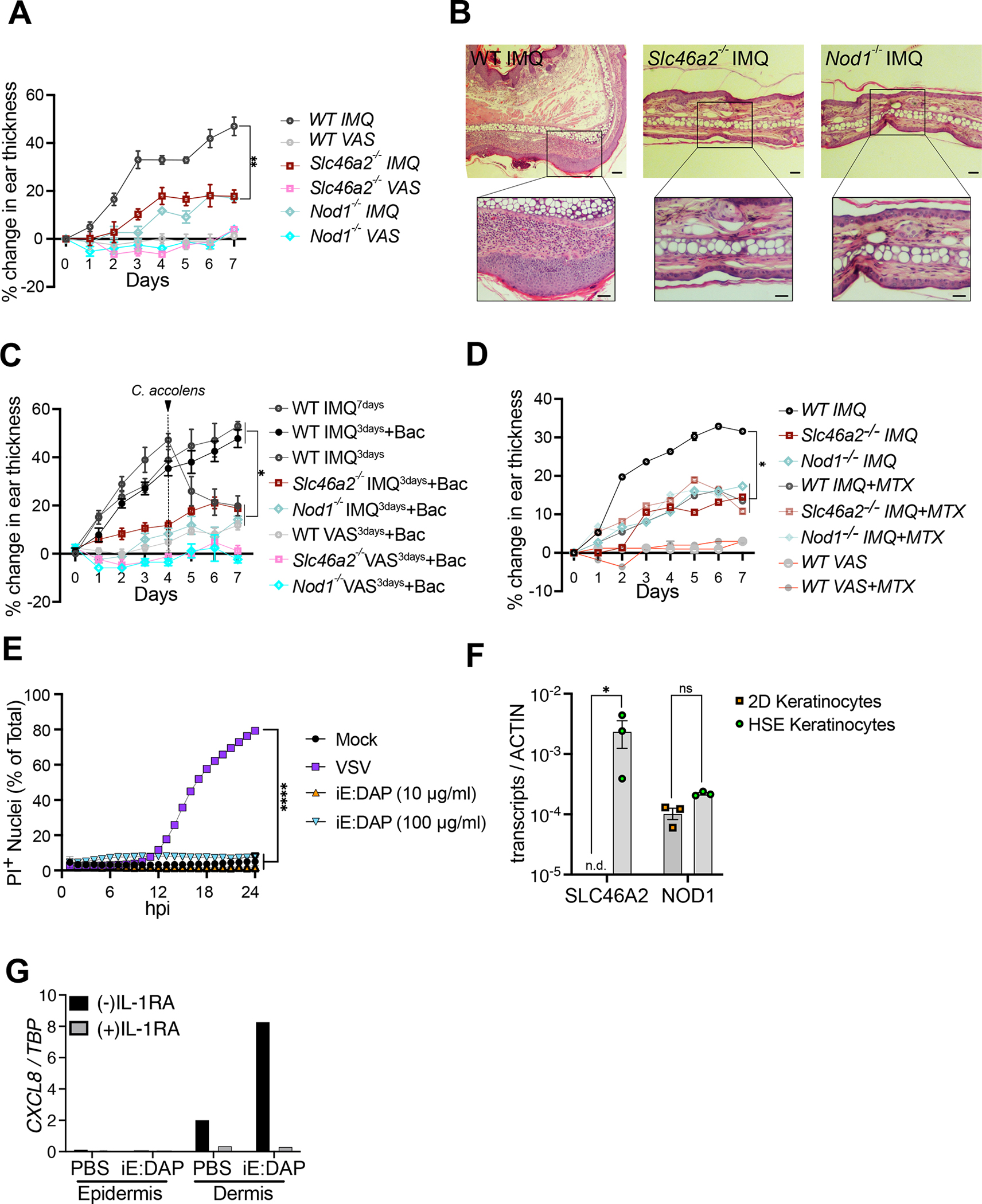
Cytosolic innate immune sensing is critical for protecting barrier tissues. NOD1 and NOD2 are cytosolic sensors of small peptidoglycan fragments (muropeptides) derived from the bacterial cell wall. These muropeptides enter cells, especially epithelial cells, through unclear mechanisms. We previously implicated SLC46 transporters in muropeptide transport in Drosophila immunity. Here, we focused on Slc46a2, which was highly expressed in mammalian epidermal keratinocytes, and showed that it was critical for the delivery of diaminopimelic acid (DAP)-muropeptides and activation of NOD1 in keratinocytes, whereas the related transporter Slc46a3 was critical for delivering the NOD2 ligand MDP to keratinocytes. In a mouse model, Slc46a2 and Nod1 deficiency strongly suppressed psoriatic inflammation, whereas methotrexate, a commonly used psoriasis therapeutic, inhibited Slc46a2-dependent transport of DAP-muropeptides. Collectively, these studies define SLC46A2 as a transporter of NOD1-activating muropeptides, with critical roles in the skin barrier, and identify this transporter as an important target for anti-inflammatory intervention.

Keywords: NOD1; SLC transporters; inflammation; innate immunity; methotrexate; muropeptide; psoriasis.


Conflict of interest statement

Declaration of interests A provisional patent on targeting SLC46s to inhibit inflammation in psoriasis and other auto-inflammatory diseases has been filed by some of the authors (N.S., R.B., and M.H.O.). T.-D.K. consults for Pfizer.

Figures

Fig. 1.
Fig. 1.. Slc46a2 is required for neutrophil recruitment in response to NOD1 stimulation in the mouse peritoneum and skin
(A) Neutrophil recruitment in the peritoneum 3 h after intraperitoneal injection of 100 μl iE-DAP (30 μM) or MDP (10 μM), shown as a percent of CD45+ cells, in wild type, Slc46a2−/−, Nod1−/−, Slc46a3−/− or Nod2−/− mice. See also Figure S1A–F. (B) Neutrophil recruitment to the skin 3 h after intradermal injection of 10 μl iE-DAP (30 μM) or MDP (10 μM), shown as a percent of CD45+ cells, in wild type, Slc46a2−/−, Nod1−/−, Slc46a3−/− or Nod2−/− mice. See also Figure S1G–H. (C) Images of FACS sorted GR1+ neutrophils 3 h after iE-DAP challenge from WT mouse skin. Cells prepared using cytospin and stained with Giemsa stain show multilobed nuclei. (D) Neutrophil recruitment to the skin shown as a percent of CD45+ cells, 3 h after intradermal challenged with 10 μl different DAP-type muropeptides, TCT (8 μM), iE-DAP (30 μM), or Tri-DAP (25 μM), comparing wild type and Slc46a2−/− animals. (E) Neutrophil recruitment measured 3 h after topical association of tape-stripped pinnae skin with C. accolens, shown as a percent of CD45+ cells, in wild type, Slc46a2−/−, or Nod1−/− animals. See also Figure S2A. (F) Recruitment of neutrophils to pinnae 3 h after intradermal injection of 10 μl of 8 μM TCT or PBS in WT, Slc46a2−/−, Myd88−/− or Pycard−/− (Asc-deficient) mice, shown as a percent of CD45+ cells. (G) Recruitment of neutrophils to pinnae 3 h after intradermal injection of 10 μl of 8 μM TCT or PBS in WT, Slc46a2−/−, Il1r1−/−, or Il1a−/− & Il1b−/−, shown as a percent of CD45+ cells. See also Figure S2B and S2C. (H) Recruitment of neutrophils after 3 h of intradermal injection of 10 μl of 30 μM iE-DAP in WT, Il1a−/−, or Nod1−/− mice, shown as a percent of CD45+ cells. See also Figure S2D. Genotypes are indicated on all panels. Comparisons with two-way ANOVA with Tukey’s multiple comparisons test to determine significance. **** P < 0.0001; *** P < 0.001; ** P < 0.01; * P < 0.05; ns, not significant. Each dot represents an individual animal, data pooled from two to four separate trials, except for panel C which shows a representative image from three independent experiments. The scale bar is 10μM. See also Figures S1 & S2
Fig. 2.
Fig. 2.. Primary mouse epidermal keratinocytes respond to DAP-muropeptides via Slc46a2 and Nod1
(A) Primary keratinocytes from wild type, Slc46a2−/−, or Nod1−/− mice were isolated and cultured ex vivo, stimulated with 30 μM iE DAP for 24 h, and supernatants were assayed for IL-1α cytokine production by ELISA. See also Figure S2E–F and S2I, where other cytokines were similarly analyzed. (B) Neutrophil recruitment to the peritoneum of naïve wild type mice after IP injection of conditioned media from WT, Slc46a2−/−, Nod1−/− or Il1a−/− keratinocytes stimulated with 30 μM iE-DAP for 24 h, or unstimulated as a control, shown as a percent of CD45+ cells. See also Figure S2G and S2H. (C) Using live cell imaging, WT, Slc46a2−/− and Nod1−/− keratinocyte permeabilization was measured with a Sytox dye uptake assay over a 24 h time course following 30 μM iE-DAP treatment. Additionally, WT keratinocytes were pre-treated with pan-caspase inhibitor zVAD-fmk (10μm). An equal volume of DMSO was added to the media as vehicle control. (D) Similar to (C) using live cell imaging, WT, Slc46a2−/− and Ripk2−/− keratinocyte permeabilization was measured with a Sytox dye uptake assay over a 24 h time course following 30 μM iE-DAP treatment. Vehicle control was media alone. (E) Immunoprecipitation-immunoblot of lysates from iE-DAP challenged keratinocytes probed for phospho- RIPK2, total RIPK2, and β-actin from whole cell lysates. (F) Neutrophil recruitment to pinnae 3 h after intradermal injection of 10μl of 30 μM iE-DAP in WT, Slc46a2−/−, Casp1−/− and Gasdmd−/−, shown as a percent of CD45+ cells. (G) Similar to (C), Sytox uptake was measured in WT, Slc46a2−/−, Nod1−/−, or Casp1−/− keratinocytes for 24 hours after challenge with 30 μM iE-DAP. Vehicle control was media alone. (H) Sytox dye uptake by keratinocytes over 24h following 30 μM iE-DAP challenge in WT, Slc46a2−/−, and Gasdmd−/−. WT keratinocytes were also treated with the Gasdermin inhibitor DMF (50 μM). An equal volume of DMSO was added to the media as vehicle control. Genotypes are indicated on all panels. Panels A displays data from 3 independent measurements and is representative of at least 3 separate trials, error bars represent standard error of the mean (SEM) and analyzed by two-way ANOVA and Tukey’s multiple comparisons tests. For panels B & F each dot represents an individual mouse, data pooled from 2 separate trials, analyzed by two-way ANOVA and Tukey’s multiple comparisons tests. In panels C, D, G, & H each data point represents the mean with SEM of at least three independent measurements, and are representative of 2 to 4 separate trials, analyzed by one-way ANOVA and Tukey’s multiple comparisons tests. Panel E shows a representative image from three independent experiments. **** P < 0.0001; *** P < 0.001; ** P < 0.01; * P < 0.05; ns, not significant. See also Figures S2.
Fig. 3.
Fig. 3.. DAP-muropeptide transport requires Slc46a2 and is blocked by methotrexate
(A) Confocal images of WT, Slc46a2−/− or Nod1−/− keratinocytes after 1 h treatment with 30 μM “click-iE-DAP” or left untreated. Cells were fixed and then visualized with click-reacted Calflour488-azide. See also Figure S3A and S3B. (B) Using live cell imaging, WT, Slc46a2−/− and Nod1−/− keratinocyte permeabilization was measured with a Sytox dye uptake assay over a 24 h time course following challenge with either 30 μM naked iE-DAP or lipid nanoparticles (NP) loaded with iE-DAP. See also Figure S3C. (C) Similar to (B), Sytox uptake was measured in WT keratinocytes stimulated with 30 μM iE-DAP plus increasing concentrations of MTX, or vehicle, for 24hrs time course. Slc46a2−/− keratinocytes, without MTX, were included as an additional control. An equal volume of DMSO was added to the media as vehicle control. See also Figure S3D. (D) Confocal images of WT, Slc46a2−/− or Nod1−/− keratinocytes after 1 h challenge with 30 μM “click-iE-DAP” or with 30μM “click-iE DAP” plus 250 μM methotrexate (MTX). Cells were fixed and then visualized with click-reacted Calflour488-azide. See also Figure S3E. (E) Similar to (B), Sytox dye uptake using WT, Slc46a2−/− and Nod1−/−keratinocytes over 24h following stimulation with naked iE-DAP plus 250 μM MTX, or lipid nanoparticles (NP) loaded with iE-DAP, with or without 250μM MTX. See also Figure S3F. (F) Similar to (C), Sytox uptake was measured in WT keratinocytes treated with increasing concentrations of SSZ and challenged with 30 μM iE-DAP. See also Figure S3G. Genotypes are indicated on all panels. In panels B, C, E, and F each data point shown as a mean plus SEM of at least three independent results, representative of at least 2 to 4 separate trials, analyzed by one-way ANOVA and Tukey’s multiple comparisons tests. Panels A and D are representative images from at least three independent experiments. **** P < 0.0001; *** P < 0.001; ** P < 0.01; * P < 0.05; ns, not significant. The scale bar is 10μM. See also Figure S3 and Figure S4 for compound structures.
Fig. 4.
Fig. 4.. In SLC46-transfected cells, fand anti-folates inhibit iE-DAP transport to varying degrees
(A) HEK293T cells were transiently transfected with empty vector (EV), Slc46a1, Slc46a2, or Slc46a3 expression plasmids, and challenged with 30 μM click-iE-DAP with or without folates/antifolates. 1 hr after the challenge, cells were fixed, and then iE-DAP entry was visualized by confocal microscopy following reaction with CalFluor488-Azide. SLC46A2 more effectively transported iE-DAP, compared to SLC46A3, while SLC46A1 was inactive. MTX (250 μM) blocked iE-DAP transport through SLC46A2 or SLC46A3 whereas another antifolate, pemetrexed (PTX, 250 μM) and folic acid (FA) (250 μM) interfered with SLC46A2 mediated transport while MDP (30 μM) more potently inhibited SLC46A3 transport. (B) Mean fluorescence intensity computed from images similar to panel (A) across three independent replicates. Each dot in the graph represents fluorescence from a cell. N.I. indicates no inhibitor. In panel A, representative images are shown from at least three independent experiments. Panel B uses a two-way ANOVA test and Tukey’s multiple comparisons test to determine significance. **** P < 0.0001; *** P < 0.001; ** P < 0.01; * P < 0.05; ns, not significant. The scale bar is 10μM. For inhibitor structures, see Figure S4.
Fig. 5.
Fig. 5.. Slc46a2−/− and Nod1−/− mice are resistant to IMQ-induced psoriatic inflammation
(A) 5% imiquimod (IMQ) was topically applied with daily for 7 days to pinnae to induce psoriasis and ear inflammation was quantified daily by caliper measurement. Contralateral pinnae were treated with Vaseline (VAS) as vehicle control. The mean thickness of the pinnae and SEM are plotted. (B) H&E-stained histology images of IMQ-treated pinnae on day 7 from the experiment in (A). Genotypes are indicated on all panels. Vaseline control treated pinnae shown in Figure S5A. (C) IMQ was applied for only 3 days, and then pinnae were treated daily for 3 days with topical application of live C. accolens (107 CFU/ml), except for controls with either a full 7 days or just 3 days of IMQ treatment. Contralateral pinnae were treated with VAS as vehicle control or VAS with C. accolens. The mean thickness and SEM of the pinnae is plotted. See also Figure S5B–S5D. (D) is similar to (A) except IMQ was applied daily along with 50 μl of 250 μM MTX. The mean thickness and SEM of the pinnae is plotted. See also Figure S5E and S5F. (E) Propidium Iodide (PI) uptake assay using primary human foreskin keratinocytes challenged with indicated iE-DAP doses or VSV infection (MOI 10) as a positive control. iE-DAP treatment did not induce cell permeabilization in human keratinocytes. (F) Expression analysis of SLC46A2 and NOD1 in human keratinocytes grown in 2D culture and 3D skin organoids (HSE). (G) Induction of CXCL8 in skin organoid epidermal and dermal layers after PBS or iE-DAP challenge of the epidermis in the presence and absence of IL-1 receptor inhibitor (IL-1RA). High expression of CXCL8 was observed in the dermis following the iE-DAP challenge of the epidermis, and this was blocked by IL-1RA. See also Figure S5G and S5H. In panels A, C, D, and E each data point displays the mean and SEM from 5 animals in each group, data representative of 2 to 4 separate trials. Significance was analyzed by one-way ANOVA with Tukey’s multiple comparisons tests. Panels B and G are representatives of three independent experimental results. For panel F, each dot represents an independent experiment result, with bar displaying mean and SEM indicated, analyzed by a two-way ANOVA and Tukey’s multiple comparisons tests. **** P < 0.0001; *** P < 0.001; ** P < 0.01; * P < 0.05; ns, not significant. The scale bar is 100μM. See also Figure S5.

Comment in

References

    1. Ragland SA, and Kagan JC (2021). Cytosolic detection of phagosomal bacteria-Mechanisms underlying PAMP exodus from the phagosome into the cytosol. Mol Microbiol 116, 1420–1432. 10.1111/mmi.14841. - DOI - PMC - PubMed
    1. Rathinam VA, and Fitzgerald KA (2011). Cytosolic surveillance and antiviral immunity. Curr Opin Virol 1, 455–462. 10.1016/j.coviro.2011.11.004. - DOI - PMC - PubMed
    1. Okude H, Ori D, and Kawai T (2020). Signaling Through Nucleic Acid Sensors and Their Roles in Inflammatory Diseases. Front Immunol 11, 625833. 10.3389/fimmu.2020.625833. - DOI - PMC - PubMed
    1. Swanson KV, Deng M, and Ting JP (2019). The NLRP3 inflammasome: molecular activation and regulation to therapeutics. Nat Rev Immunol 19, 477–489. 10.1038/s41577-019-0165-0. - DOI - PMC - PubMed
    1. Johnston EL, Heras B, Kufer TA, and Kaparakis-Liaskos M (2021). Detection of Bacterial Membrane Vesicles by NOD-Like Receptors. Int J Mol Sci 22. 10.3390/ijms22031005. - DOI - PMC - PubMed

Publication types