. 2023 Jun;299(6):104758.
doi: 10.1016/j.jbc.2023.104758. Epub 2023 Apr 26.

Peptidoglycan recognition in Drosophila is mediated by LysMD3/4

Affiliations

Peptidoglycan recognition in Drosophila is mediated by LysMD3/4

Mark Snee et al. J Biol Chem. 2023 Jun.

Abstract

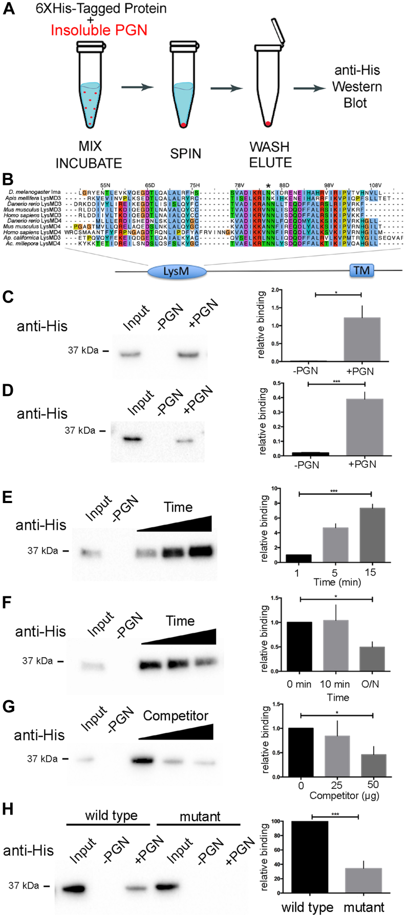
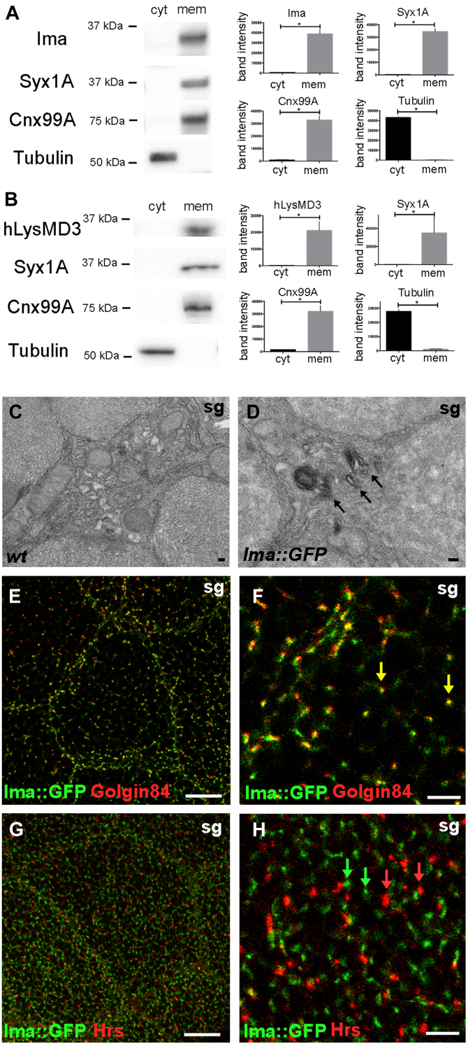
Microbial recognition is a key step in regulating the immune signaling pathways of multicellular organisms. Peptidoglycan, a component of the bacterial cell wall, exhibits immune stimulating activity in both plants and animals. Lysin motif domain (LysMD) family proteins are ancient peptidoglycan receptors that function in bacteriophage and plants. This report focuses on defining the role of LysMD-containing proteins in animals. Here, we characterize a novel transmembrane LysMD family protein. Loss-of-function mutations at the lysMD3/4 locus in Drosophila are associated with systemic innate immune activation following challenge, so we refer to this gene as immune active (ima). We show that Ima selectively binds peptidoglycan, is enriched in cell membranes, and is necessary to regulate terminal innate immune effectors through an NF-kB-dependent pathway. Hence, Ima fulfills the key criteria of a peptidoglycan pattern recognition receptor. The human Ima ortholog, hLysMD3, exhibits similar biochemical properties. Together, these findings establish LysMD3/4 as the founding member of a novel family of animal peptidoglycan recognition proteins.

Keywords: Drosophila; NF-kappa B; human; innate immunity; lysin motif domain; pattern recognition receptor; peptidoglycan.


Conflict of interest statement

Conflict of interest The authors declare that they have no conflicts of interest with the contents of this article.

Figures

Figure 1
Figure 1
LysMD proteins bind gram-negative peptidoglycan. A, an affinity-binding assay was used to measure the interaction between recombinant LysMD-containing proteins and insoluble Gram-negative peptidoglycan (PGN). B, sequence comparison of LysM domains from different species showing regions of amino acid conservation. CH, Western blots were probed with anti-His antibodies to detect 6XHis-tagged proteins and signal intensity was quantified. Two percent of the total 6XHis-tagged protein present in each binding reaction was directly loaded on the gel alone, as a positive control for detection of 6XHis-tagged protein (Input). C, 6XHis-Ima was incubated in the presence or absence of PGN (±PGN); n = 3 independent trials. Values normalized to input. D, 6XHis-humanLysMD3 was incubated in the presence or absence of PGN (±PGN); n = 5 independent trials. Values normalized to input. E, 6XHis-Ima was incubated in the presence or absence of PGN (±PGN) for different periods of time. The effect of varying incubation time on the extent of PGN binding was measured; n = 3 independent trials. Values normalized to binding at 1 min. F, 6XHis-Ima was incubated in the presence or absence of PGN (±PGN). The effect of increasing wash time prior to elution on the extent of PGN binding was measured; n = 3 independent trials. Values normalized to binding at 0 min dissociation. G, 6XHis-Ima was incubated in the presence or absence of PGN (±PGN). The effect of increasing soluble competitor on PGN binding was measured; n = 4 independent trials. Values normalized to 0 μg competitor. H, 6XHis-Ima and 6XHis-Ima[N85A] point mutant (asterisk in B) were incubated in the presence or absence of PGN (±PGN). The effect of mutating a single conserved residue in the LysM domain on PGN binding was measured; n = 6 independent trials. Values normalized to WT binding. ∗p < 0.05, ∗∗∗p < 0.001, unpaired Students t test; Error bars, SE. ima, immune active; LysMD, lysin motif domain.
Figure 2
Figure 2
LysMD proteins localize to cell membranes.A and B, biochemical separation of whole cell lysates from adult flies into cytoplasmic (cyt) and membrane (mem) subfractions. Fractions were analyzed for protein components using Western blotting; Syx1A, plasma membrane marker; Cnx99A, ER membrane marker; Tubulin, cytoplasm marker. Mann-Whitney, ∗p < 0.05. Band intensity is plotted in arbitrary fluorescence units. A, WT flies; anti-Ima; n = 4 independent trials. B, the human ima homolog, hlysMD3, was expressed in WT Drosophila using the ubiquitous da-gal4 promoter driving the UAS-hlysMD3 transgene; anti-hLysMD3; n = 4 independent trials. C and D, distribution of Ima-GFP using immuno-EM. Arrows show stacked Golgi membranes in larval salivary gland (sg) cells. The scale bar represents 100 nm. EH, colocalization of Ima-GFP with endomembrane markers using immunohistocytochemistry and confocal microscopy in larval salivary gland (sg) cells. Golgin-84, marker of Golgi rims; Hrs, early endosome. E and G, the scale bar represents 20 μm. F and H, the scale bar represents 5 μm. ER, endoplasmic reticulum; ima, immune active; LysMD, Lysin motif domain.
Figure 3
Figure 3
LysMD proteins protect against gram-negative immune challenge. A pathogen challenge model was used to test ima function in response to Pseudomonas entomophila exposure in vivo. A and B, mock treatment, dotted lines. Pe treatment, solid lines. w1118, (wt); w1118; imaΔ/imaΔ; da-gal4/+, (imaΔ); w1118; imaΔ/imaΔ; da-gal4/UAS-ima, (imaΔ; da>UAS-ima); w1118; imaΔ/imaΔ; da-gal4/UAS-hLysMD3 (imaΔ; da>UAS-hLysMD3). A, survival of adult flies homozygous for the imaΔ mutation in the presence (blue) and absence (red) of a WT UAS-ima transgene under the control of the ubiquitous da promoter. n ≥ 248, three independent trials. p < 0.0001, log-rank test. B, survival of adult flies homozygous for the imaΔ mutation in the presence (blue) and absence (red) of a UAS-hLysMD3 transgene under the control of the ubiquitous da promoter. n ≥ 353, three independent trials. p < 0.0001, log-rank test. C, qRT-PCR was used to measure transcriptional changes in WT and homozygous imaΔ whole adult flies to a panel of innate immune effector genes 24 h after Pe exposure. n ≥ 6. ∗p < 0.05, ∗∗p < 0.01, ∗∗∗p < 0.001, unpaired Student’s t test using Welch’s correction. Relative expression indicates normalization to WT mock-treated condition. D, confocal microscopy was used to visualize the distribution of Ima-GFP signal in adult midgut enterocytes (green, grayscale middle panel). Samples were colabeled for the medial Golgi marker P120 (red, grayscale right panel) and for DNA (blue). E, survival of adult flies homozygous for the imaΔ mutation in the presence (blue) and absence (red) of a UAS-ima transgene under the control of the gut enterocyte Myo1A promoter. Mock treatment, dotted lines. Pe treatment, solid lines. w1118, (wt); w1118; imaΔ, Myo1A-gal4/imaΔ(imaΔ); w1118; imaΔ, Myo1A-gal4/imaΔ; UAS-ima/+, (imaΔ, Myo1A>UAS-ima). n ≥ 220, three independent trials. p < 0.0001, log-rank test. F, qRT-PCR was used to measure transcriptional changes in isolated adult midguts dissected from WT and homozygous imaΔ flies on a panel of innate immune effector genes 24 h after Pe exposure. n ≥ 6. ∗p < 0.05, ∗∗p < 0.01, ∗∗∗p < 0.001, unpaired Student’s t test using Welch’s correction. Relative expression indicates normalization to WT mock-treated condition. Error bars, SE. The scale bar represents 10 μm. ima, immune active; LysMD, Lysin motif domain; qRT-PCR, quantitative reverse transcription PCR.
Figure 4
Figure 4
ima mutants interact genetically with Imd immune signaling pathway components. A and B, pathogen challenge model was used to test genetic interactions between ima and Imd pathway components in response to Pe exposure. A, survival analysis. w1118, (wt); w1118; imaΔ/imaΔ(w; imaΔ); y1, w1118/w1118; imaΔ/imaΔ(yw/w; imaΔ); y1, w67C23, PGRP-LE112/w1118; imaΔ/imaΔ(PGRP-LE112, yw/w; imaΔ); w1118; imaΔ/imaΔ; PGRP-LCΔ5/+ (w; imaΔ; PGRP-LCΔ5/+); w1118; imaΔ/imaΔ; RelE20/+ (w; imaΔ; RelE20/+). Mock treatment, dotted lines. Pe treatment, solid lines. n ≥ 139, three independent trials. p < 0.0001, log-rank test. B, AMP analysis. qRT-PCR was used to measure transcriptional changes of the AMPs dptA and cecA1 in adults 24 h after Pe exposure. w1118, (wt); w1118; imaΔ/imaΔ(w; imaΔ); y1, w1118; imaΔ/imaΔ(yw; imaΔ); y1, w67C23, PGRP-LE112; imaΔ/imaΔ(yw, PGRP-LE112; imaΔ); w1118; imaΔ/imaΔ; PGRP-LCΔ5/PGRP-LCΔ5(w; imaΔ; PGRP-LCΔ5); w1118; imaΔ/imaΔ; PGRP-LC1/PGRP-LC1(w; imaΔ; PGRP-LC1); w1118; imaΔ/imaΔ; RelE20/RelE20 (w; imaΔ; RelE20). n ≥ 3. ∗∗∗p < 0.001, ∗∗∗∗p < 0.0001, NS not significant, Student’s t test using Welch’s correction. Relative expression indicates normalization to WT mock treated condition. C, Relish immunostaining was used to measure Relish nuclear fluorescence relative to untreated wt levels in fat bodies (fb) from wt, imaΔ or RelE20 mutants (w1118; RelE20) 1 h after systemic Pe or Ecc15 challenge. n ≥ 3. ∗p < 0.05, ∗∗p < 0.01, ∗∗∗p < 0.001, ∗∗∗∗p < 0.0001, NS not significant, Student’s t test using Welch’s correction. Relative nuclear fluorescence is normalized to untreated WT condition. The scale bar represents 100 μm. AMA, antimicrobial peptides; AMPs, antimicrobial peptides; ima, immune active; Imd, immune deficiency; PGRP, peptidoglycan recognition proteins; qRT-PCR, quantitative reverse transcription PCR.

References

    1. Schumann P. Peptidoglycan structure. Methods Microbiol. 2011;38:101–129.
    1. Turner R.D., Vollmer W., Foster S.J. Different walls for rods and balls: the diversity of peptidoglycan. Mol. Microbiol. 2014;91:862–874. - PMC - PubMed
    1. Vollmer W., Seligman S.J. Architecture of peptidoglycan: more data and more models. Trends Microbiol. 2009;18:59–66. - PubMed
    1. Schleifer K.H., Kandler O. Peptidoglycan types of bacterial cell walls and their taxonomic implications. Bacteriol. Rev. 1972;36:407–477. - PMC - PubMed
    1. Vollmer W. Structural variation in the glycan strands of bacterial peptidoglycan. FEMS Microbiol. Rev. 2008;32:287–306. - PubMed

Publication types

LinkOut - more resources