. 2023 Apr;11(4):e006718.
doi: 10.1136/jitc-2023-006718.

Vector Aided Microenvironment programming (VAMP): reprogramming the TME with MVA virus expressing IL-12 for effective antitumor activity

Affiliations

Vector Aided Microenvironment programming (VAMP): reprogramming the TME with MVA virus expressing IL-12 for effective antitumor activity

Laura Seclì et al. J Immunother Cancer. 2023 Apr.

Abstract

Background: Tumor microenvironment (TME) represents a critical hurdle in cancer immunotherapy, given its ability to suppress antitumor immunity. Several efforts are made to overcome this hostile TME with the development of new therapeutic strategies modifying TME to boost antitumor immunity. Among these, cytokine-based approaches have been pursued for their known immunomodulatory effects on different cell populations within the TME. IL-12 is a potent pro-inflammatory cytokine that demonstrates striking immune activation and tumor control but causes severe adverse effects when systemically administered. Thus, local administration is considered a potential strategy to achieve high cytokine concentrations at the tumor site while sparing systemic adverse effects.

Methods: Modified Vaccinia Ankara (MVA) vector is a potent inducer of pro-inflammatory response. Here, we cloned IL-12 into the genome of MVA for intratumoral immunotherapy, combining the immunomodulatory properties of both the vector and the cargo. The antitumor activity of MVA-IL-12 and its effect on TME reprogramming were investigated in preclinical tumor models. RNA sequencing (RNA-Seq) analysis was performed to assess changes in the TME in treated and distal tumors and the effect on the intratumoral T-cell receptor repertoire.

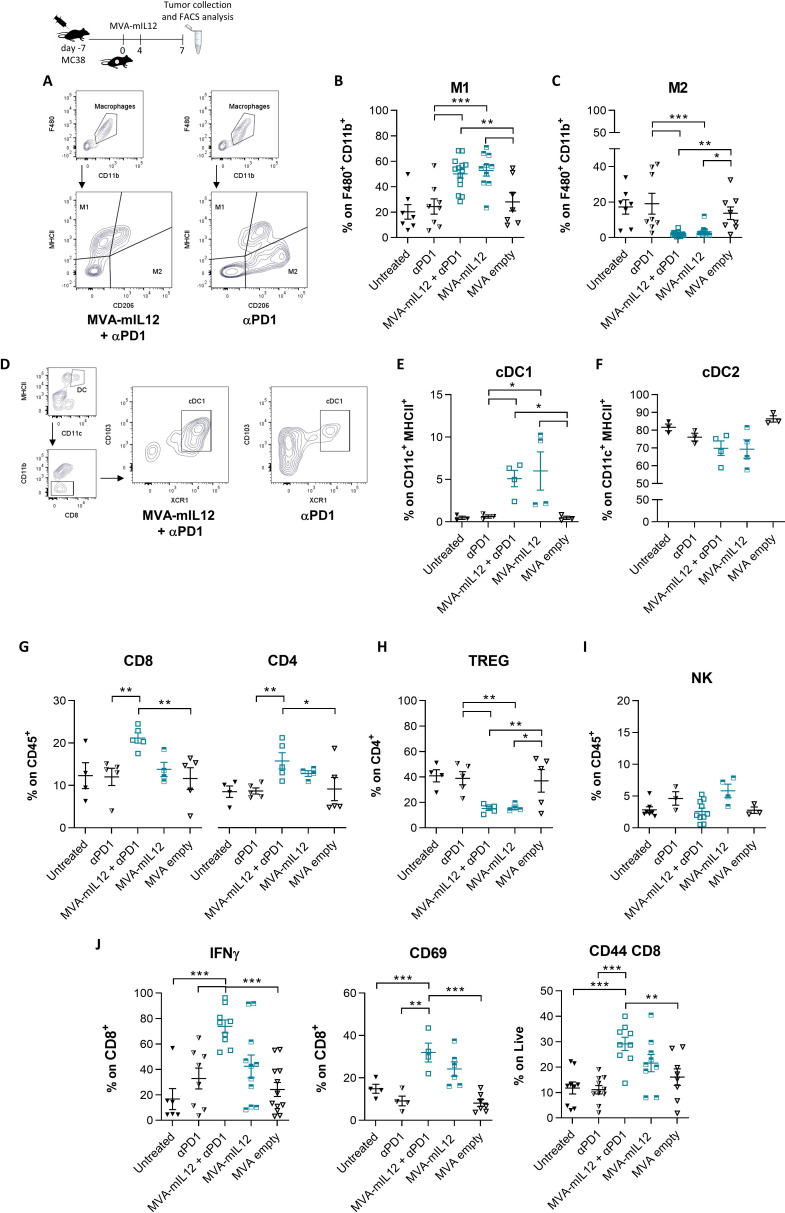
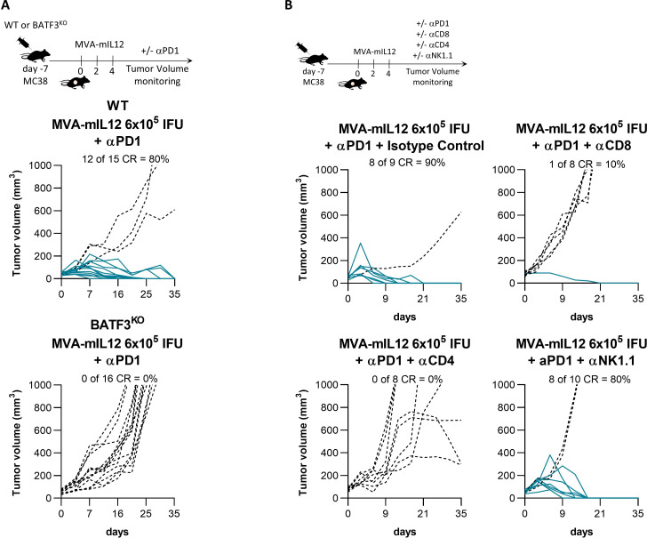
Results: Intratumoral injection of MVA-IL-12 resulted in strong antitumor activity with the complete remission of established tumors in multiple murine models, including those resistant to checkpoint inhibitors. The therapeutic activity of MVA-IL-12 was associated with very low levels of circulating cytokine. Effective TME reprogramming was demonstrated on treatment, with the reduction of immunosuppressive M2 macrophages while increasing pro-inflammatory M1, and recruitment of dendritic cells. TME switch from immunosuppressive into immunostimulatory environment allowed for CD8 T cells priming and expansion leading to tumor attack.

Conclusions: Intratumoral administration of MVA-IL-12 turns immunologically 'cold' tumors 'hot' and overcomes resistance to programmed cell death protein-1 blockade.

Keywords: cytokines; immunomodulation; immunotherapy; lymphocytes, tumor-infiltrating; tumor microenvironment.


Conflict of interest statement

Competing interests: ES is founder of Nouscom. AMD, LS, LI, LN, MDL, GC, GL, EM, IG, GS, FT, SA, GR are employees of Nouscom.

Figures

Figure 1
Figure 1
Potent efficacy of intratumoral MVA-mIL-12 in different syngeneic preclinical models. (A) Schematic structure of MVA-mIL-12. (B) HeLa cells were infected at MOI 5 with MVA-mIL-12 or MVA empty and supernatants were collected at different time points to quantify IL-12 by ELISA. (C) In vivo levels of IL-12. C57BL/6 mice were challenged subcutaneously (SC) with MC38 cells. After 7 days, mice with established tumors were randomized, treated intratumoral (IT) with MVA-mIL-12 (6×10⁵ IFU), and serum was collected at different time points post injection. The amount of IL-12 was measured by ELISA in tumor tissue and serum (n≥3). (D) C57BL/6 mice were challenged SC with MC38 cells. After 7 days, mice with established tumors were randomized and treated IT three times with MVA-mIL-12 at 107 IFU alone or in combination with αPD-1, with MVA-mIL-12 at 6×10⁵ IFU alone or in combination with α-PD-1 or α-PD-1 as control. Tumor volume was monitored over time. Lines in the graphs represent each individual tumor (full lines, responder tumors showing a complete response (CR); dot lines, non-responder tumors). Percentages on the graphs indicate the rate of CR. (E) Tumor volumes are shown as mean±SEM with n≥6 (day 25). (F) Mice considered complete responders were re-challenged with MC38, 40 days after tumor challenge. Tumor volumes post re-challenge are shown as mean±SEM with n=10. One-way ANOVA test with Fisher’s LSD test was performed to obtain p value. (G) Survival curves of C57BL/6 mice SC injected with B16F10 cells. After 7 days, mice with established tumors were randomized and treated IT eight times with MVA-mIL-12 at 6×10⁵ IFU or MVA empty at 6×10⁵ IFU alone or in combination with αPD-1 or αPD-1 as control. n≥6. Comparison of survival curves with Gehan-Breslow-Wilcoxon test. *, p<0.05; ***, p<0.001; ****, p<0.0001. ANOVA, analysis of variance; IFU, Infectious Unit; IL, interleukin; LSD, least significant difference; m, murine; MOI, Multiplicity of Infection; MVA, Modified Vaccinia virus Ankara; ns, not significant; PD-1, programmed cell death protein-1.
Figure 2
Figure 2
Local MVA-mIL-12 injection combined with αPD-1 exerts antitumoral activity on non-injected distant tumors. (A–F) C57BL/6 mice were challenged SC with MC38 cells on both flanks. After 7 days, mice with established tumors of similar volume were randomized. One tumor (treated) was injected IT with MVA-mIL12 at 107 IFU or 6×10⁵ in combination with αPD-1 (A, B) the other one (distal) did not receive any IT treatment (D, E). Mice treated with αPD-1 only were used as control for both flanks (C, F). Tumor volume was monitored over time. Lines in the graphs represent each individual tumor (full lines, responder tumors; dot lines, non-responder tumors). Percentages on the graphs indicate the rate of complete response (CR). n≥9. (G) C57BL/6 mice were challenged SC with MC38 cells on both flanks. After 7 days, mice with established tumors of similar volume were randomized. One tumor (treated) was injected IT with MVA-mIL12 6×10⁵ and the other one was left untreated (distal). ELISA was performed on both tumors: blue dots represent treated tumors, violet dots untreated (n=3). IFU, Infectious Unit; IL, interleukin; IT, intratumoral; m, murine; MVA, Modified Vaccinia virus Ankara; PD-1, programmed cell death protein-1; SC, subcutaneous.
Figure 3
Figure 3
The TME is re-shaped by intratumoral treatment with MVA-mIL12. C57BL/6 mice were challenged SC with MC38 cells. After 7 days, mice with established tumors were randomized and treated IT as in the scheme with αPD-1, MVA-IL-12 at 6×10⁵ IFU, the combination of MVA-IL-12 (6×10⁵ IFU) with αPD-1 or with MVA empty (6×10⁵ IFU). Tumors from all the groups were collected and analyzed by Flow Cytometry. n≥3. In (A) representative contour plot of macrophages subpopulations; in (B) the frequency of MHCII+ CD206 M1 macrophages, in (C) CD206+ MHCII M2 macrophages; in (D) representative contour plot of cDC1; in (E) graph represents CD103+ XCR1+ cDC1, in (F) XCR1 SIRP+ cDC2, in (G) CD8+ and CD4+ lymphocytes, in (H) TREG CD4+ FOXP3+, in (I) NK cells, in (J) CD8+ IFN-γ+, CD8+ CD69+ and memory CD8+ CD44+ lymphocytes. Data are presented as median values+SEM; n≥3 mice. One-way ANOVA with the Fisher LSD test was performed to obtain p values. *, p<0.05; **, p<0.005; ***, p<0.001. ANOVA, analysis of variance; cDC1, conventional type 1 DC; DC, dendritic cell; FACS, Fluorescence-Activated Cell Sorter; IFN, interferon; IFU, Infectious Unit; IL, interleukin; IT, intratumoral; LSD, least significant difference; m, murine; MHC, Major Histocompatibility Complex; MVA, Modified Vaccinia virus Ankara; NK, natural killer; PD-1, programmed cell death protein-1; SC, subcutaneous; TME, tumor microenvironment; TREG, Regulatory T cells.
Figure 4
Figure 4
The efficacy of MVA-mIL-12 + αPD-1 is dependent on cDC1, CD4 and CD8. (A) C57BL/6 WT or BATF3KO mice were challenged SC with MC38 cells. After 7 days, mice with established tumors were randomized and treated IT with MVA-mIL-12 (6×10⁵ IFU) and αPD-1. Lines in the graphs represent each individual tumor (full lines, responder tumors; dot lines, non-responder tumors). Percentages on the graphs indicate the rate of efficacy. (B) C57BL/6 mice were challenged SC with MC38 cells. After 7 days, mice with established tumors were randomized and treated IT with MVA-mIL-12 (6×10⁵ IFU) and αPD-1 in combination or not with isotype control, αCD8, αCD4 or αNK1.1 depletion antibody. Lines in the graphs represent each individual tumor (full lines, responder tumors; dot lines, non-responder tumors). Percentages on the graphs indicate the rate of efficacy. CR, complete response; IFU, Infectious Unit; IL, interleukin; IT, intratumoral; m, murine; MVA, Modified Vaccinia virus Ankara; NK, natural killer; PD-1, programmed cell death protein-1; SC, subcutaneous; WT, wild-type.
Figure 5
Figure 5
MVA-mIL-12 induces expansion and diversification of the intratumoral TCR repertoire and affects immune-related TME signature. MC38 tumor bearing mice were treated with MVA-IL-12 (6×10⁵ IFU IT; d0, d4) with or without αPD-1, MVA empty, αPD-1 or left untreated. Tumors were harvested at day 7 and RNA-Seq was performed. (A) Bar charts reporting the number of TCR-β clones detected by RNA-Seq. Each bar represents a TCR-β individual clonotype. On the vertical axis the number of copies of each clonotype is reported. A representative mouse TCR-β repertoire for each group is shown. (B) Mean (+SEM) number of TCR clonotypes in each group (n=3). One-way ANOVA test with Fisher’s LSD test was performed to obtain p value. *, p<0.05; **, p<0.005; ***, p<0.001. (C) Heatmap representing the differentially expressed genes (red: up-regulated; blue: down-regulated) detected by RNA-Seq on tumors of mice for each group of treatment versus untreated (n≥3) (median log2 fold change ≤1 or >1; consensus of 3 methods; Benjamini-Hochberg corrected p value<0.05). (D) Violin plots of select genes. Genes found differentially expressed when compared with untreated are indicated with an asterisk (*), while a cross (×), or a dot (·) indicates genes found differentially expressed versus αPD-1 and MVA empty, respectively. ANOVA, analysis of variance; IL, interleukin; IT, intratumoral; LSD, least significant difference; m, murine; MVA, Modified Vaccinia virus Ankara; PD-1, programmed cell death protein-1; TCR, T-cell receptor; TPM, Transcripts per Million; RNA-Seq, RNA sequencing.
Figure 6
Figure 6
MVA-mIL-12 reshapes the TME of both treated and distant tumors. (A) Mean (+SEM) number of intratumoral TCR clonotypes in treated and distal MC38 tumors (bilateral tumor model) (n=3). Treated tumors were injected IT with MVA-mIL-12 at 6×10⁵ IFU (d0, d4) in combination with αPD-1, while the distant tumors did not receive any IT treatment. One-way ANOVA test with Fisher’s LSD test was performed to obtain p value. *, p<0.05; (B) Representative bar chart reporting the total number of T-cell clones detected in treated and distal tumors. Shared clonotypes are represented with the same color. Gray clones are unique for each group. (C) Frequency of shared TCR between treated and distal tumors treated with MVA-mIL-12 and αPD-1 compared with the untreated group. (D) Bar chart showing the number of DEG (red: upregulated; blue; downregulated) detected by RNA-Seq on treated/distal tumors and untreated controls compared with each other. (E) Venn diagram displaying the overlap among the lists of DEGs obtained in each comparison. (F) Heatmap showing the expression values (TPM) of immune-related genes of interest detected in treated and distal tumors of mice receiving MVA-mIL-12 and αPD-1 versus untreated controls (left and right tumors). The annotation columns indicate DEGs in a specific comparison (median log2 fold change of at least ±1; consensus of three different methods Benjamini-Hochberg corrected p value in each method <0.05). (G) MC38 bilateral tumor bearing mice were treated with the combination of MVA-mIL-12 and αPD-1, MVA empty and αPD-1 or left untreated. Treated and distal tumors were collected and analyzed by RT-PCR. One-way ANOVA test with Fisher’s LSD test was performed to obtain p value. *, p<0.05; **, p<0.005; ***, p<0.001. ANOVA, analysis of variance; DEG, differentially expressed gene; IFU, Infectious Unit; IL, interleukin; IT, intratumoral; LSD, least significant difference; m, murine; MVA, Modified Vaccinia virus Ankara; PD-1, programmed cell death protein-1; RNA-Seq, RNA sequencing; TCR, T-cell receptor; TME, tumor microenvironment; TPM, Transcripts per Million.
Figure 7
Figure 7
Working model of MVA-IL-12. Intratumoral administration of M-IL-12 resulted in TME reprogramming, reduction of immunosuppressive M2 macrophages while increasing pro-inflammatory M1, and recruitment of dendritic cells. This switch from immunosuppressive into immunostimulatory TME allows CD8 T cells to exert cytolytic activity against the tumor. IL, interleukin; MVA, Modified Vaccinia virus Ankara; TME, tumor microenvironment.

Similar articles

Cited by

References

    1. Schoenfeld AJ, Hellmann MD. Acquired resistance to immune checkpoint inhibitors. Cancer Cell 2020;37:443–55. 10.1016/j.ccell.2020.03.017 - DOI - PMC - PubMed
    1. Binnewies M, Roberts EW, Kersten K, et al. . Understanding the tumor immune microenvironment (TIME) for effective therapy. Nat Med 2018;24:541–50. 10.1038/s41591-018-0014-x - DOI - PMC - PubMed
    1. Labani-Motlagh A, Ashja-Mahdavi M, Loskog A. The tumor microenvironment: a milieu hindering and obstructing antitumor immune responses. Front Immunol 2020;11:940. 10.3389/fimmu.2020.00940 - DOI - PMC - PubMed
    1. Hinshaw DC, Shevde LA. The tumor microenvironment innately modulates cancer progression. Cancer Res 2019;79:4557–66. 10.1158/0008-5472.CAN-18-3962 - DOI - PMC - PubMed
    1. Bejarano L, Jordāo MJC, Joyce JA. Therapeutic targeting of the tumor microenvironment. Cancer Discov 2021;11:933–59. 10.1158/2159-8290.CD-20-1808 - DOI - PubMed

Supplementary concepts