. 2023 Apr 28;13(1):7005.
doi: 10.1038/s41598-023-34225-5.

Visual perceptual load and processing of somatosensory stimuli in primary and secondary somatosensory cortices

Affiliations

Visual perceptual load and processing of somatosensory stimuli in primary and secondary somatosensory cortices

Antje Peters et al. Sci Rep. .

Abstract

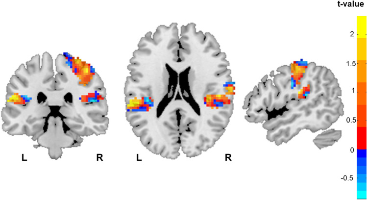
Load theory assumes that neural activation to distractors in early sensory cortices is modulated by the perceptual load of a main task, regardless of whether task and distractor share the same sensory modality or not. While several studies have investigated the question of load effects on distractor processing in early sensory areas, there is no functional magnetic resonance imaging (fMRI) study regarding load effects on somatosensory stimuli. Here, we used fMRI to investigate effects of visual perceptual load on neural responses to somatosensory stimuli applied to the wrist in a study with 44 participants. Perceptual load was manipulated by an established sustained visual detection task, which avoided simultaneous target and distractor presentations. Load was operationalized by detection difficulty of subtle or clear color changes of one of 12 rotating dots. While all somatosensory stimuli led to activation in somatosensory areas SI and SII, we found no statistically significant difference in brain activation to these stimuli under high compared to low sustained visual load. Moreover, exploratory Bayesian analyses supported the absence of differences. Thus, our findings suggest a resistance of somatosensory processing to at least some forms of visual perceptual load, possibly due to behavioural relevance of discrete somatosensory stimuli and separable attentional resources for the somatosensory and visual modality.


Conflict of interest statement

The authors declare no competing interests.

Figures

Figure 1
Figure 1
Schematic diagram of the experimental procedure. There was one low-load and one high-load run with 50 somatosensory stimulus events and 50 null events each. The high- and low-load conditions differed in the difficulty of the tasks: The target detection with subtle color change corresponded to the high-load task, and the detection with clearly visible color change corresponded to the low-load task. There were 10 target presentations per run. The order of the runs was counterbalanced between subjects. Rotation changes are indicated with dashed white arrows for illustration purposes. Their actual occurrence was more frequent and variable in timing (see main text for details). The timeline illustrates the time points of stimulus events (orange dashes), null events (grey dashes), and visual target stimuli (black dashes).
Figure 2
Figure 2
Regions of interest where processing of somatosensory stimuli was investigated: The primary somatosensory cortex contralateral to the stimulus application SI (red) and the secondary somatosensory cortex in both hemispheres SII contralateral (blue) and SII ipsilateral (yellow). Displayed layers intersect at x, y, z = 50, − 30, 20.
Figure 3
Figure 3
Clusters of increased activity in the load contrast during target processing in the task-difficulty ROIs and visual areas for N = 27 participants. In the task-difficulty ROIs, increased activation was found under high load in the right insula (peak t-value = 5.40, p < 0.05) and the right anterior cingulate cortex (peak t-value = 4.09, p < 0.05). In visual areas, one cluster of increased activity under high load was observed in the left occipital pole (peak t-value = 4.23, p < 0.05). Displays of the clusters are accompanied by raincloud plots of the individual data points in each condition, comprising a jittered scatter plot of the cluster average of the betas per subject, a box-and-whisker plot, and a violin plot of the same data.
Figure 4
Figure 4
Clusters of increased activity in the stimulus compared to the no stimulus condition in the somatosensory areas in the primary and secondary somatosensory cortices (displayed in Fig. 2) for N = 44 participants. In the primary somatosensory cortex, three clusters with increased activity can be found: Two corresponding to the hand area of the left wrist, where the somatosensory stimuli were applied and one in the right parietal operculum in the neighbourhood of the secondary somatosensory cortex (cluster 1: peak t-value = 3.59, cluster 2: peak t-value = 4.64, both p < 0.05). In the secondary somatosensory cortex, one cluster can be found on each side (ipsilateral: peak t-value = 4.96, contralateral: peak t-value = 6.03, both p < 0.05). Displays of the clusters are accompanied by raincloud plots of the individual data points in each condition, comprising a jittered scatter plot of the cluster average of the betas per subject, a box-and-whisker plot, and a violin plot of the same data.
Figure 5
Figure 5
Voxelwise t-maps for the stimulus × load interaction in somatosensory areas (contralateral SI, contra-, and ipsilateral SII) for exploratory reasons. In this figure, the untresholded t-values per voxel for the contrast (low load (stimulus–no stimulus)—high load (stimulus–no stimulus)) are shown. While descriptively increased t-values are seen, please note that the permutation analysis did not reveal significant effects and that Bayes factors for the averaged activity in somatosensory areas support the null hypothesis.

References

    1. Lavie N. Perceptual load as a necessary condition for selective attention. J. Exp. Psychol. Hum. Percept. Perform. 1995;21:451–468. doi: 10.1037/0096-1523.21.3.451. - DOI - PubMed
    1. Lavie N, Tsal Y. Perceptual load as a major determinant of the locus of selection in visual attention. Percept. Psychophys. 1994;56:183–197. doi: 10.3758/BF03213897. - DOI - PubMed
    1. Lavie N. Distracted and confused?: Selective attention under load. Trends Cogn. Sci. 2005;9:75–82. doi: 10.1016/j.tics.2004.12.004. - DOI - PubMed
    1. Lavie N. Attention, distraction, and cognitive control under load. Curr. Dir. Psychol. Sci. 2010;19:143–148. doi: 10.1177/0963721410370295. - DOI
    1. Brockhoff L, Schindler S, Bruchmann M, Straube T. Effects of perceptual and working memory load on brain responses to task-irrelevant stimuli: Review and implications for future research. Neurosci. Biobehav. Rev. 2022;135:104580. doi: 10.1016/j.neubiorev.2022.104580. - DOI - PubMed

Publication types