Review
. 2023 Jun;7(6):439-455.
doi: 10.1038/s41570-023-00483-0. Epub 2023 Apr 17.

Miniaturizing chemistry and biology using droplets in open systems

Affiliations
Review

Miniaturizing chemistry and biology using droplets in open systems

Yuting Zeng et al. Nat Rev Chem. 2023 Jun.

Abstract

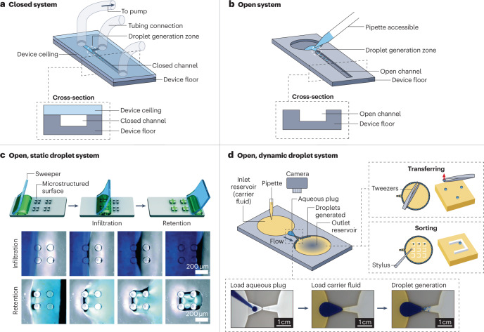
Open droplet microfluidic systems manipulate droplets on the picolitre-to-microlitre scale in an open environment. They combine the compartmentalization and control offered by traditional droplet-based microfluidics with the accessibility and ease-of-use of open microfluidics, bringing unique advantages to applications such as combinatorial reactions, droplet analysis and cell culture. Open systems provide direct access to droplets and allow on-demand droplet manipulation within the system without needing pumps or tubes, which makes the systems accessible to biologists without sophisticated setups. Furthermore, these systems can be produced with simple manufacturing and assembly steps that allow for manufacturing at scale and the translation of the method into clinical research. This Review introduces the different types of open droplet microfluidic system, presents the physical concepts leveraged by these systems and highlights key applications.


Conflict of interest statement

A.B.T. has ownership in Stacks to the Future. S.T. and E.B. have ownership in Stacks to the Future, Tasso and Salus Discovery. Y.Z., J.W.K., T.L.v.N., W-c.T. and J.B. have no conflicts of interest.

Figures

Fig. 1
Fig. 1. Comparing closed and open channel droplet microfluidic systems.
An overview of closed and open droplet systems in which we subdivide open droplet systems into two categories: static and dynamic. a, A schematic and cross-sectional view of a closed microfluidic system in which the channel is accessible only through tubing connection ports. In this setup, droplets are generated using active pumps connected through tubing. b, A schematic and cross-sectional view of an open microfluidic system in which there is no ceiling, making the channel accessible. c, A static open droplet system that uses a sweeping method to load droplets onto the platform. Top: the stages in the sweeping process, which traps droplets within the microstructures on the platform surface. Bottom: images of the platform during the infiltration and retention stages. d, A schematic of a dynamic open droplet system that generates droplets autonomously by constricting the open channel. Bottom inset: photographs of the stages of droplet generation in the open device. Top right inset: examples of droplet manipulation using tweezers or a stylus made from polytetrafluoroethylene. Part c is reprinted with permission from ref. , Royal Society of Chemistry. Part d is adapted from ref. under a Creative Commons licence CC BY 4.0.
Fig. 2
Fig. 2. Forces acting at droplet interfaces.
Key physical considerations and theory that aid the design of open droplet microfluidic systems. a, Asymmetry of molecular interactions at an air–liquid interface results in surface tension. The yellow and green arrows indicate the interactions between air and liquid molecules, respectively. b, Sketches of a sharp (left) or smeared (right) interface, corresponding to high or low surface tension, respectively. c, The Young–Dupré law for a sessile liquid droplet (L1) immersed in an immiscible liquid (L2) on a solid surface (S), where γ is the surface tension and θ is the contact angle. d, The Neumann triangle for the contact of two liquid phases (L1 and L2) and a gas phase (G). e, The dispersed phase (that is, the droplet, shown in yellow and orange) can float on (left) or be immersed in (right) a carrier fluid (green). The different locations of the droplet were calculated using the Surface Evolver software. f, Different flow behaviours of the carrier fluid (green) in a rounded- or rectangular-bottomed open channel. Capillary filaments can form in rectangular-bottomed channels but not rounded-bottomed ones. The three rounded-bottomed channels show how the aspect (width to height) ratio of the channel affects the meniscus shape of the flow. g, A schematic (left) and photo (right) of the formation of capillary filaments by the carrier fluid in the corners of a rectangular channel, where 2α is the corner interior angle. h, Example of two-phase flow in a channel in which the dispersed liquid phase (that is, droplets and plugs) does not wet the walls,, produced using the Surface Evolver software. i, Photos of a two-phase open channel flow with an aqueous plug (yellow) dispersed in a carrier fluid, 1-nonanol (blue). Liquids were dyed with food colouring for visualization. Part e is adapted with permission from ref. , John Wiley & Sons. Part f (left) is adapted with permission from ref. , IOP Publishing. Part f (right) is adapted with permission from ref. , Wiley-Scrivener. Part g (left) is adapted with permission from ref. , IOP Publishing. Part g (right) is adapted from ref. under a Creative Commons licence CC BY 4.0. Part h is adapted with permission from ref. , John Wiley & Sons. Part i (left) is reprinted with permission from ref. , IOP Publishing. Part i (right) is reprinted with permission from ref. , American Chemical Society.
Fig. 3
Fig. 3. Examples of droplet manipulation.
We divide droplet manipulation techniques into four categories that are typically used in droplet systems and are important for downstream applications. a, Droplet generation. Left: a schematic of acoustophoretic printing in which droplet generation is controlled by balancing the capillary force, gravity, acoustic force and radiation pressure. Right: a large-area image made from droplets produced by rasterized acoustophoretic printing. b, Droplet transportation and sorting. The use of magnetic beads to transport (orange), split (green), release (blue) and rotate (yellow) 250-μl droplets. The images show the state of the system at different times, t, into the process. The grey arrows indicate the direction that the robots are travelling in. c, Droplet splitting. The schematic for parallel plate droplet splitting. Droplets containing cells are first spotted onto a glass plate and allowed to incubate. Samples of the cell culture supernatants are extracted by applying a separate plate from above to which the droplets adhere, which is then removed to split the droplets while the cells remain in the droplets on the bottom plate owing to the effect of gravity. d, Droplet extraction. Images (top) and schematics (bottom) of the extraction of a single droplet using pipette aspiration. Part a is adapted from ref. , AAAS. Part b is reprinted with permission from ref. , AAAS. Part c is adapted with permission from ref. , American Chemical Society. Part d is adapted with permission from ref. , PNAS.
Fig. 4
Fig. 4. Open droplet microfluidics for synthesis and combinatorial reactions.
An illustration of common workflows in open droplet microfluidic systems that minimize reagent consumption and enable parallel reactions. a, Magnetically actuated robots (that is, two steel beads), indicated by the red arrows, controlled by a magnetic control system, are programmed to manipulate droplets precisely for use in a sequential acid–base neutralization reaction. The robot is used to transport a daughter droplet from the NaOH droplet to the indicator droplet, which changes from colourless to purple, indicating that it is now alkaline. The robot then transports a daughter droplet from the HCl droplet to the indicator droplet, which returns to colourless as it is neutralized. b, An on-chip solution-based platform for compound synthesis and cellular screening (chemBIOS). A library of lipidoids is generated on-chip by sandwiching together slide A, which contains droplets of amines, and slide B, which contains mixtures of thiolactone and pyridyl disulfide derivatives dissolved in dimethyl sulfoxide (DMSO). Sandwiching the slides initiates the reaction between the reagents (shown in the expansions) to produce a microarray of different lipidoids. c, An automated microfluidic nanolitre-droplet gradient system for high-throughput screening. The three steps are aspirating the sample, aspirating the diluent to form the gradient and generating the droplet. This process leads to a sequence of droplets with a concentration gradient. d, A gradient droplet array for high-throughput screening of cellular responses to varying combinations of chemical and mechanical cues visualized with fluorescent dyes. Part a is reprinted with permission from ref. , AAAS. Part b is adapted from ref. under a Creative Commons licence CC BY 4.0. Part c is adapted with permission from ref. , American Chemical Society. Part d is reprinted with permission from ref. , Royal Society of Chemistry.
Fig. 5
Fig. 5. Three-dimensional cell culture and spheroid formation in open droplet systems.
Spheroids are common biomimetic culture models and open microfluidic systems enable more sophisticated fluidic manipulation of hanging drop networks. a, Liquid droplets containing cells created on a polydimethylsiloxane (PDMS) substrate. The hydrophobic nature of PDMS produces rounded droplets, and the addition of extracellular matrix components such as collagen creates an environment that is similar to those experienced by cells in the body, which encourages spheroid formation. b, A fluidically connected hanging drop network used for parallel spheroid formation. A top-down view with a close-up image of the open channels (top) and a side view (bottom). c, An integrated medium reservoir enables long-term cultures (up to 30 days) in 3D open droplet systems. Top: a cross-sectional view of the device. Bottom: fluorescence images of spheroids formed in this platform show that the cells remain viable and maintain their shape throughout a 30-day culture period; cells were stained with calcein AM and propidium iodide to indicate living (green) and dead (red) cells, respectively. d, Top: droplet merging results in multi-spheroid architectures. Bottom left: fluorescence microscopy images of spheroids containing two different cell types 24 h after merging — HEK 293T stained with a green fluorescent dye and HeLa expressing red fluorescent protein (RFP). Bottom right: scanning electron microscopy image of a multi-spheroid architecture containing HepG2 cells 24 h after merging. Part a is adapted from ref.  under a Creative Commons licence CC BY 4.0. Part b is reprinted from ref. , Springer Nature Limited. Part c is adapted with permission from ref. , Elsevier. Part d is adapted with permission from ref. , Wiley.
Fig. 6
Fig. 6. Droplet analysis in open systems.
a, A multimicrochannel superwettable microspine chip for detecting prostate-specific antigen (PSA). The chip has detection zones that are pre-functionalized with biotinylated PSA antibodies (Ab1-biotin). The droplet containing target molecules and fluorescein isothiocyanate-labelled PSA antibodies (Ab2-FITC) is spontaneously transported to the detection zones to form a sandwich immunoassay for the fluorescent detection of PSA. b, The evaporation enrichment of analytes on superhydrophilic microwells with superhydrophobic substrates. Top: analytes are concentrated after liquid evaporation. Bottom: the application of the system as a biosensor for ultra-trace DNA samples. The superhydrophilic microwell is functionalized with capture DNA (middle), which only allows specific target DNA to be captured, concentrated and detected by the fluorescent probe DNA (left), but does not recognize non-complementary DNA (right). c, Illustration of a workflow for open microfluidic gel electrophoresis. The nanolitre DNA sample droplet was first spotted and dehydrated on a planar substrate (1). The droplet was then rehydrated with a liquid gel line of sieving gel matrix (2) and covered with mineral oil (3). Finally, an electric field and a detector were used to perform electrophoresis (4). Bottom: a photo of the system under oil without electrodes. d, Schematic of the push–pull probe design with two open microfluidic channels for scanning electrochemical microscopy imaging including the working electrode (WE) and the counter and reference electrodes (CE/RE). The insets show the two probes and a USB drive for size comparison (top) and a schematic of the push–pull probe for imaging (bottom). Part a is adapted with permission from ref. , American Chemical Society. Part b is adapted with permission from ref. , Wiley. Part c is adapted with permission from ref. , Wiley. Part d is adapted with permission from ref. , American Chemical Society.
Fig. 7
Fig. 7. Cell analysis in open droplet systems.
Open droplet microfluidic platforms enable more flexibility and control in cell culture experiments, and many of the platforms are compatible with conventional in vitro experimental readouts. a, Left: a schematic of an under-oil open microfluidic system used to study the growth dynamics and microcolony motility of wild-type Flavobacterium johnsoniae with physical and optical access. Right: a composite bright-field and fluorescent Concanavalin A (ConA) lectin-stained image of a microdrop obtained with this system 12 h after the culture growth was initiated. b, Left: a schematic of the oil-immersed lossless total analysis system (OIL-TAS) used to perform integrated RNA extraction and detection of SARS-CoV-2. The detection step uses loop-mediated isothermal amplification (LAMP). Right: images of a unit of the OIL-TAS device before and after extraction in which the wells were coloured for visualization. c, Top: a reconfigurable multilayer suspended cell-culture system (‘stacks’) for studying spatiotemporal paracrine signalling across different culture microenvironments. Bottom: immunocytochemistry images show the difference in morphology of the endothelial structures (green) in the endothelial–fibroblast mixture layer when macrophages are absent (left) and present (right) in the top layer. d, Single-cell proteomic analysis using a nanolitre-scale oil–air–droplet chip with a self-aligning monolithic device. The pretreatment of the sample involves the addition of RapiGest (RG), tris(2-carboxyethyl)-phosphine (TCEP), iodoacetamide (IAA), endoproteinase Lys-C, trypsin (Try) and formic acid (FA) to extract proteins from a single cell and digest them into peptides. The sample is then injected into a capillary liquid chromatography column for subsequent liquid chromatography tandem mass spectrometry analysis. The inset shows microscopy images of droplets containing a single cell. Part a is adapted from ref. under a Creative Commons licence CC BY 4.0. Part b is adapted from ref. under a Creative Commons licence CC BY 4.0. Part c is reprinted from ref. , Springer Nature Limited. Part d is adapted from ref. , American Chemical Society.

References

    1. Sun H, et al. A suspending-droplet mode paper-based microfluidic platform for low-cost, rapid, and convenient detection of lead(II) ions in liquid solution. Biosens. Bioelectron. 2018;99:361–367. doi: 10.1016/j.bios.2017.07.073. - DOI - PubMed
    1. Benz M, Molla MR, Böser A, Rosenfeld A, Levkin PA. Marrying chemistry with biology by combining on-chip solution-based combinatorial synthesis and cellular screening. Nat. Commun. 2019;10:2879. doi: 10.1038/s41467-019-10685-0. - DOI - PMC - PubMed
    1. Yu J, et al. Reconfigurable open microfluidics for studying the spatiotemporal dynamics of paracrine signalling. Nat. Biomed. Eng. 2019;3:830–841. doi: 10.1038/s41551-019-0421-4. - DOI - PMC - PubMed
    1. Kaigala GV, Lovchik RD, Delamarche E. Microfluidics in the ‘open space’ for performing localized chemistry on biological interfaces. Angew. Chem. Int. Edn. 2012;51:11224–11240. doi: 10.1002/anie.201201798. - DOI - PubMed
    1. Berthier, J., Brakke, K. A. & Berthier, E. Open Microfluidics (Wiley-Scrivener, 2016).

Publication types