

Differentiated Papillary NUT Carcinoma: An Unexpected, Deceptively Bland Presentation of a Sinonasal Carcinoma
- PMID: 37118352
- PMCID: PMC10513967
- DOI: 10.1007/s12105-023-01554-w
Differentiated Papillary NUT Carcinoma: An Unexpected, Deceptively Bland Presentation of a Sinonasal Carcinoma
Abstract
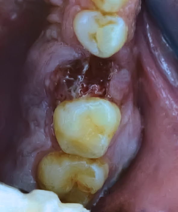
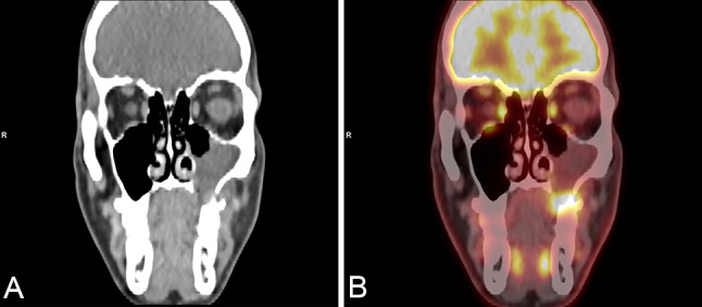
Background: In recent years, the list of tumor entities in the sinonasal tract has significantly expanded, requiring advanced diagnostic testing. We report the case of a 32-year-old patient with an unusual NUT carcinoma originating in the maxillary sinus, which showed extensive well-differentiated, papillary squamous morphology, similar to the spectrum of the recently described DEK::AFF2 fusion-associated carcinoma.
Methods: We performed immunohistochemical and molecular studies including EBV- and HPV-testing, as well as DNA/RNA next generation sequencing.
Results: The tumor showed predominantly exophytic papillary growth with mature squamous differentiation. An additional component harbored atypical, less differentiated basaloid tumor cells with infiltration of the adjacent stroma. Conspicuous inflammation was evident. There was no evidence of HPV DNA or EBV RNA. Next-generation sequencing revealed a NUT::NSD3 gene fusion corresponding to ("speckled-type") immunopositivity of NUT in the tumor cells.
Conclusions: We describe a NUT::NSD3 gene fusion-associated NUT carcinoma of the sinonasal tract with a deceptively well-differentiated papillary growth pattern, thus expanding the morphological spectrum of this typically poorly differentiated neoplasm.
Keywords: DEK::AFF2 carcinoma; EBV- and HPV-associated carcinomas; NUT carcinoma; Undifferentiated as well as SWI/SNF complex deficient sinonasal carcinomas.
© 2023. The Author(s).
Conflict of interest statement
The authors declare that they have no conflict of interest. The authors have no financial or non-financial interests that are directly or indirectly related to the work submitted for publication to disclose.
Figures

References
-
- WHO Classification of Tumours Editorial Board. Head and neck tumours [Internet; beta version ahead of print]. Lyon (France): International Agency for Research on Cancer; 2022 [cited 2022 12 19]. (WHO classification of tumours series, 5th ed.; vol. 9). Available from: https://tumourclassification.iarc.who.int/chapters/52.
-
- Kuo YJ, Lewis JS, Jr, Zhai C, Chen YA, Chernock RD, Hsieh MS, Lan MY, Lee CK, Weinreb I, Hang JF. DEK-AFF2 fusion-associated papillary squamous cell carcinoma of the sinonasal tract: clinicopathologic characterization of seven cases with deceptively bland morphology. Mod Pathol. 2021;34:1820–1830. doi: 10.1038/s41379-021-00846-2. - DOI - PubMed
-
- Rooper LM, Agaimy A, Dickson BC, Dueber JC, Eberhart CG, Gagan J, Hartmann A, Khararjian A, London NR, MacMillan CM, Palsgrove DN, Nix JS, Sandison A, Stoehr R, Truong T, Weinreb I, Bishop JA. DEK-AFF2 carcinoma of the sinonasal region and skull base: detailed clinicopathologic characterization of a distinctive entity. Am J Surg Pathol. 2021;45:1682–1693. doi: 10.1097/PAS.0000000000001741. - DOI - PubMed
-
- Udager AM, Rolland DCM, McHugh JB, Betz BL, Murga-Zamalloa C, Carey TE, Marentette LJ, Hermsen MA, DuRoss KE, Lim MS, Elenitoba-Johnson KSJ, Brown NA. High-Frequency targetable EGFR mutations in sinonasal squamous cell carcinomas arising from inverted sinonasal papilloma. Cancer Res. 2015;75:2600–2606. doi: 10.1158/0008-5472.CAN-15-0340. - DOI - PMC - PubMed
Publication types
MeSH terms
Substances
LinkOut - more resources
Full Text Sources
Miscellaneous
