

Metastatic lung tumor from hepatocellular carcinoma with tumor thrombus invasion in the pulmonary vein: a case report
- PMID: 37118823
- PMCID: PMC10148522
- DOI: 10.1186/s13019-023-02230-4
Metastatic lung tumor from hepatocellular carcinoma with tumor thrombus invasion in the pulmonary vein: a case report
Abstract
Background: Metastatic lung tumor with a tumor thrombus in the peripheral pulmonary vein is very rare. We present a case of a metastatic lung tumor from hepatocellular carcinoma (HCC) with tumor thrombus invasion in the pulmonary vein that was diagnosed preoperatively and underwent complete resection by segmentectomy.
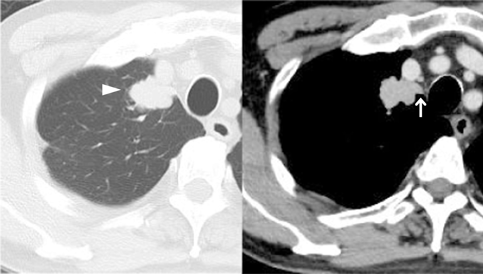
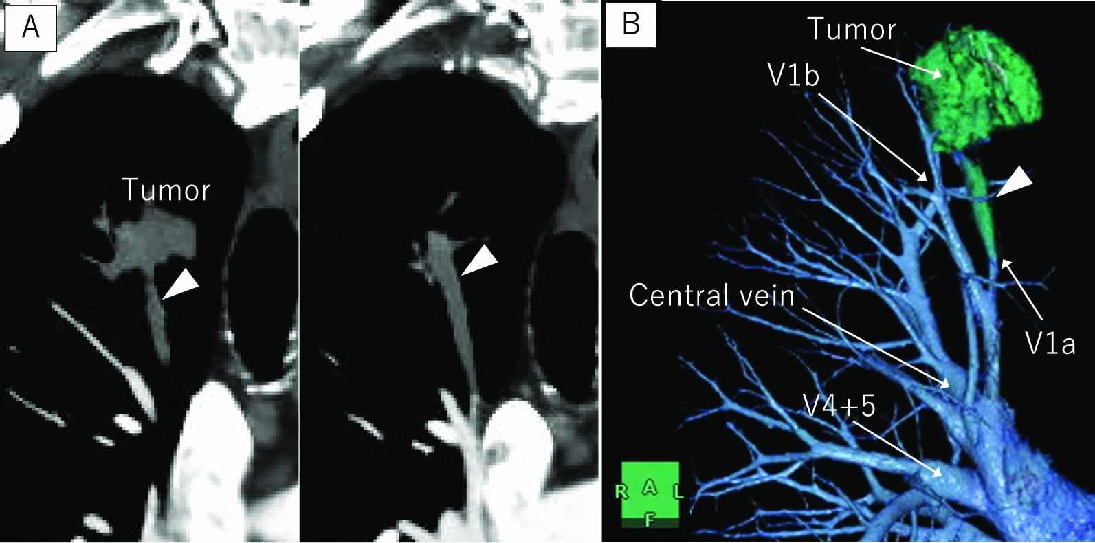
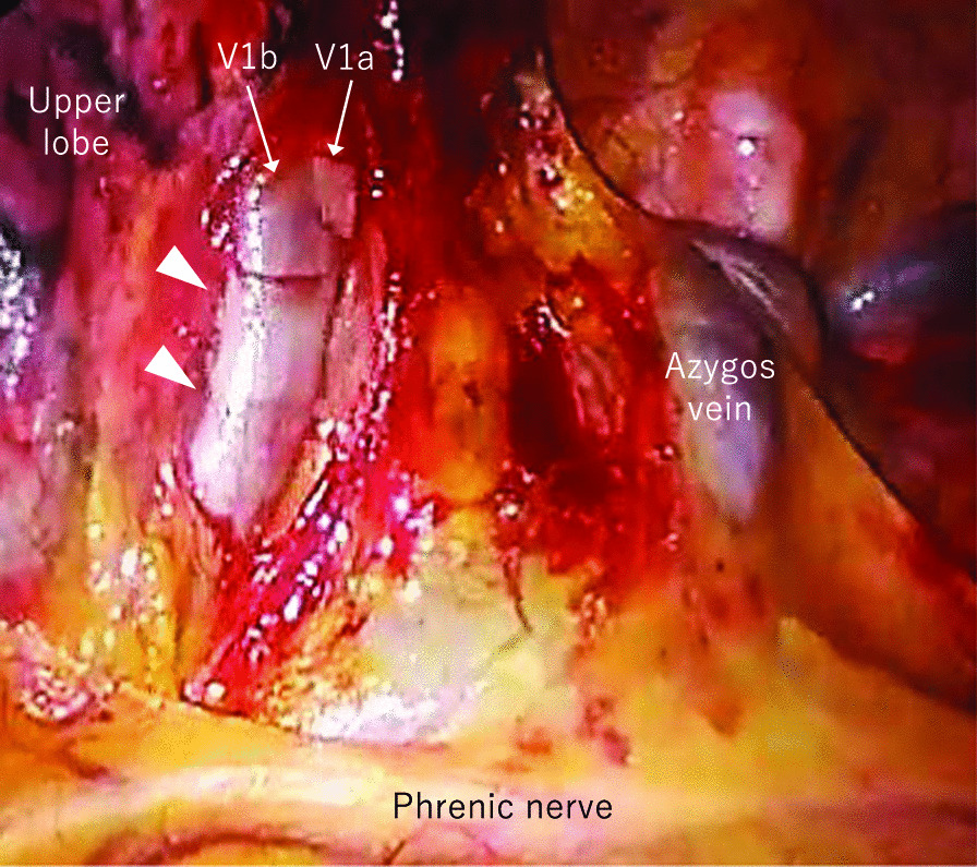
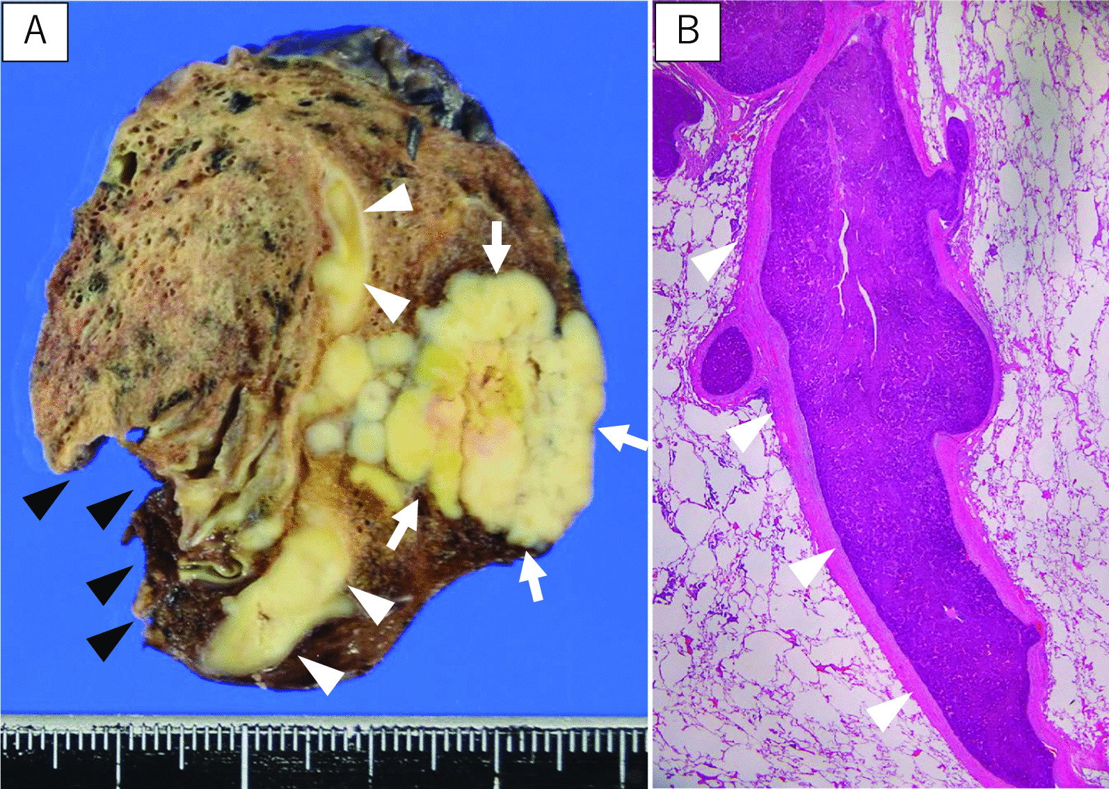
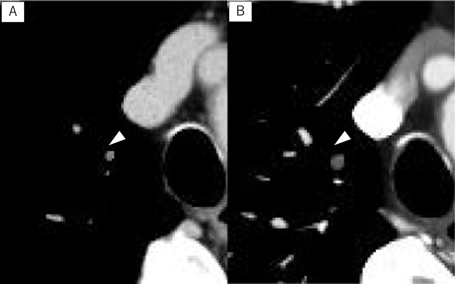
Case presentation: A 77-year-old man underwent laparoscopic lateral segment hepatectomy for HCC eight years ago. Protein induced by vitamin K absence or antagonist-II remained elevated from two years ago. Contrast-enhanced chest computed-tomography (CT) showed a 27 mm nodule in the right apical segment (S1). He was pathologically diagnosed with a metastatic lung tumor from HCC via transbronchoscopic biopsy. We planned to perform right S1 segmentectomy. Before surgery, contrast-enhanced CT in the pulmonary vessels phase for three-dimensional reconstruction showed that the tumor extended into the adjusting peripheral pulmonary vein, and we diagnosed tumor thrombus invasion in V1a. The surgery was conducted under 3-port video-assisted thoracic surgery. First, V1 was ligated and cut. A1 and B1 were cut. The intersegmental plane was cut with mechanical staplers. Pathological examination revealed moderately-differentiated metastatic HCC with tumor thrombus invasions in many pulmonary veins, including V1a. No additional postoperative treatments were performed.
Conclusions: As malignant tumors tend to develop a tumor thrombus in the primary tumor, it might be necessary to perform contrast-enhanced CT in the pulmonary vessel phase to check for a tumor thrombus before the operation for metastatic lung tumors.
Keywords: Hepatocellular carcinoma; Metastatic lung tumor; Segmentectomy; Three-dimensional computed tomography; Tumor thrombus.
© 2023. The Author(s).
Conflict of interest statement
The authors declare that they have no competing interests.
Figures
References
-
- Senthilinathan S, Memon K, Lewandowski RJ, Kulik L, Mulcahy MF, Riaz A, et al. Extrahepatic metastases occur in a minority of hepatocellular carcinoma patients treated with locoregional therapies: analyzing patterns of progression in 285 patients. Hepatology. 2012;55:1432–1442. doi: 10.1002/hep.24812. - DOI - PMC - PubMed
Publication types
MeSH terms
LinkOut - more resources
Full Text Sources
Medical
Research Materials
