Review
. 2023 Jun;42(2):367-388.
doi: 10.1007/s10555-023-10105-2. Epub 2023 Apr 29.

Oncohistones and disrupted development in pediatric-type diffuse high-grade glioma

Affiliations
Review

Oncohistones and disrupted development in pediatric-type diffuse high-grade glioma

Jennifer K Ocasio et al. Cancer Metastasis Rev. 2023 Jun.

Abstract

Recurrent, clonal somatic mutations in histone H3 are molecular hallmarks that distinguish the genetic mechanisms underlying pediatric and adult high-grade glioma (HGG), define biological subgroups of diffuse glioma, and highlight connections between cancer, development, and epigenetics. These oncogenic mutations in histones, now termed "oncohistones", were discovered through genome-wide sequencing of pediatric diffuse high-grade glioma. Up to 80% of diffuse midline glioma (DMG), including diffuse intrinsic pontine glioma (DIPG) and diffuse glioma arising in other midline structures including thalamus or spinal cord, contain histone H3 lysine 27 to methionine (K27M) mutations or, rarely, other alterations that result in a depletion of H3K27me3 similar to that induced by H3 K27M. This subgroup of glioma is now defined as diffuse midline glioma, H3K27-altered. In contrast, histone H3 Gly34Arg/Val (G34R/V) mutations are found in approximately 30% of diffuse glioma arising in the cerebral hemispheres of older adolescents and young adults, now classified as diffuse hemispheric glioma, H3G34-mutant. Here, we review how oncohistones modulate the epigenome and discuss the mutational landscape and invasive properties of histone mutant HGGs of childhood. The distinct mechanisms through which oncohistones and other mutations rewrite the epigenetic landscape provide novel insights into development and tumorigenesis and may present unique vulnerabilities for pHGGs. Lessons learned from these rare incurable brain tumors of childhood may have broader implications for cancer, as additional high- and low-frequency oncohistone mutations have been identified in other tumor types.

Keywords: Diffuse hemispheric glioma; Diffuse midline glioma; Epigenetic; H3 K27M; H3G34-mutant; H3K27-altered; Oncohistones; Pediatric diffuse glioma.


Conflict of interest statement

Statements and Declarations

Authors have no conflicts of interest to disclose.

Conflict of Interest Statement: On behalf of all authors, the corresponding author states that there is no conflict of interest.

Figures

Fig. 1:
Fig. 1:. Canonical and non-canonical histone H3 are differentially regulated during development.
a) Schematic illustrating the five amino acids that differ between histones H3.1, H3.2 and H3.3 (white font in red squares). Frequently mutated sites are denoted with asterisks. Histones b) H3.1/2 and c) H3.3 are deposited into nucleosomes at different genomic locations and at different points in the cell cycle through distinct chaperones. d) Schematic distribution of histones H3.1/2 (light green) and H3.3 (dark green) across chromosomes demonstrate enrichment at specific genomic sites and DNA processes (figure adapted from [177]). H3.1 is broadly deposited throughout the chromosome while H3.3 deposition is enriched at telomeres, centromeres, regulatory elements, actively transcribed regions, and in regions that have undergone DNA repair.
Fig. 2:
Fig. 2:. Overview of pediatric high-grade gliomas.
a) Graphs depicting the age of onset in years across pediatric high-grade glioma subgroups with the median age for each sex denoted above the graph and the percentage of patients of each sex denoted below the graph. The dotted horizontal line represents the threshold for pediatric classification at 21 years old. The asterisks indicate significant sex differences. b) Cartoon brains illustrate common tumor locations (marked in shades of red) within each subgroup. The darker shades indicate regions with higher incidence for the associated subgroup. c) Graphs depicting the most common nonsynonymous genetic mutations associated with each tumor subgroup, excluding amplifications and losses. d) Kaplan-Meier curves depicting overall survival rates across subgroups with the median survival indicated in months in the top right corner of each graph. Graphs and survival curves generated with published data [4, 6, 7, 17, 21, 24, 25, 178].
Fig. 3:
Fig. 3:. Oncohistones dysregulate histone methylation patterns.
a) PRC2 deposits methyl groups (red circles) at H3K27 in wild-type cells. H3 K27M causes global loss of H3K27me3 while H3 G34R/V increases H3K27me3 at active enhancers. b) SETD2 and SETD5 deposit the third methyl group at H3K36 (dark red circles), which can be removed by KDM4 (KDM4A, KDM4B, and KDM4D). H3K36me3 is unaffected in H3 K27-altered tumors while H3 G34R/V causes a loss of H3K36me3 in cis.
Fig. 4:
Fig. 4:. Oncohistones produce widespread epigenetic consequences.
H3 K27-altered tumors exhibit a) altered deposition of specific epigenetic marks, b) activation of bivalent genes and c) reactivation of endogenous retroviral elements. a) H3 K27-altered tumors demonstrate global loss of H3K27me3 (left) but focally retain the mark at specific loci. Conversely, H3 K27-altered tumors exhibit spreading of H3K36me2 (right) across the genome. b) Bivalent promoters are characterized by the combined presence of the activating mark H3K4me3 (blue diamond) and the repressive mark H3K27me3 (red circle) and are poised for induced gene expression. The global loss of H3K27me3 in H3 K27-altered tumors releases bivalency and activates gene expression. H3 K27M may also recruit MLL1 to deposit H3K4me3 at distal regions. c) Repeat elements, such as endogenous retroviral elements (ERVs) normally retain H3K27me3 and are not expressed. H3 K27-altered tumors promote CBP/P300 recruitment at ERVs, resulting in H3K27ac (dark blue circle) deposition and priming ERVs for expression.
Fig. 5:
Fig. 5:. DNA methylation classification of common childhood and adult brain tumors.
tSNE projections of tumor methylation classes from published brain malignancy methylation datasets. a) tSNE projection of full reference cohort (n = 2751) with high grade gliomas encircled. b) tSNE projection of high-grade gliomas (n=1259). Samples are shaped based on the study from which they originated [23, 68, 77]. The methylation classes are grouped by tumor type and color-coded (pHGG: pediatric high-grade glioma; GBM: glioblastoma multiforme; LGG: low grade glioma; EPN: ependymoma; ATRT: atypical teratoid rhabdoid tumors; MB: medulloblastoma). tSNE projections were generated from 1-variance weighted Pearson correlation between samples, which were calculated from normalized methylation beta values for the 35000 most variable probes.
Fig. 6:
Fig. 6:. Cooperating mutations differ between oncohistone-driven tumors.
Although many cooperating mutations are shared across oncohistone-driven tumors, some occur at much higher frequency in combination with specific oncohistones. Cooperating mutations with selective high frequency associations with histones a) H3.1/2 and b and c) H3.3 are denoted with asterisks. (TF: transcription factor)
Fig. 7:
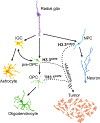
Fig. 7:. Cell states during development may be uniquely susceptible to oncohistone-driven transformation into tumors.
Radial glial cells give rise to transit amplifying neural progenitor cells (NPCs) and glial progenitors. Neurogenesis driven by rapid NPC proliferation and differentiation into mature neurons precedes gliogenesis, where glial progenitors like intermediate glial cells (iGCs) and oligodendrocyte precursor cells (OPCs) differentiate into astrocyte or oligodendrocyte lineages respectively. The introduction of oncohistones at specific points in development, such as H3 G34R/V in interneuron progenitors or H3 K27M at different stages of early glial development, can cooperate with other acquired mutations to transform cells into tumors. Solid arrows show normal development. Dotted arrows show where specific mutations can lead to tumorigenesis.

References

    1. Ostrom QT, Price M, Ryan K, Edelson J, Neff C, Cioffi G, et al., CBTRUS Statistical Report: Pediatric Brain Tumor Foundation Childhood and Adolescent Primary Brain and Other Central Nervous System Tumors Diagnosed in the United States in 2014–2018. 2022, NLM (Medline). p. iii1–iii38. - PMC - PubMed
    1. Pajtler KW, Wen J, Sill M, Lin T, Orisme W, Tang B, et al. (2018). Molecular heterogeneity and CXorf67 alterations in posterior fossa group A (PFA) ependymomas. Acta Neuropathol, 136(2), 211–226. 10.1007/s00401-018-1877-0 - DOI - PMC - PubMed
    1. Schwartzentruber J, Korshunov A, Liu XY, Jones DT, Pfaff E, Jacob K, et al. (2012). Driver mutations in histone H3.3 and chromatin remodelling genes in paediatric glioblastoma. Nature, 482(7384), 226–31. 10.1038/nature10833 - DOI - PubMed
    1. Sturm D, Witt H, Hovestadt V, Khuong-Quang DA, Jones DT, Konermann C, et al. (2012). Hotspot mutations in H3F3A and IDH1 define distinct epigenetic and biological subgroups of glioblastoma. Cancer Cell, 22(4), 425–37. 10.1016/j.ccr.2012.08.024 - DOI - PubMed
    1. Wu G, Broniscer A, McEachron TA, Lu C, Paugh BS, Becksfort J, et al. (2012). Somatic histone H3 alterations in pediatric diffuse intrinsic pontine gliomas and non-brainstem glioblastomas. Nat Genet, 44(3), 251–3. 10.1038/ng.1102 - DOI - PMC - PubMed

Publication types