Review
. 2023 May 18;83(10):1552-1572.
doi: 10.1016/j.molcel.2023.04.009. Epub 2023 Apr 28.

PARPs and ADP-ribosylation: Deciphering the complexity with molecular tools

Affiliations
Review

PARPs and ADP-ribosylation: Deciphering the complexity with molecular tools

Morgan Dasovich et al. Mol Cell. .

Abstract

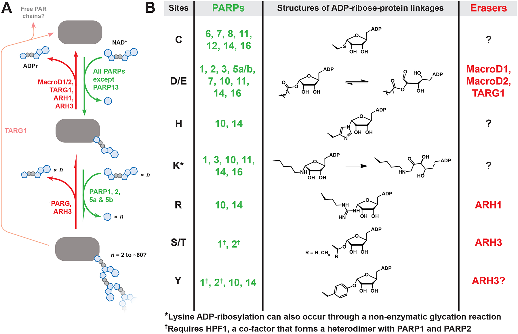
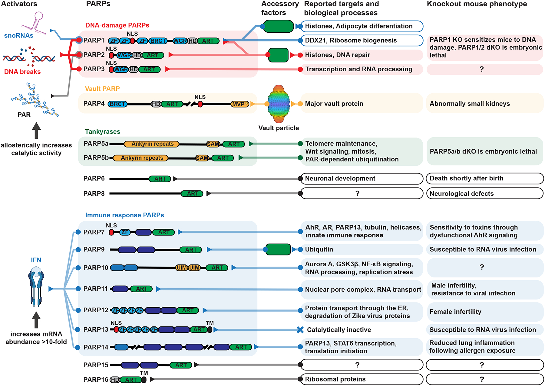
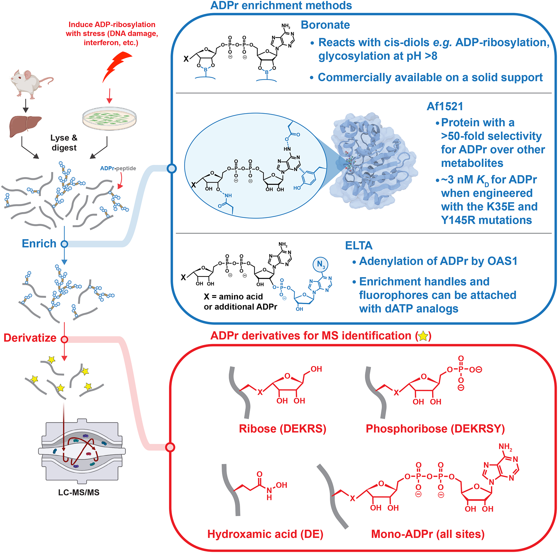
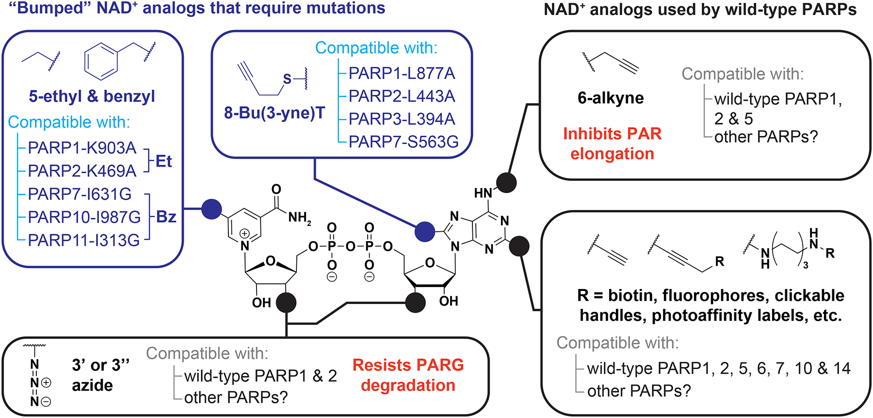
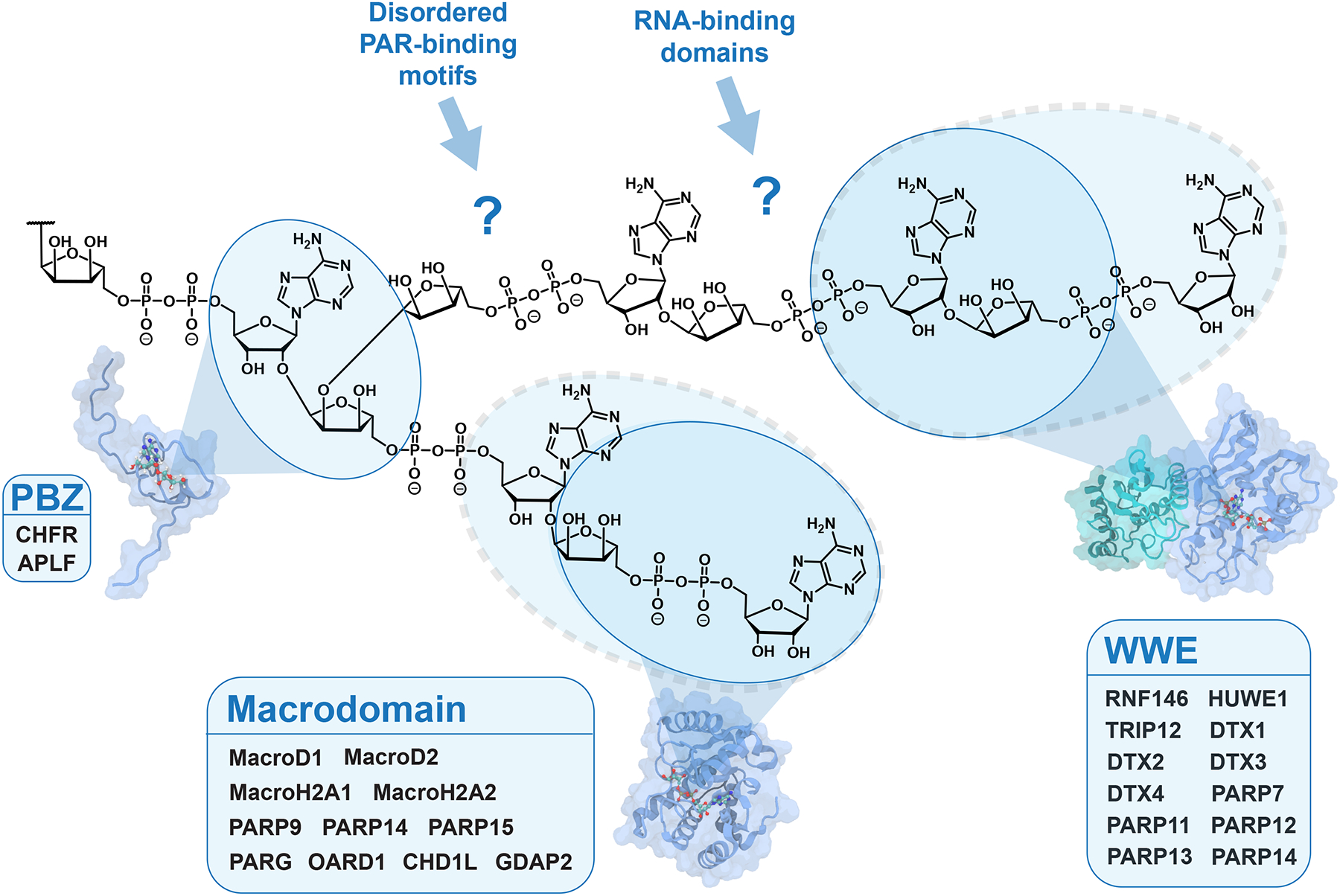
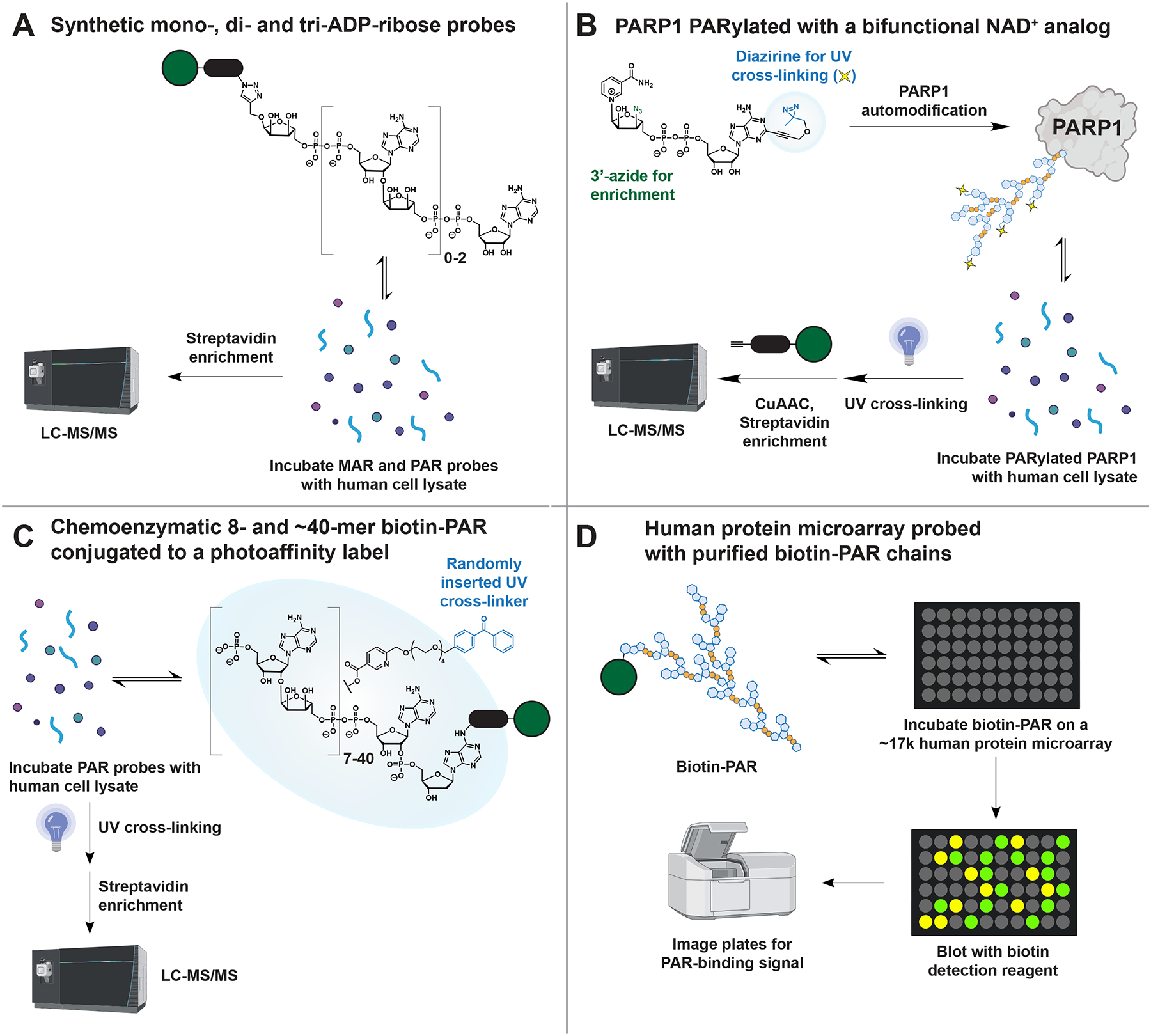
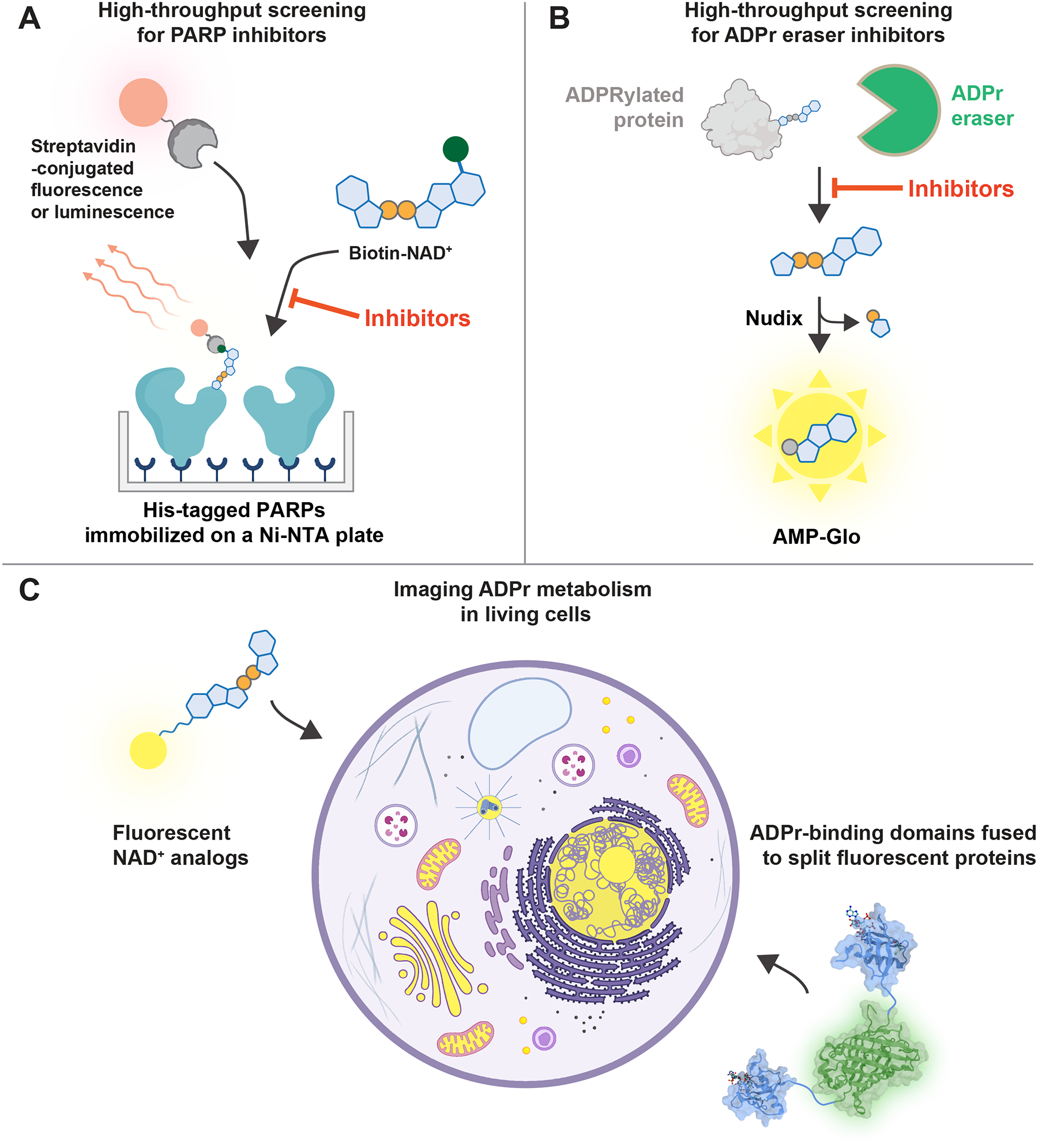
PARPs catalyze ADP-ribosylation-a post-translational modification that plays crucial roles in biological processes, including DNA repair, transcription, immune regulation, and condensate formation. ADP-ribosylation can be added to a wide range of amino acids with varying lengths and chemical structures, making it a complex and diverse modification. Despite this complexity, significant progress has been made in developing chemical biology methods to analyze ADP-ribosylated molecules and their binding proteins on a proteome-wide scale. Additionally, high-throughput assays have been developed to measure the activity of enzymes that add or remove ADP-ribosylation, leading to the development of inhibitors and new avenues for therapy. Real-time monitoring of ADP-ribosylation dynamics can be achieved using genetically encoded reporters, and next-generation detection reagents have improved the precision of immunoassays for specific forms of ADP-ribosylation. Further development and refinement of these tools will continue to advance our understanding of the functions and mechanisms of ADP-ribosylation in health and disease.

Keywords: ADP-ribose biosensor; ADP-ribosylation; ADP-ribosylome; PAR-binding proteins; PARPs; chemical biology; drug development; proteomics.


Conflict of interest statement

Declaration of interests A.K.L.L. holds a patent related to the ELTA technology used for labeling ADP-ribosylated molecules.

Figures

Figure 1.
Figure 1.. The chemical diversity of human PARP metabolism.
(A) PAR metabolism involves four steps: 1) transfer of a single ADP-ribose from NAD+ to the target protein, resulting in mono-ADP-ribose (MAR). 2) Further ADPr transfers onto MAR, producing linear and branched poly(ADP-ribose) (PAR). 3) PAR degradation, which is primarily carried out by poly(ADP-ribose) glycohydrolase (PARG). Notably, human PARG does not remove the final ADP-ribose attached to the protein, thereby converting PAR to MAR. 4) Cleavage of the ADP-ribose-protein bond by various “eraser” enzymes, each with a preference for specific functional groups. Note: ARH3 can also degrade PAR, although it is less efficient than PARG and unable to remove branch points. (B) The chemical diversity of ADPr-protein bonds synthesized by human PARPs, with known site preferences listed. See Data S1 for related references.
Figure 2.
Figure 2.. An overview of the PARP family, including domains, targets, and phenotypes.
Domain abbreviations: ZF = zinc finger, NLS = nuclear localization signal, BRCT = BRCA1 C-terminus, WGR = the three most conserved amino acids in this DNA-binding domain, HD = helical domain, ART = ADP-ribosyltransferase, MVPID = major vault protein interacting domain, SAM = sterile alpha motif, WWE = the three most conserved amino acids in this PAR-binding domain, Macro = macrodomain, RRM = RNA recognition motif, UIM = ubiquitin interacting motif, TM = transmembrane. See Data S1 for related references.
Figure 3.
Figure 3.. Key steps in ADP-ribosylomics workflows.
Following protein extraction from cultured cells or tissues and tryptic digestion, ADP-ribosylated peptides are enriched with a variety of methods (blue). Enriched peptides contain heterogeneous ADP-ribose modifications (i.e., MAR and PAR), which are then derivatized to a single, homogenous mass (red) to simplify the interpretation of mass spectra. For each derivative, the amino acids searched in published experiments are in paratheses, though ribose and phosphoribose could be on any amino acid. The structure of Af1521 is from reference .
Figure 4.
Figure 4.. NAD+ analogs used to study ADP-ribosylation.
Analogs with substitutions to the N6, C2, and 3’-OH positions can be used with wild-type PARP proteins, whereas substitutions to the C5 and C8 positions require mutations in the NAD+-binding pocket to confer analog sensitivity.
Figure 5.
Figure 5.. Protein domains that bind to ADP-ribosylated molecules.
The PAR-binding zinc finger (PBZ), WWE domain, and macrodomain are well-defined folds that recognize specific forms of ADP-ribosylation. The blue circles indicate the binding preferences for each domain based on structural data, with dashed circles indicating variations in binding preference within the family. For instance, most macrodomains bind ADP-ribose, MARylated proteins, and/or the terminal ADPr of PAR, except for CHD1L (ALC1), which specifically binds PAR. Several RNA-binding domains and intrinsically disordered PAR-binding motifs are reported to bind PAR, but how these domains engage PAR is unclear due to a lack of structural data. The structures of Af1521, RNF146 RING-WWE, and APLF PBZ are from references , , and , respectively.
Figure 6.
Figure 6.. Techniques for identifying MAR and PAR readers with chemically and enzymatically synthesized probes.
(A) Kliza et al., (B) Lam et al., (C) Dasovich et al., and (D) Kang et al., have developed complementary methods that together have identified 1,918 MAR- and PAR-binding candidate proteins.
Figure 7.
Figure 7.. Methods for visualizing ADPr in vitro and in vivo.
(A) A high-throughput ADP-ribosylation assay identifies PARP inhibitors by measuring the number of biotin-ADPr modifications. (B) A high-throughput ADP-ribosylhydrolase assay identifies inhibitors by converting hydrolyzed ADP-ribose into luminescence. (C) Small molecule and genetically encoded methods for imaging ADP-ribosylation dynamics in living cells. The structures of RNF146 WWE and split GFP are from references and , respectively.

References

    1. Krishnakumar R, & Kraus WL (2010). The PARP Side of the Nucleus: Molecular Actions, Physiological Outcomes, and Clinical Targets. Mol. Cell, 39, 8–24. 10.1016/j.molcel.2010.06.017 - DOI - PMC - PubMed
    1. Pascal JM (2018). The comings and goings of PARP-1 in response to DNA damage. DNA Repair (Amst.) 71, 177–182. 10.1016/j.dnarep.2018.08.022 - DOI - PMC - PubMed
    1. Schuller M & Ahel I (2022). Beyond protein modification: the rise of non-canonical ADP-ribosylation. Biochem. J 479, 463–477. 10.1042/BCJ20210280 - DOI - PMC - PubMed
    1. Lüscher B, Bütepage M, Eckei L, Krieg S, Verheugd P, & Shilton BH (2018). ADP-Ribosylation, a Multifaceted Posttranslational Modification Involved in the Control of Cell Physiology in Health and Disease. Chem. Rev 118, 1092–1136. 10.1021/acs.chemrev.7b00122 - DOI - PubMed
    1. Perina D, Mikoč A, Ahel J, Ćetković H, Žaja R, & Ahel I (2014). Distribution of protein poly(ADP-ribosyl)ation systems across all domains of life. DNA Repair (Amst.), 23, 4–16. 10.1016/j.dnarep.2014.05.003 - DOI - PMC - PubMed

Publication types

MeSH terms

Substances

LinkOut - more resources