. 2023 Apr 29;14(4):296.
doi: 10.1038/s41419-023-05813-0.

Targeting fatty acid oxidation via Acyl-CoA binding protein hinders glioblastoma invasion

Affiliations

Targeting fatty acid oxidation via Acyl-CoA binding protein hinders glioblastoma invasion

Ceren Duman et al. Cell Death Dis. .

Abstract

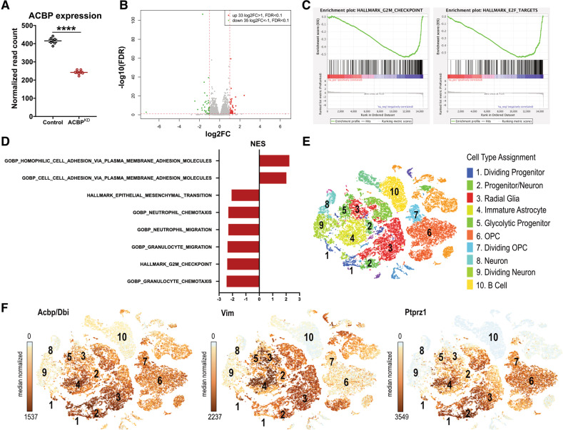
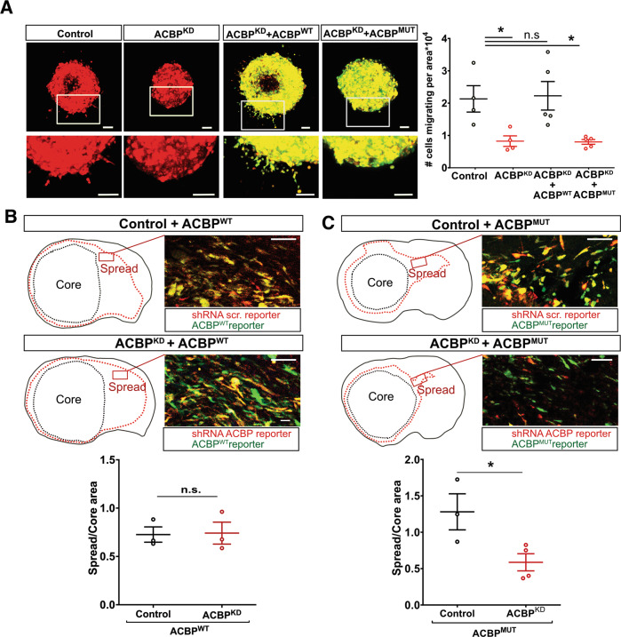
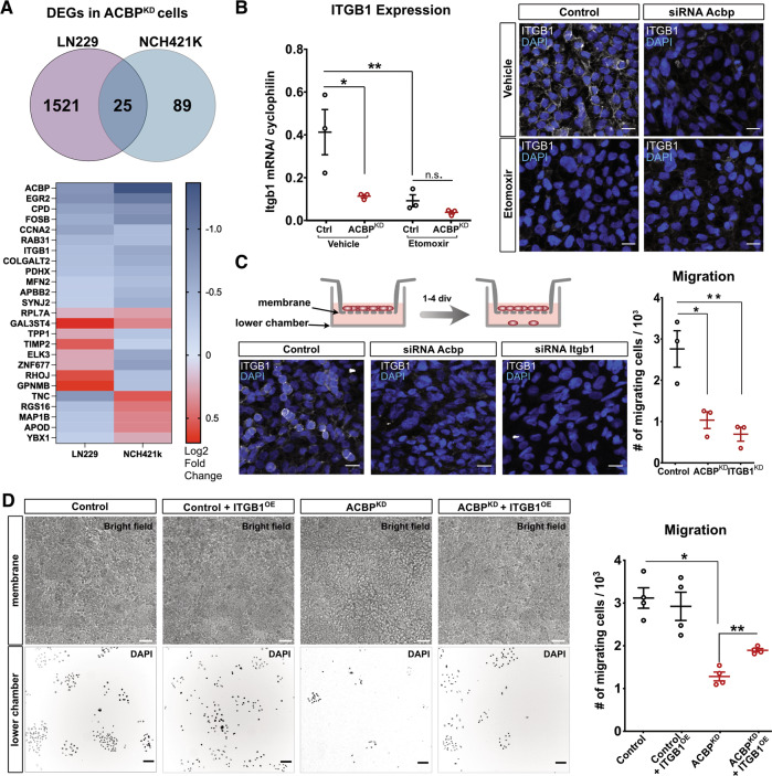
The diffuse nature of Glioblastoma (GBM) tumors poses a challenge to current therapeutic options. We have previously shown that Acyl-CoA Binding Protein (ACBP, also known as DBI) regulates lipid metabolism in GBM cells, favoring fatty acid oxidation (FAO). Here we show that ACBP downregulation results in wide transcriptional changes affecting invasion-related genes. In vivo experiments using patient-derived xenografts combined with in vitro models demonstrated that ACBP sustains GBM invasion via binding to fatty acyl-CoAs. Blocking FAO mimics ACBPKD-induced immobility, a cellular phenotype that can be rescued by increasing FAO rates. Further investigation into ACBP-downstream pathways served to identify Integrin beta-1, a gene downregulated upon inhibition of either ACBP expression or FAO rates, as a mediator for ACBP's role in GBM invasion. Altogether, our findings highlight a role for FAO in GBM invasion and reveal ACBP as a therapeutic vulnerability to stall FAO and subsequent cell invasion in GBM tumors.


Conflict of interest statement

SV and EN are employees of Bayer AG. RL is employee of Nuvisan GmbH.

Figures

Fig. 1
Fig. 1. ACBP downregulation affects cell migration and invasion transcriptional pathways in GBM cells.
A LN229 GBM cells were transduced with lentiviruses expressing either shRNA scrambled (Control) or shRNA1 against ACBP (ACBPKD) and collected one week later for bulk mRNAseq. The graph shows normalized number of reads for ACBP in each condition (mean ± SEM, n = 6 samples per group, unpaired t-test ****p < 0.0001). B Volcano plot of RNAseq results comparing Control vs ACBPKD global expression levels. C Gene set enrichment analysis (GSEA) of cell division-related pathways (left: Hallmark G2M Checkpoint, right: Hallmark E2F Targets) downregulated in ACBPKD cells.D Gene Set enrichment analysis in Control vs ACBPKD cells. Normalized Enrichment Score (NES) negative values indicate pathways enriched in Control cells, while positive NES values show pathways enriched in ACBPKD cells. All pathways displayed have FDR q-val < 0.05.E tSNE plot showing primary GBM tumor cell clustering with cell type annotations [22] obtained from single-cell mRNA sequencing data available at https://gbm.cells.ucsc.edu. F Feature plots of ACBP and invasion-associated genes Vimentin and Ptprz1.
Fig. 2
Fig. 2. ACBP knockdown impairs GBM cell migration and invasion in vitro and in vivo.
A Left: LN229 cells were transfected with either Control or ACBP siRNA. Upon reaching confluency, coverslips were scratched with a pipette tip to generate an empty cell area and cells were imaged every 10 min for a total of 10 h. Images show the initial and final timepoint, dashed lines indicate the border of the empty cell area at t = 0. Scale bars: 50 μm. Right: Quantification of the area covered by LN229 Control and ACBPKD migrating cells after 10 h (mean ± SEM, n = 3 experiments per group, unpaired two-tailed t-test *p < 0.05). See also Supplementary movies 1, 2. B Top: NCH421k tumorspheres expressing either shRNA control or shRNA1 ACBP (ACBPKD) were plated in Matrigel and imaged over time at the indicated time points. Enlarged areas show examples of invasive cells exiting the spheres and invading the 3D matrix in Control conditions. Cells from ACBPKD tumorspheres remained mostly stationary. Scale bars: 20 μm. Bottom: quantification of the number of invading cells, normalized by the sphere size for Control and ACBPKD groups (mean ± SEM, n = 4–31 spheres per group, Mixed-effect analysis p < 0.0001, Sidak’s multiple comparisons test *p < 0.05, **p < 0.01, ****p < 0.0001). C Left: NCH421k tumorspheres expressing either shRNA control or shRNA1 ACBP were plated in Matrigel and imaged 14 days later. Pictures show representative examples of tumorspheres from each group. Scale bars: 50 μm. Right: Quantification of the number of cells invading the 3D matrix from Control and ACBPKD tumorspheres, normalized by the sphere size for shRNA1 and shRNA2 sequences (mean ± SEM, n = 17–24 spheres per group, two-tailed t-test with Welch’s correction ***p < 0.001 for shRNA1, and mean ± SEM, n = 26–27 spheres per group, Mann-Whitney two-tailed test ****p < 0.0001 for shRNA2). D NSG mice were xenotransplanted with a mix of Control and ACBPKD (shRNA1) NCH421K cells and sacrificed 35 days post-surgery. Left: Confocal picture of a tumor showing Control (red) and ACBPKD (green) cells, overview and enlarged area. Core, intermediate and spread areas are separated with dashed lines. White and gray arrows point at Control and ACBPKD invasive cells, respectively, migrating in the spread area. Scale bar: 50 µm. Right: Control/ACBPKD cell ratio in core, intermediate and spread area (mean ± SEM, n = 4 tumors, one-way ANOVA with adjusted p-value for multiple comparisons ***p < 0.0005).
Fig. 3
Fig. 3. ACBP requires binding to acyl-CoA to support GBM invasion.
A Left: Representative pictures of NCH421k tumorspheres expressing either shRNA Control (+ TdTomato), shRNA1 ACBP (ACBPKD + TdTomato), ACBPKD + bovine ACBP wildtype (ACBPWT + EGFP) or ACBPKD + bovine ACBP low binding mutant (ACBPMUT + EGFP) plated in Matrigel. Boxed areas are enlarged below the respective images. Scale bars: 50 μm. Right: Quantification of the number of cells invading the extracellular matrix in each group, normalized by the sphere size (mean ± SEM, n = 4–5 spheres per group, one-way ANOVA with adjusted p-value for multiple comparisons *p < 0.05). B NSG mice were xenotransplanted with shRNA Control or shRNA1 ACBPKD NCH421K cells co-expressing bovine ACBPWT. Mice were sacrificed when first moderate symptoms were observed (median survival 48 and 53 days for Control + ACBPWT and ACBPKD + ACBPWT, respectively, n = 4–6 mice per group, reported in [11]). Top: Outlines of representative whole brain coronal sections with core and spread tumor areas delineated with black and red dotted lines, respectively. Pictures belong to the indicated brain areas and show fluorescently-labeled tumor cells (shRNA Control and shRNA ACBP in red, ACBPWT in green). Scale bars: 100 μm. Bottom: Quantification of spread/core area ratios for each experiment (mean ± SEM, n = 3 mice per group, unpaired two-tailed t-test p = 0.91). C NSG mice were xenotransplanted with shRNA Control or ACBPKD NCH421K cells co-expressing bovine ACBPMUT. Mice were sacrificed when first moderate symptoms were observed (median survival 41 and 64 days for Control + ACBPMUT and ACBPKD + ACBPMUT, respectively, n = 4–6 mice per group, reported in [11]). Top: Outlines of representative whole brain coronal sections with core and spread tumor areas delineated with black and red dotted lines, respectively. Pictures belong to the indicated brain areas and show fluorescently-labeled tumor cells (shRNA Control and shRNA ACBP in red, ACBPMUT in green). Scale bars: 100 μm. Bottom: Quantification of spread/core area ratios for each experiment (mean ± SEM, n = 3–4 mice per group, unpaired two-tailed t-test *p = 0.03).
Fig. 4
Fig. 4. ACBPKD-induced invasion deficit is mimicked by inhibition of fatty acid oxidation and rescued by octanoate.
A Left: Representative pictures of NCH421k Control (shRNA scrambled + TdTomato) and ACBPKD (shRNA1 ACBP + EGFP) tumorspheres treated with vehicle, FAO-inhibitor Etomoxir (40 µM) or Octanoate (1 mM). Arrowheads point at invasive cells exiting the spheres. Scale bars: 50 μm. Right: Quantification of the number of cells invading the extracellular matrix in each group, normalized by sphere size (mean ± SEM, n = 6–14 spheres per group, one-way ANOVA with adjusted p-value for multiple comparisons ***p < 0.0005, ****p < 0.0001). B Top: Experimental timeline for in vivo Etomoxir injections in mice bearing either Control or ACBPKD NCH421K tumors. Bottom: Outlines of representative whole brain coronal sections with core and spread tumor areas delineated with black and red dotted lines, respectively. Pictures belong to the indicated brain areas and show fluorescently-labeled tumor cells (white) and DAPI signal (blue). St: striatum, C.C.: corpus callosum, Cx: cortex. Scale bars: 200 µm. C Quantification of spread/core area ratios per brain in each experimental group (mean ± SEM, n = 3–5 mice per group, one-way ANOVA with adjusted p-value for multiple comparisons *p < 0.5, **p < 0.01).
Fig. 5
Fig. 5. ACBPKD and FAO inhibition downregulate invasion-related genes.
A Top: Venn diagram of differentially expressed genes in LN229 and NCH421K ACBPKD cells compared to control cells (calculated with DESeq2, n = 5 and 4 samples per condition for LN229 and NCH421K, respectively, adjusted p-value <0.01). Bottom: List of common genes differentially expressed in ACBPKD cells (both cell types) with color-coded log2 Fold changes. B LN229 Control or ACBPKD cells were treated with either vehicle or Etomoxir (40 μM) for 5 days. Left: qRT-PCR data showing Itgb1 fold expression changes (mean ± SEM, n = 3 samples per group, one-way ANOVA with adjusted p-value for multiple comparisons *p < 0.05, **p < 0.01). Right: Immunostainings showing ITGB1 protein expression in each condition (ITGB1 channel was acquired with exactly the same microscopy parameters in all cases). Scale bars: 20 μM.C LN229 cells were transfected with either Control, Acbp or Itgb1 siRNAs and plated at confluency for transwell assays. Pictures show representative images of ITGB1 immunostainings in each condition (ITGB1 channel was acquired with exactly the same microscopy parameters in all cases). Scale bars: 20 μM. Transwell migration assay results for Control, Acbp or Itgb1 knockdown conditions (mean ± SEM, n = 3 samples per group, one-way ANOVA with adjusted p-value for multiple comparisons). D LN229 cells were transfected with either Control or Acbp siRNAs and transduced with either Control or ITGB1-overexpressing lentivirus. Cells from each group were plated at confluency for transwell assays. Pictures show representative bright field images of the upper (membrane) chamber and DAPI-stained migrating cells in the lower chambers, 4 days after plating. Scale bars: 50 μM (top) and 100 μM (bottom). Transwell migration assay results for each group (mean ± SEM, n = 4 samples per group, one-way ANOVA with Welch’s correction, adjusted p-value for multiple comparisons *p < 0.05, **p < 0.01).

References

    1. Louis DN, Ohgaki H, Wiestler OD, Cavenee WK, Burger PC, Jouvet A, et al. The 2007 WHO classification of tumours of the central nervous system. Acta Neuropathol. 2007;114:97–109. doi: 10.1007/s00401-007-0243-4. - DOI - PMC - PubMed
    1. Stupp R, Mason WP, van den Bent MJ, Weller M, Fisher B, Taphoorn MJB, et al. Radiotherapy plus concomitant and adjuvant temozolomide for glioblastoma. N Engl J Med. 2005;352:987–96. doi: 10.1056/NEJMoa043330. - DOI - PubMed
    1. Claes A, Idema AJ, Wesseling P. Diffuse glioma growth: a guerilla war. Acta Neuropathol. 2007;114:443–58. doi: 10.1007/s00401-007-0293-7. - DOI - PMC - PubMed
    1. Lam KHB, Leon AJ, Hui W, Lee SCE, Batruch I, Faust K, et al. Topographic mapping of the glioblastoma proteome reveals a triple-axis model of intra-tumoral heterogeneity. Nat Commun. 2022;13:116–29. doi: 10.1038/s41467-021-27667-w. - DOI - PMC - PubMed
    1. Darmanis S, Sloan SA, Croote D, Mignardi M, Chernikova S, Samghababi P, et al. Single-cell RNA-seq analysis of infiltrating neoplastic cells at the migrating front of human glioblastoma. Cell Rep. 2017;21:1399–410. doi: 10.1016/j.celrep.2017.10.030. - DOI - PMC - PubMed

Publication types