. 2023 Apr 29;14(1):2488.
doi: 10.1038/s41467-023-38202-4.

Virus diversity, wildlife-domestic animal circulation and potential zoonotic viruses of small mammals, pangolins and zoo animals

Affiliations

Virus diversity, wildlife-domestic animal circulation and potential zoonotic viruses of small mammals, pangolins and zoo animals

Xinyuan Cui et al. Nat Commun. .

Abstract

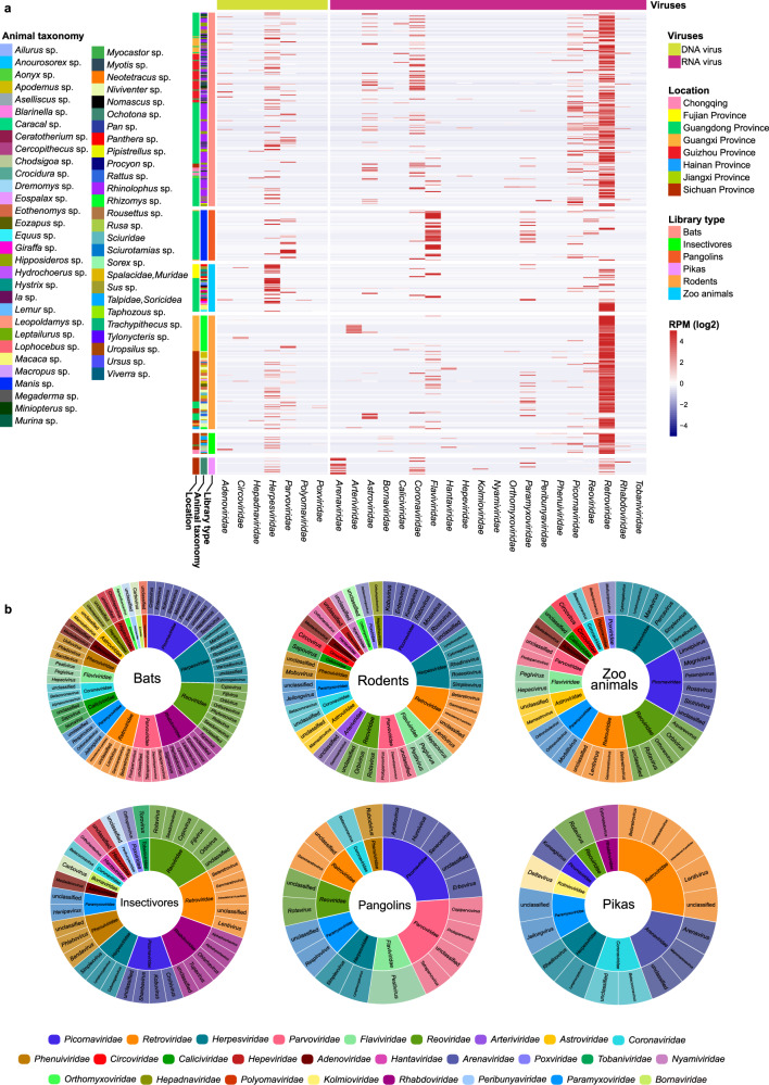
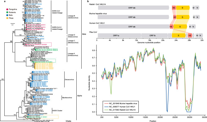
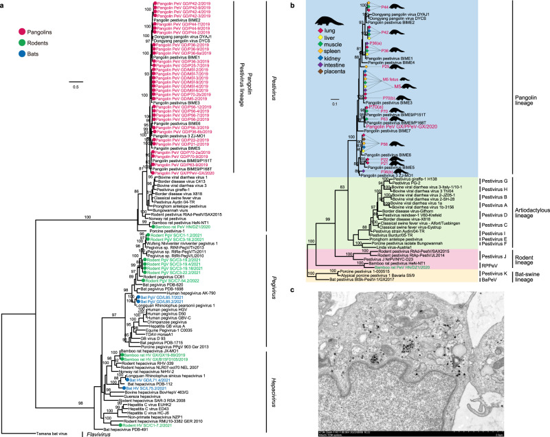
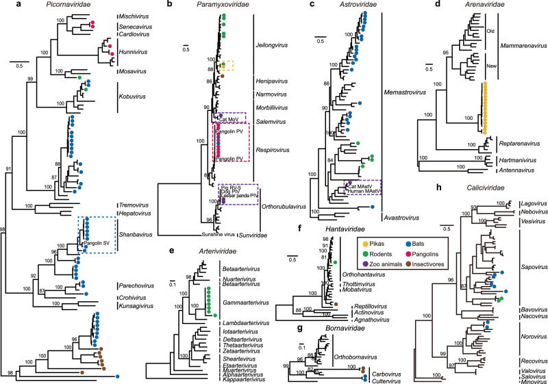
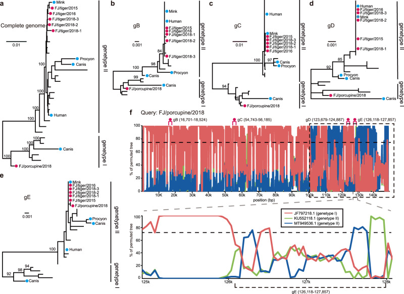
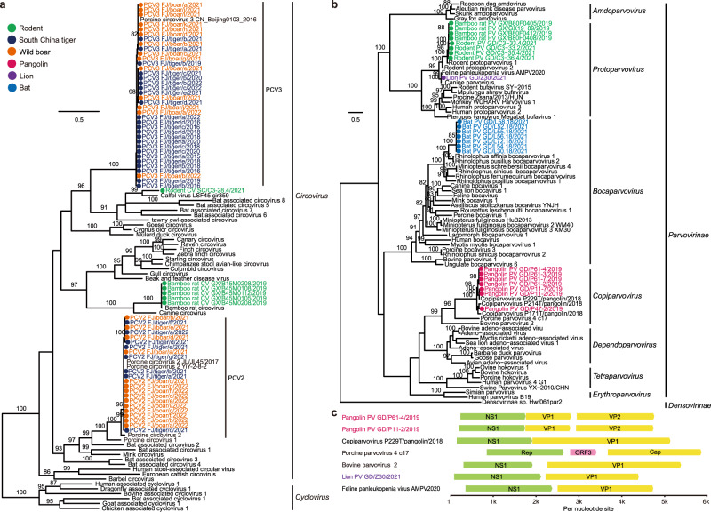
Wildlife is reservoir of emerging viruses. Here we identified 27 families of mammalian viruses from 1981 wild animals and 194 zoo animals collected from south China between 2015 and 2022, isolated and characterized the pathogenicity of eight viruses. Bats harbor high diversity of coronaviruses, picornaviruses and astroviruses, and a potentially novel genus of Bornaviridae. In addition to the reported SARSr-CoV-2 and HKU4-CoV-like viruses, picornavirus and respiroviruses also likely circulate between bats and pangolins. Pikas harbor a new clade of Embecovirus and a new genus of arenaviruses. Further, the potential cross-species transmission of RNA viruses (paramyxovirus and astrovirus) and DNA viruses (pseudorabies virus, porcine circovirus 2, porcine circovirus 3 and parvovirus) between wildlife and domestic animals was identified, complicating wildlife protection and the prevention and control of these diseases in domestic animals. This study provides a nuanced view of the frequency of host-jumping events, as well as assessments of zoonotic risk.


Conflict of interest statement

The authors declare no competing interests.

Figures

Fig. 1
Fig. 1. Overall view of the viral reads in the meta-transcriptomic data.
a Heatmap based on normalized sequence reads of 27 families of mammalian viruses in each pooled sample and their Chinese province of sampling. Names of mammalian viral families are presented in the text row at the bottom. b Overview of virus classifications, from family to genus, of the viruses identified in bats, rodents, pikas, insectivores, pangolins, and zoo animals in this study. Different families are labeled in different colors.
Fig. 2
Fig. 2. Phylogenetic diversity of the coronaviruses identified here.
a Phylogeny of the Coronaviridae based on amino acid sequences of the RdRp gene. Viruses identified in this study are color-coded according to the animal from which they were sampled. The scale bar depicts the number of amino acid substitutions per site. The tree was rooted with δ-CoVs. b Comparison of the genomic organization and similarity plot of Pika-CoV and related viruses. Predicted ORFs are indicated. Parameters for the similarity plots are: window, 500 bp; step, 100 bp.
Fig. 3
Fig. 3. Phylogenetic diversity of flaviviruses identified here.
a Phylogeny of the Flaviviridae based on amino acid sequences of the NS5 gene. Viruses identified in this study are color-coded according to the animal from which they were sampled. The tree was rooted with Tamana bat virus. Scale bar depicts the number of amino acid substitutions per site. b Phylogeny of pestiviruses based on the amino acid sequences of the NS5 gene. Representatives of Pestivirus A-K are incorporated. The tree was rooted with Pestivirus K and BaPeV. Viruses detected in the different tissues of the pangolins are labeled in different colors. c Viral particles of pangolin pestivirus, grown in Vero E6 cell culture, seen by transmission electron microscopy image. Scale bar, 2 μm. The experiment was performed independently in triplicate with similar results.
Fig. 4
Fig. 4. Phylogeny of other major viral families identified in this study.
a Picornaviridae, b Paramyxoviridae, c Astroviridae, d Arenaviridae, e Arteriviridae, f Hantaviridae, g Bornaviridae, and h Caliciviridae. Phylogeny of a Picornaviridae, d Arenaviridae, e Arteriviridae, f Hantaviridae, and g Bornaviridae based on the amino acid sequences of the replicase domain of RdRp. Phylogeny of b Paramyxoviridae, c Astroviridae, and h Caliciviridae based on the amino acid sequences of the Large protein, the capsid protein precursor and VP1, respectively. Numbers (>80) above branches are percentage bootstrap values for the major nodes. The scale bars depict the number of amino acid substitutions per site. Detailed trees are showed in Supplementary Fig. 3.
Fig. 5
Fig. 5. Phylogenetic history and recombination in pseudorabies viruses (PRVs).
The phylogenetic trees were estimated based on a full genome sequences, and the b gB, (c) gC, d gD, and e gE genes, utilizing the best-fit nucleotide substitution model obtained by IQ-TREE (Minh et al., 2020). All trees were midpoint rooted. Numbers (>80) above branches are percentage bootstrap values for the major nodes. The scale bar represents the number of substitutions per site. Red circles indicate the six tiger- and porcupine-isolated PRVs generated in this study. f Similarity plot of the full-length PRV genome of porcupine PRV against sequences of a genotype I strain (JF797218.1) and two genotype II strains (KU552118.1 and MT949536.1). The parameters for the similarity plots are: window, 200 bp; step, 50 bp.
Fig. 6
Fig. 6. Maximum likelihood trees of the circoviruses and parvoviruses based on the amino acid sequences of the NS1 gene.
a Circoviruses, rooted with the cycloviruses. b Parvoviruses, rooted with the Densovirinae sp. Both phylogenetic trees were estimated with the best-fit substitutional model obtained by IQ-TREE (Minh et al., 2020) with 1000 bootstrap replicates. Numbers (>80) above branches are percentage bootstrap values for the associated nodes. c Genomic organization of the parvoviruses. Predicted ORFs are indicated.

References

    1. Jones KE, et al. Global trends in emerging infectious diseases. Nature. 2008;451:990–993. doi: 10.1038/nature06536. - DOI - PMC - PubMed
    1. Allen T, et al. Global hotspots and correlates of emerging zoonotic diseases. Nat. Commun. 2017;8:1124. doi: 10.1038/s41467-017-00923-8. - DOI - PMC - PubMed
    1. Gibb R, et al. Zoonotic host diversity increases in human-dominated ecosystems. Nature. 2020;584:398–402. doi: 10.1038/s41586-020-2562-8. - DOI - PubMed
    1. Calisher CH, Childs JE, Field HE, Holmes KV, Schountz T. Bats: important reservoir hosts of emerging viruses. Clin. Microbiol. Rev. 2006;19:531–545. doi: 10.1128/CMR.00017-06. - DOI - PMC - PubMed
    1. Hayman DT. Bats as viral reservoirs. Annu. Rev. Virol. 2016;3:77–99. doi: 10.1146/annurev-virology-110615-042203. - DOI - PubMed

Publication types