. 2023 May 30;42(5):112432.
doi: 10.1016/j.celrep.2023.112432. Epub 2023 Apr 28.

Identifying the core genome of the nucleus-forming bacteriophage family and characterization of Erwinia phage RAY

Affiliations

Identifying the core genome of the nucleus-forming bacteriophage family and characterization of Erwinia phage RAY

Amy Prichard et al. Cell Rep. .

Abstract

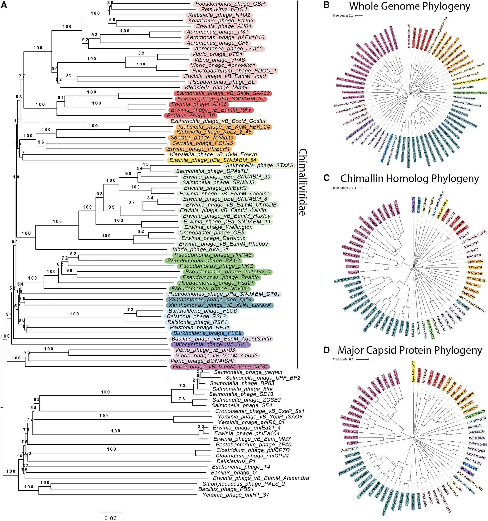
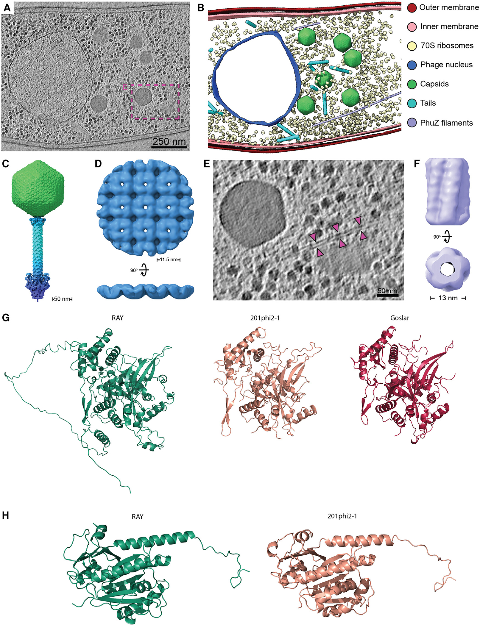
We recently discovered that some bacteriophages establish a nucleus-like replication compartment (phage nucleus), but the core genes that define nucleus-based phage replication and their phylogenetic distribution were still to be determined. Here, we show that phages encoding the major phage nucleus protein chimallin share 72 conserved genes encoded within seven gene blocks. Of these, 21 core genes are unique to nucleus-forming phage, and all but one of these genes encode proteins of unknown function. We propose that these phages comprise a novel viral family we term Chimalliviridae. Fluorescence microscopy and cryoelectron tomography studies of Erwinia phage vB_EamM_RAY confirm that many of the key steps of nucleus-based replication are conserved among diverse chimalliviruses and reveal variations on this replication mechanism. This work expands our understanding of phage nucleus and PhuZ spindle diversity and function, providing a roadmap for identifying key mechanisms underlying nucleus-based phage replication.

Keywords: CP: Microbiology; Chimallin; Erwinia; PhuZ; core genome; cytoskeleton; nuclear shell; phage; phage nucleus; phage tubulin.


Conflict of interest statement

Declaration of interests K.P. and J.P. have an equity interest in Linnaeus Bioscience Incorporated and receive income. The terms of this arrangement have been reviewed and approved by the University of California, San Diego, in accordance with its conflict-of-interest policies.

Figures

Figure 1.
Figure 1.. Phylogenetic comparison of Chimalliviridae
(A) Phylogenetic tree of the Chimalliviridae and related phages. The Chimalliviridae appear to form one clade, separate from msRNAP-encoding phages, ViPTree-predicted relatives, and other phages with large genomes color coded by predicted genus (Table S2) to improve readability. (B) Phylogenetic tree based on whole-genome comparison of 66 representative chimallin-encoding phages (Table S1). (C and D) Phylogenetic tree based on the protein sequences of the (C) chimallin homologs and (D) major capsid proteins from the 66 phages used in our analysis. Trees were colored in iTOL by predicted genus (Table S2). Some of these predicted genera are not fully consistent with current ICTV classification, but for maximum readability and consistency of the colors, only the genera predicted by VICTOR are color coded.
Figure 2.
Figure 2.. Core genome determination
(A) A genome alignment of Erwinia phage RAY, Escherichia phage Goslar, Pseudomonas phage ΦKZ, and Bacillus phage PBS1. PBS1 was included for comparison since it encodes the unique msRNAP but does not encode chimallin. The highly conserved regions with more than 3 genes are annotated in colors as different blocks. Core genes that are not part of blocks are colored black. While in chimalliviruses the core genes are conserved in seven blocks, in PBS1, some of these genes are present but are dispersed across its genome rather than being encoded in blocks. Goslar gp188 and ΦKZ gp055 are homologous to each other and to two RAY genes, gp223 (orange) and gp070 (black). The full list of genes and blocks can be found in Table S3. (B) A circular bar plot of RAY’s genome with heights of bars corresponding to the percentage of phage that contain genes homologous to each RAY gene. Certain regions are more variable (right of circle), while other regions are highly conserved. Colors denote blocks and asterisks denote unique genes in (A) and (B). (C) Histogram showing the frequency of conservation of each RAY protein among chimalliviruses. The left peak indicates RAY-specific proteins (conserved in less than 10% of chimalliviruses), and the right peak indicates core proteins (conserved in 90% or more chimalliviruses).
Figure 3.
Figure 3.. RAY phage nucleus formation
(A) DAPI localization showing the presence of a bright staining consistent with phage nucleus formation. Cells were imaged during midinfection between 60 and 75 minutes post infection (mpi). (B) GFP-ChmARAY surrounds the bright DAPI zones, consistent with the morphology of a phage nucleus. (C) H-NS-GFP localizes to the DNA outside of the phage nucleus. (D) H-NS-GFP moves away from midcell during the course of infection (Video S1). (E) The phage nucleus rotates during infection (Video S2). Time-lapse images were taken 4 s apart. The white arrow shows a segment of the nuclear shell that was tracked to determine rotation speed. (F) Violin plot showing distribution of rotation speeds of individual measured RAY nuclei using either a chimallin (ChmA) (n = 25) or major capsid protein (MCP) (n = 17) GFP tag to track rotation. Scale bar is 1 μm; magenta is membrane stain FM4–64, cyan is DNA stain DAPI, green is GFP, and grayscale is brightfield. Cells were imaged between 60 and 75 mpi (midinfection) unless labeled otherwise.
Figure 4.
Figure 4.. Localization of proteins encoded by RAY
(A) Putative RNA (nvRNAP β subunit 1 gp002, nvRNAP β subunit 2 gp248, nvRNAP β′ subunit 1 gp223, and nvRNAP β′ subunit 2 gp249) and DNA (DNA polymerase gp220) polymerases co-localize with phage DNA in the phage nucleus. (B) RAY gp116 is a homolog of HslUV superfamily heat shock proteases, but other nuclear-localized phage proteins are homologs of known DNA-associated proteins such as proteins involved in recombination (UvsX gp150 and DprA gp153) and helicases (non-virion superfamily 2 helicase gp250 and DnaB-like replicative helicase gp315). (C) Cytoplasmic proteins are not predicted to be involved with DNA replication, recombination, or transcription. These include an SspB homolog gp039, RtcB homolog gp048, putative exonuclease gp064, thymidylate kinase gp311, and putative XRE superfamily transcriptional regulator gp094. (D) The MCP gp317 and tail sheath gp179 are structural components of the RAY virion that localize near the periphery of the phage nucleus. Puncta can be seen near the membrane in the MCP fusion, possibly because capsids are assembled there or, alternatively, as part of an aggregate formed due to overexpression of gp317-GFP. (E) Virion proteins (vRNAP β subunit 2 gp154, vRNAP β′ subunit 1 gp163, vRNAP β′ subunit 2 gp270, virion-associated superfamily 2 helicase gp131, and head protein of unknown function gp299) can be seen with similar localizations as the MCP and sometimes (in the case of gp270 and gp299) have visible DAPI co-localized with them. For all images, scale bar is 1 μm; magenta is membrane stain FM4–64, cyan is DNA stain DAPI, and green is GFP.
Figure 5.
Figure 5.. RAY PhuZ homolog
(A) PhuZRAY has conserved tubulin motifs. T5 is a structural motif with a poorly conserved sequence and is not included. (B) Wild-type PhuZRAY does not polymerize spontaneously until it reaches a critical concentration, but the D198A mutant polymerizes at all levels of arabinose induction tested. (C) The average percentage of uninfected cells with PhuZ filaments plotted versus arabinose concentration. Data are represented as mean ± SEM. (D) Wild-type PhuZ filaments form with 0% arabinose during RAY infections. The nucleus is normally positioned near midcell (white arrows show bacterial DNA, and gold arrows show phage nuclear DNA). (E) When the D198A mutant PhuZ is expressed, midcell localization of the phage nucleus during infection is not as common as with wild-type PhuZ (white arrows show bacterial DNA, and gold arrows show phage nuclear DNA). (F) When only one bacterial nucleoid is present, the phage nucleus positioning histogram has a wide distribution for wild-type PhuZ (blue), and the D198A mutant PhuZ (orange) does not appear to strongly affect positioning. (G) When two bacterial nucleoids are present, the wild-type PhuZ has a strong nucleus positioning bias toward midcell (blue), and the D198A mutant PhuZ has a weaker positioning bias toward midcell (orange). For all microscopy images (B, D, and E), scale bar is 1 μm; magenta is membrane stain FM4–64, cyan is DNA stain DAPI, and green is GFP.
Figure 6.
Figure 6.. Cryoelectron tomography and structural analysis
(A) Slice through a cryoelectron tomogram of a RAY-infected Erwinia amylovora cell at approximately 105–110 mpi. Scale bar is 250 nm. (B) Segmentation of the tomogram in (A). (C) Composite RAY virion reconstruction. (D) Orthogonal views of the RAY chimallin lattice reconstruction. (E) Enlarged view of region boxed in (A) with magenta arrows pointing to a putative RAY PhuZ filament. Scale bar is 50 nm. (F) Reconstruction of the putative RAY PhuZ filaments showing the five-stranded structure and hollow lumen. (G) AlphaFold v.2.1.0 structure of RAY chimallin compared with experimentally determined 201φ2–1 and Goslar chimallin. (H) AlphaFold v.2.1.0 structure of RAY PhuZ compared with experimentally determined 201φ2–1 PhuZ., Accession numbers: (C) EMD-28003 (additional map), (D) EMD-28007, and (F) EMD-28008. See Figure S7 for subtomogram analysis workflows of RAY components.
Figure 7.
Figure 7.. RAY infection model
See text for details.

Update of

References

    1. Paszkowski P, Noyce RS, and Evans DH (2016). Live-cell imaging of vaccinia virus recombination. PLoS Pathog. 12, e1005824. - PMC - PubMed
    1. Tomer E, Cohen EM, Drayman N, Afriat A, Weitzman MD, Zaritsky A, and Kobiler O (2019). Coalescing replication compartments provide the opportunity for recombination between coinfecting herpesviruses. FASEB J 33, 9388–9403. - PMC - PubMed
    1. Kieser Q, Noyce RS, Shenouda M, Lin Y-CJ, and Evans DH (2020). Cytoplasmic factories, virus assembly, and DNA replication kinetics collectively constrain the formation of poxvirus recombinants. PLoS One 15, e0228028. - PMC - PubMed
    1. Trinh JT, Shao Q, Guan J, and Zeng L (2020). Emerging heterogeneous compartments by viruses in single bacterial cells. Nat. Commun. 11, 3813. - PMC - PubMed
    1. Labarde A, Jakutyte L, Billaudeau C, Fauler B, López-Sanz M, Ponien P, Jacquet E, Mielke T, Ayora S, Carballido-López R, and Tavares P (2021). Temporal compartmentalization of viral infection in bacterial cells. Proc. Natl. Acad. Sci. USA 118, e2018297118. 10.1073/pnas.2018297118. - DOI - PMC - PubMed

Publication types