. 2023 Jun 1;324(6):L849-L862.
doi: 10.1152/ajplung.00028.2023. Epub 2023 Apr 25.

IL-8 concurrently promotes idiopathic pulmonary fibrosis mesenchymal progenitor cell senescence and PD-L1 expression enabling escape from immune cell surveillance

Affiliations

IL-8 concurrently promotes idiopathic pulmonary fibrosis mesenchymal progenitor cell senescence and PD-L1 expression enabling escape from immune cell surveillance

Libang Yang et al. Am J Physiol Lung Cell Mol Physiol. .

Abstract

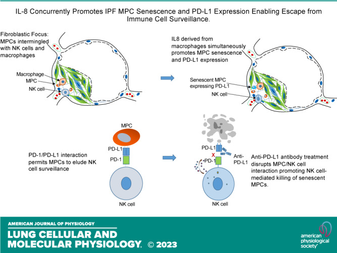
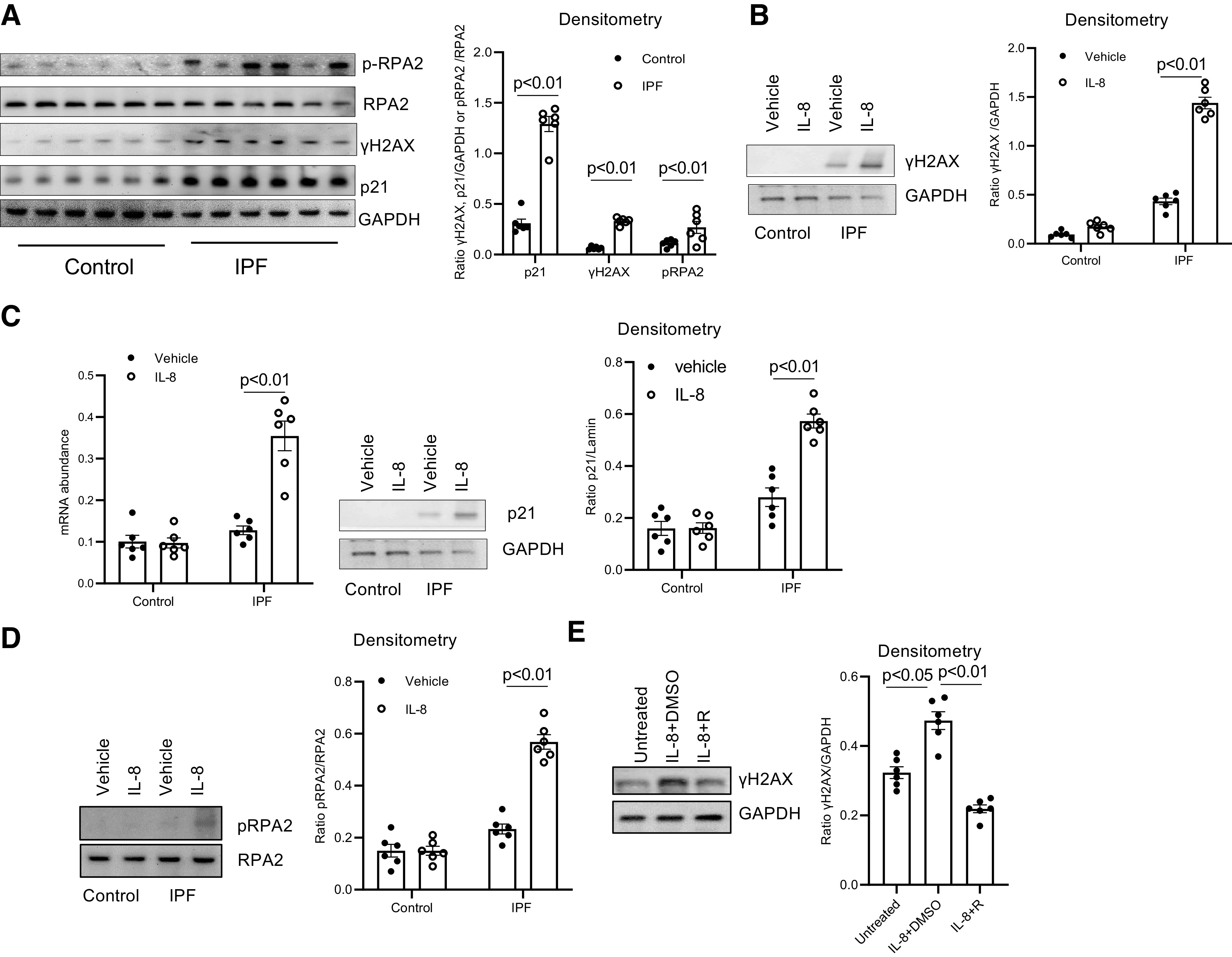
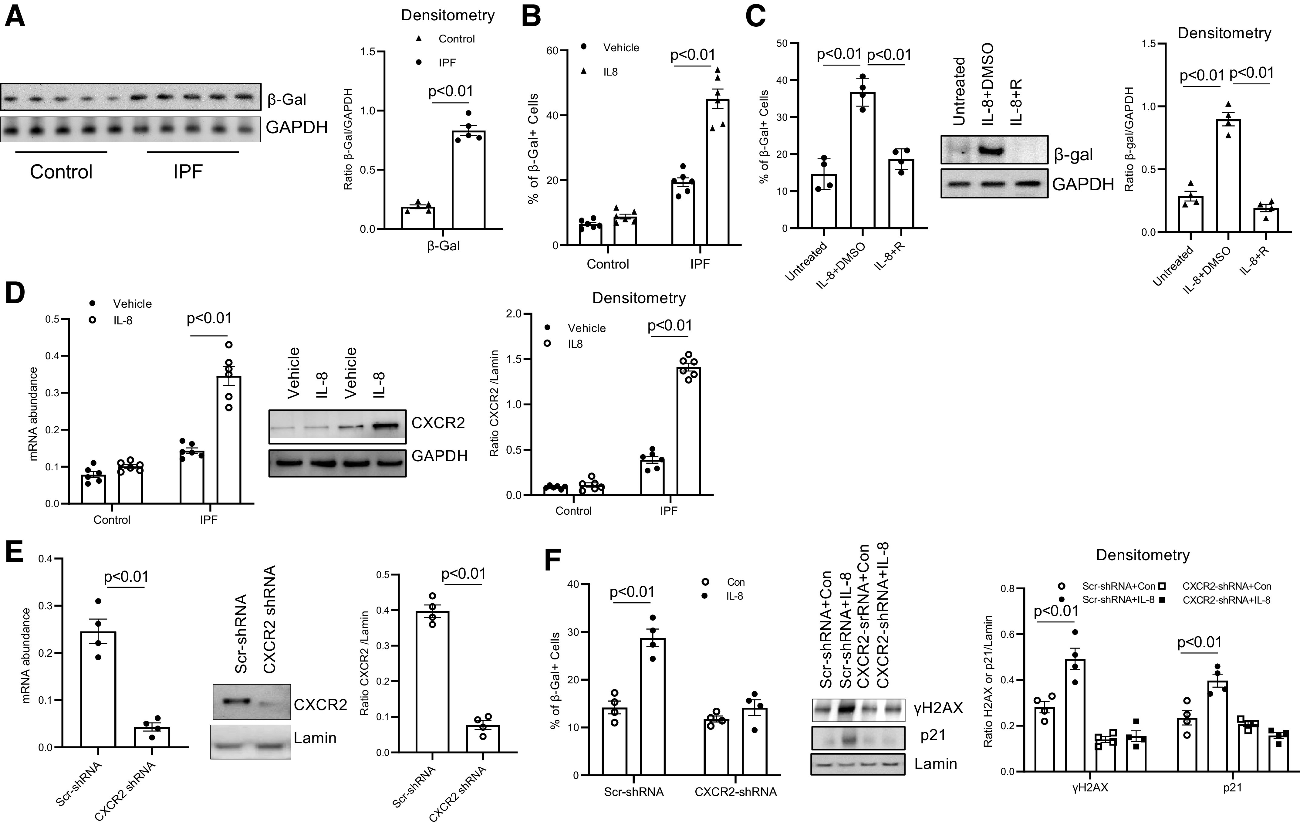
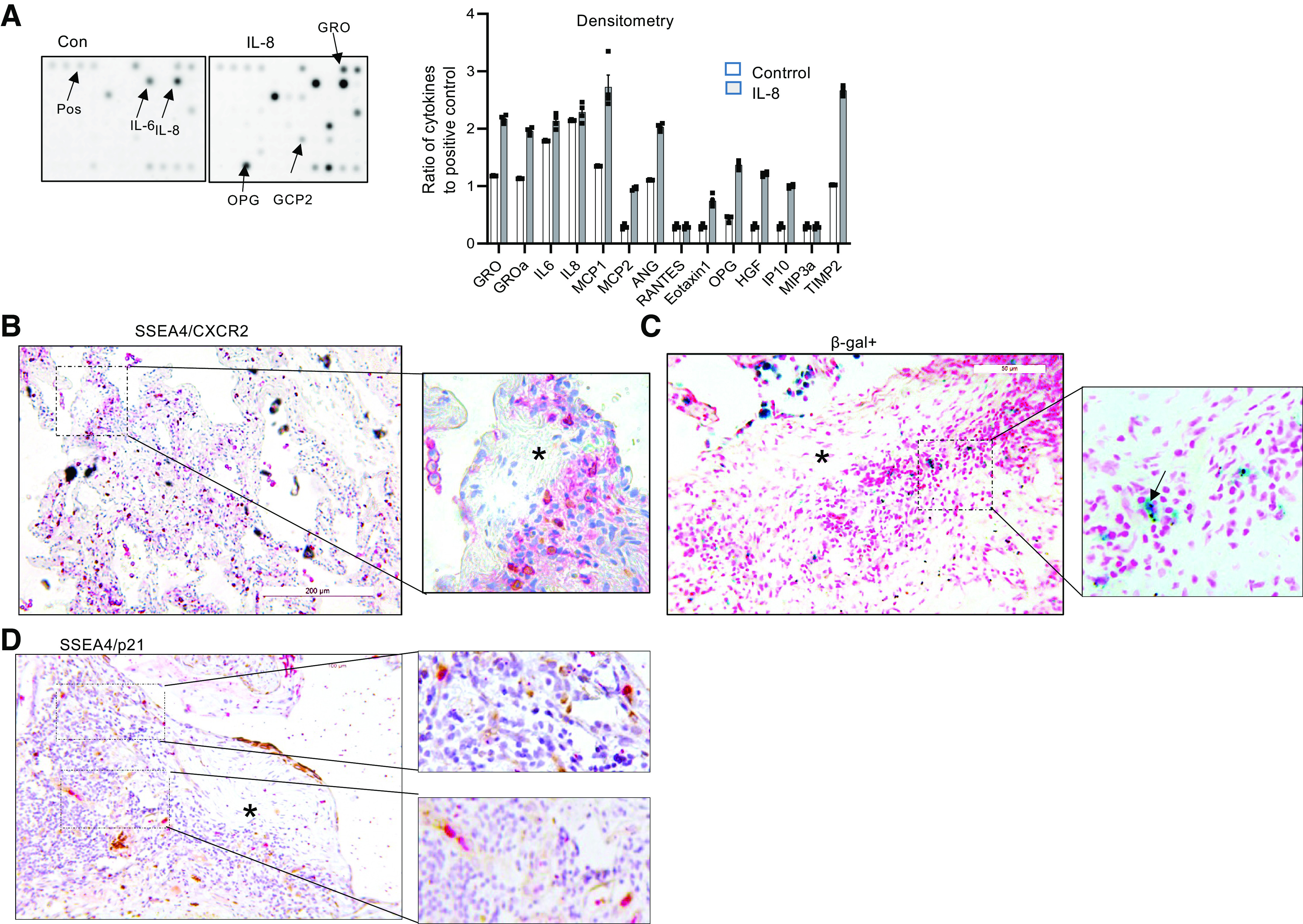
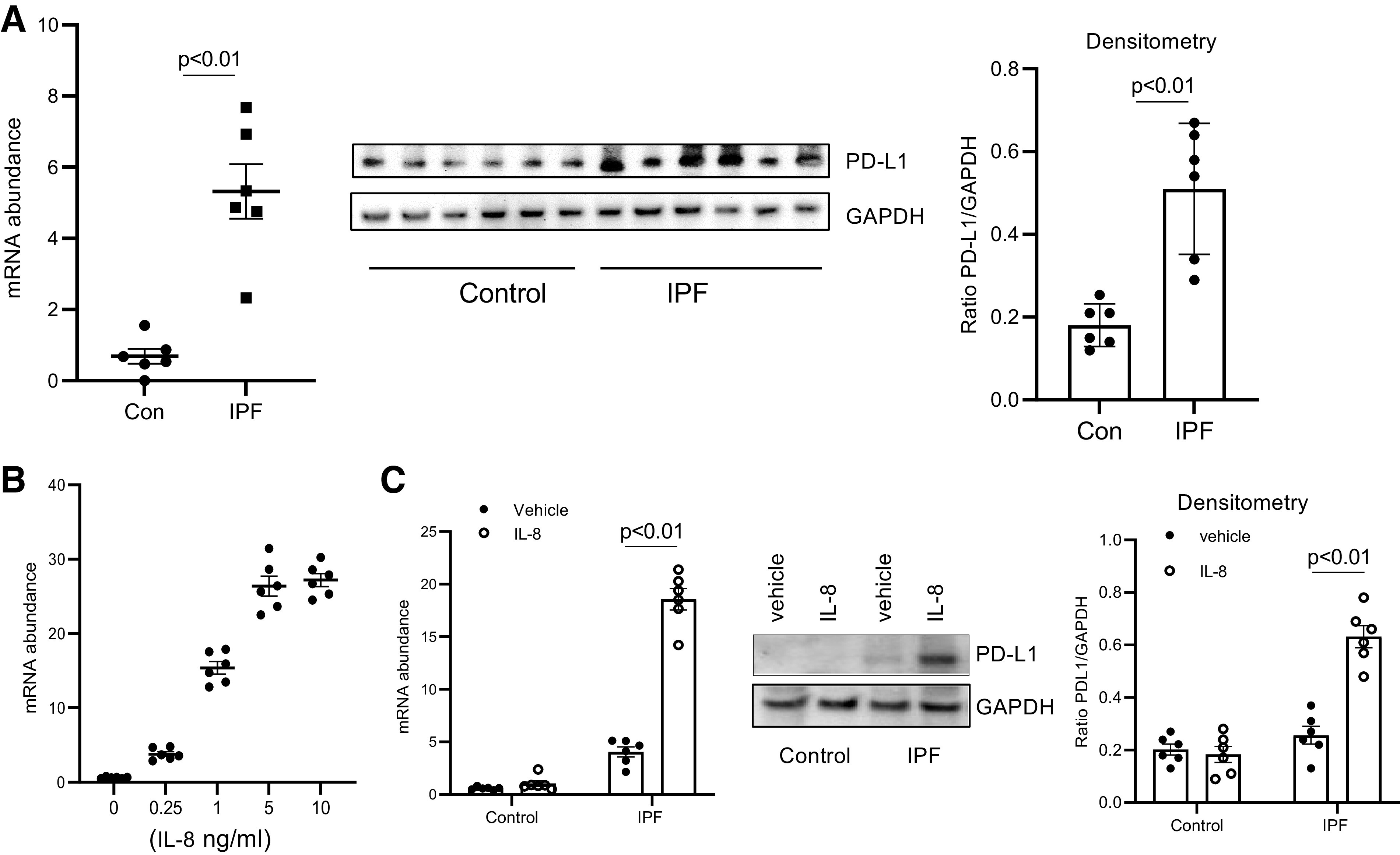
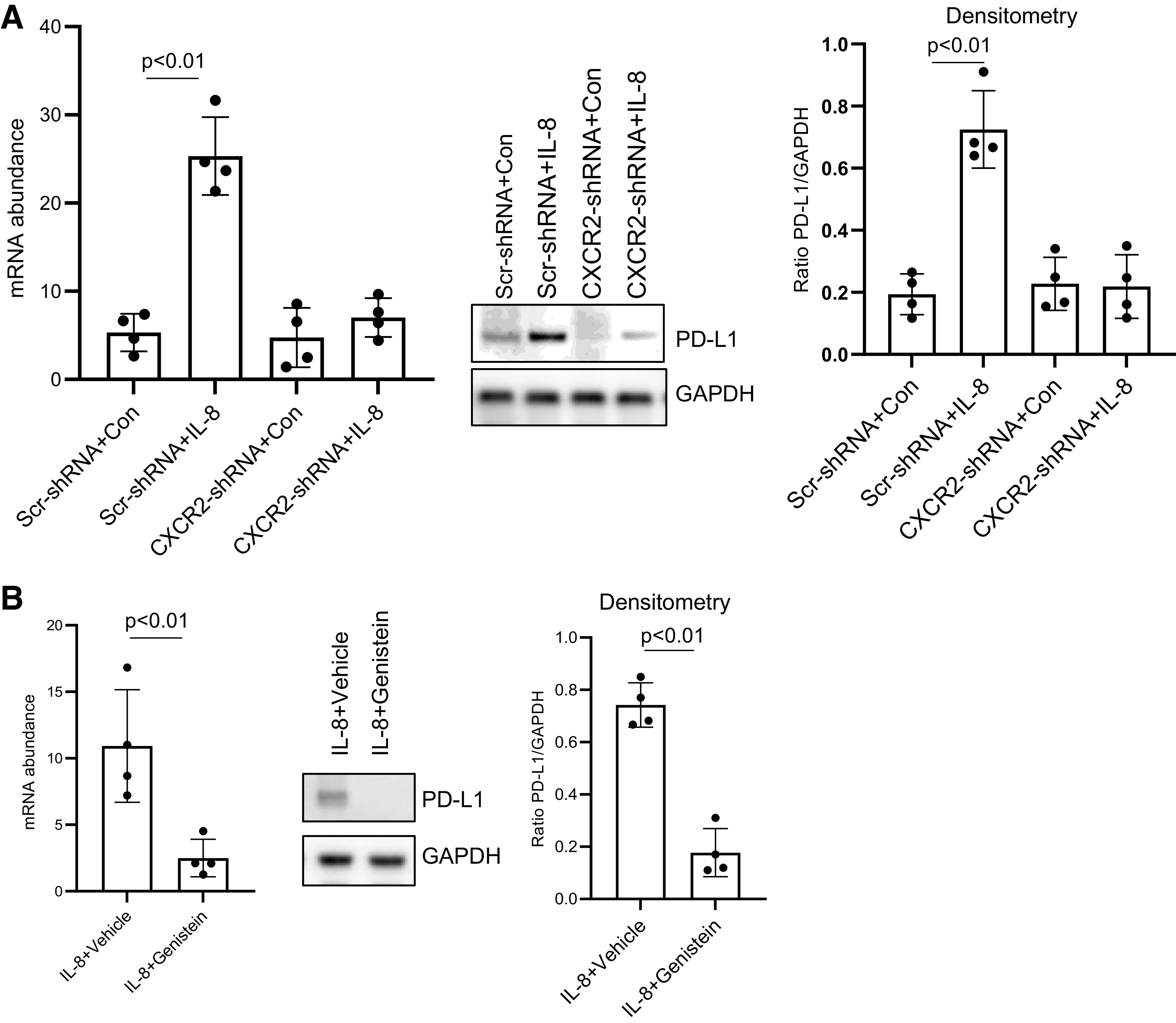
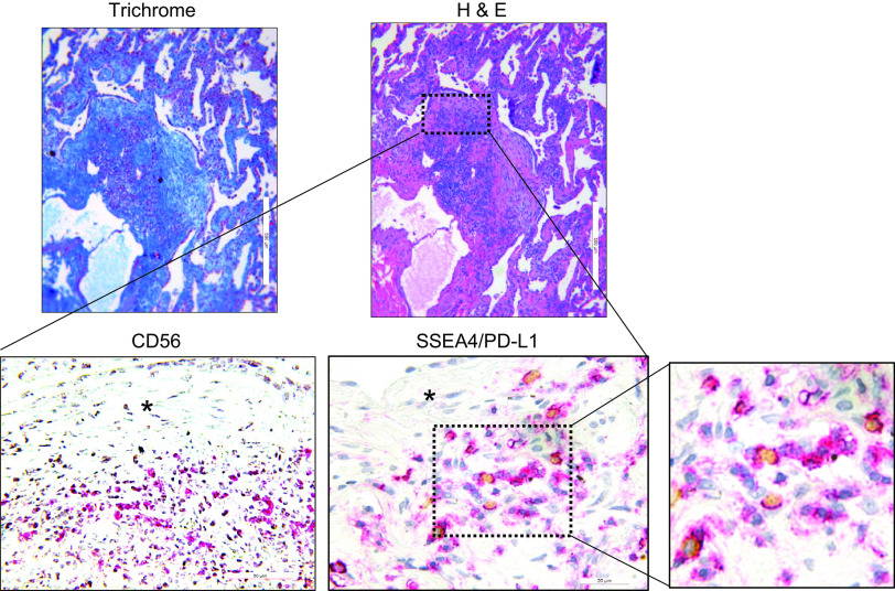
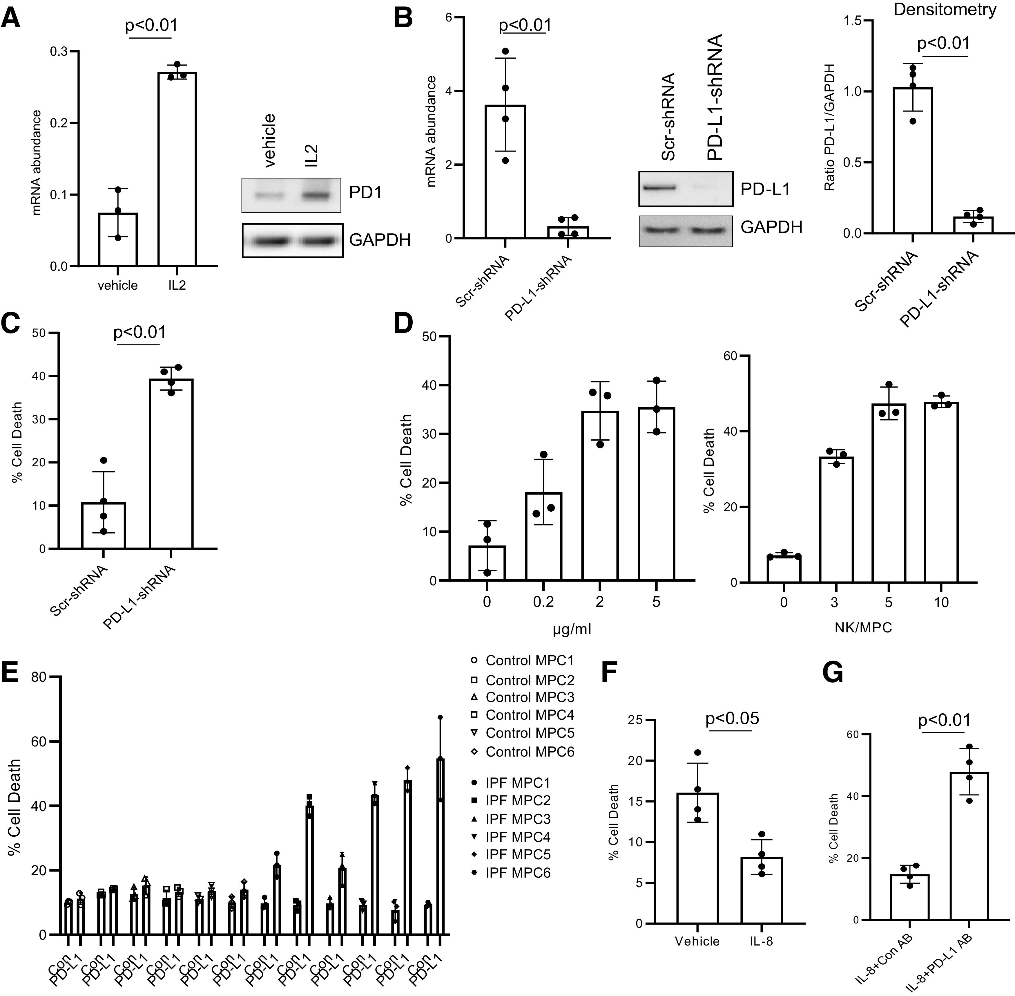
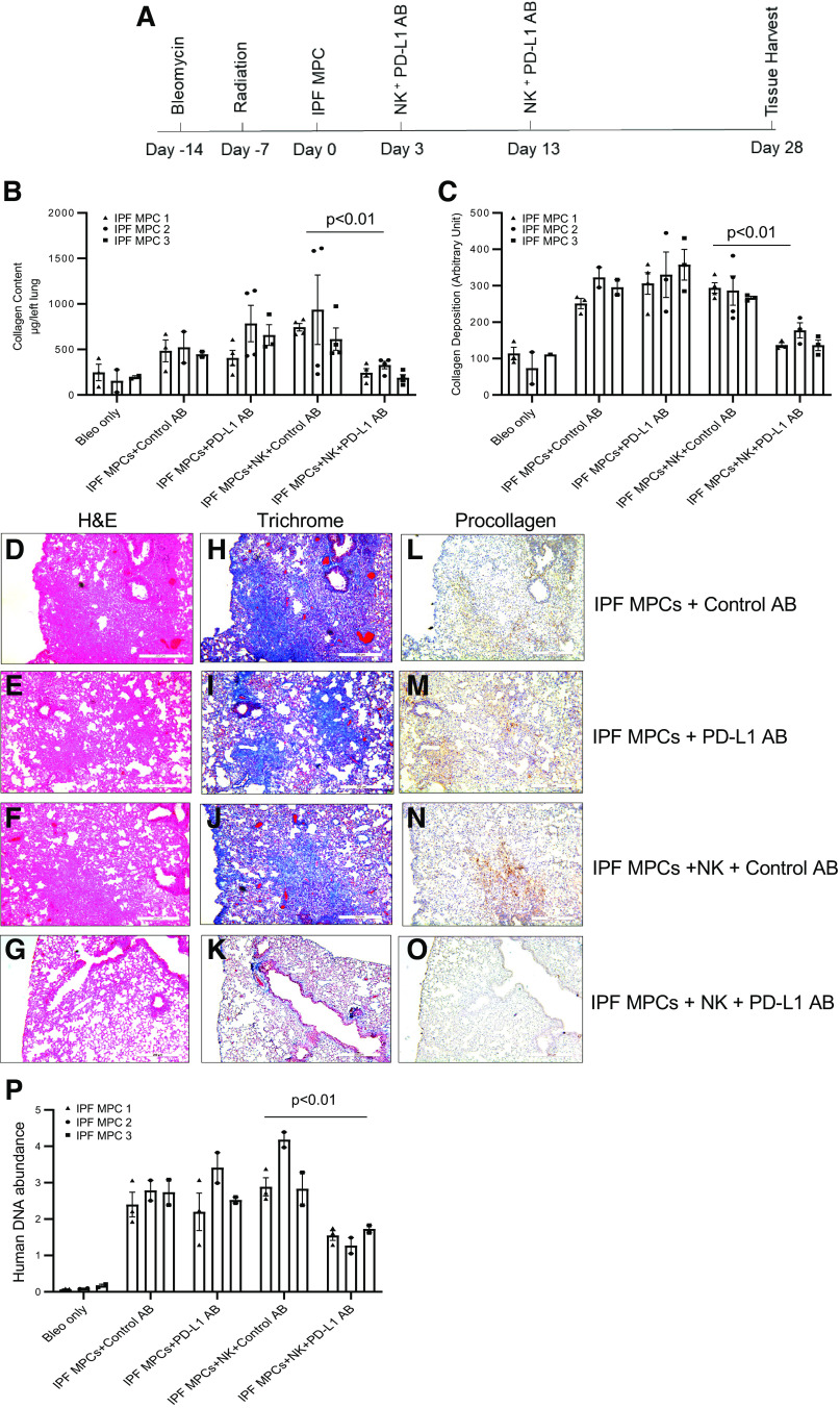
Idiopathic pulmonary fibrosis (IPF) is a progressive fibrotic lung disease. We discovered fibrogenic mesenchymal progenitor cells (MPCs) in the lungs of IPF patients that display cell-autonomous fibrogenicity and drive fibrotic progression. In a study of the IPF MPC nuclear proteome, we identified DNA damage as one of the most altered functions in IPF MPCs. In prior work we found that IL-8 drives IPF MPC self-renewal. IL-8 can promote replicative stress and DNA damage and induce senescence through the CXCR2 receptor. We hypothesized that IL-8 promotes DNA damage-mediated senescence in IPF MPCs. We show that IL-8 induces DNA damage and promotes IPF MPC senescence. We discovered that IL-8 concurrently promotes senescence and upregulation of the programmed death ligand 1 (PD-L1) in a CXCR2-dependent manner. Disruption of programmed cell death protein-1 (PD-1)-PD-L1 interaction promotes natural killer (NK) cell killing of IPF MPCs in vitro and arrests IPF MPC-mediated experimental lung fibrosis in vivo. Immunohistochemical (IHC) analysis of IPF lung tissue identified PD-L1-expressing IPF MPCs codistributing with NK cells and β-galactosidase-positive cells. Our data indicate that IL-8 simultaneously promotes IPF MPC DNA damage-induced senescence and high PD-L1 expression, enabling IPF MPCs to elude immune cell-targeted removal. Disruption of PD-1-PD-L1 interaction may limit IPF MPC-mediated fibrotic progression.NEW & NOTEWORTHY Here we show that IL-8 concurrently promotes senescence and upregulation of PD-L1 in IPF MPCs. IHC analysis identifies the presence of senescent IPF MPCs intermingled with NK cells in the fibroblastic focus, suggesting that senescent MPCs elude immune cell surveillance. We demonstrate that disruption of PD-1/PD-L1 interaction promotes NK cell killing of IPF MPCs and arrests IPF MPC-mediated experimental lung fibrosis. Disruption of PD-1/PD-L1 interaction may be one means to limit fibrotic progression.

Keywords: PD-L1; idiopathic pulmonary fibrosis; mesenchymal progenitor cells; senescence.


Conflict of interest statement

No conflicts of interest, financial or otherwise, are declared by the authors.

Figures

None
Graphical abstract
Figure 1.
Figure 1.
IL-8 induces idiopathic pulmonary fibrosis (IPF) mesenchymal progenitor cell (MPC) DNA damage. A: histone H2AX (γH2AX), p21, and phosphorylated (p) and total replication protein subunit 2 (RPA2) levels were quantified in IPF and control MPCs by Western blot analysis (left) (n = 6 IPF and control MPC cell lines each). GAPDH served as loading control. Densitometry values summarizing Western blot data are shown at right. B–D: IPF and control MPCs were treated with recombinant IL-8 (5 ng) or vehicle control (n = 6 IPF and control MPC cell lines each). B: a representative Western blot quantifying γH2AX levels in 1 IPF and 1 control MPC cell line (left). GAPDH served as loading control. Densitometry values summarizing Western blot data are shown at right. C: p21 levels were quantified by quantitative PCR (Q-PCR; left) and Western blot analysis (shown is a representative Western blot; center). Densitometry values summarizing Western blot data are shown at right. D: phosphorylated and total RPA2 levels were quantified by Western blot analysis (shown is a respresentative Western blot, left). Densitometry values summarizing the Western blot data are shown at right. E: IPF MPCs were treated with 5 ng of recombinant IL-8 in the presence of the IL-8 receptor inhibitor reparixin (IL-8 + R) or vehicle control (IL-8 + DMSO) (n = 6 IPF MPC cell lines). Cells not treated with IL-8 served as an additional control (Untreated). Shown is a representative Western blot quantifying γH2AX levels in 1 IPF MPC cell line (left). GAPDH served as loading control. Densitometry values summarizing the Western blot data are shown at right.
Figure 2.
Figure 2.
IL-8 promotes idiopathic pulmonary fibrosis (IPF) mesenchymal progenitor cell (MPC) senescence via the CXCR2 receptor. A: β-galactosidase (β-Gal) levels were quantified in IPF and control MPCs by Western blot analysis (left) (n = 5 IPF and control MPC cell lines each). GAPDH served as loading control. Densitometry values summarizing Western blot data are on right. B: IPF and control MPCs (n = 6 IPF and control MPC cell lines each) were treated with IL-8 (5 ng) or vehicle control; 72 h later the percentage of senescent cells was analyzed by β-Gal staining. C: IPF MPCs (n = 4 IPF MPC cell lines) were treated with IL-8 in the presence of the IL-8 receptor inhibitor reparixin (IL-8 + R) or vehicle control (IL-8 + DMSO). Cells not treated with IL-8 served as an additional control (Untreated). Senescence was quantified by β-Gal staining (left) and by quantification of β-Gal levels by Western blot analysis (shown is a respresentative Western blot; center). Densitometry values summarizing Western blot data are on right. D: IPF and control MPCs (n = 6 IPF and control MPC cell lines each) were treated with IL-8 or vehicle control. CXCR2 levels were quantified by quantitative PCR (Q-PCR; left) and Western blot analysis (shown is a respresentative Western blot; center). GAPDH = loading control. Densitometry values summarizing Western blot data are on right. E: IPF MPCs were transduced with CXCR2 (CXCR2-shRNA) or scrambled (Scr-shRNA) shRNA. CXCR2 levels were quantified by Q-PCR (left) and Western blot analysis (shown is a respresentative Western blot; center). Densitometry values summarizing Western blot data are on right. F: IPF MPCs transduced with CXCR2-shRNA or Scr-shRNA were treated with IL-8 or vehicle control (Con). Senescence was quantified by β-Gal staining (left), and histone H2AX (γH2AX) and p21 levels were quantified by Western blot analysis (center). Lamin = loading control. Shown is a respresentative Western blot. Densitometry values summarizing Western blot data are on right. The experiments in E and F were replicated with 4 IPF MPC cell lines.
Figure 3.
Figure 3.
IL-8 promotes idiopathic pulmonary fibrosis (IPF) mesenchymal progenitor cell (MPC) secretion of cytokines. A: cytokine array assay was performed on conditioned media derived from IPF MPCs pretreated with IL-8 or vehicle control (Con); 4 IPF MPC cell lines were used. Left: representative cytokine array demonstrating increased cytokine levels in the conditioned medium derived from IPF MPCs treated with IL-8 compared with control. Right: densitometry values summarizing cytokine array data. Each data point represents the ratio of the cytokine to the positive control in the conditioned medium from 1 IPF cell line. B–D: immunohistochemical (IHC) staining of IPF lung tissue for SSEA4 (brownish-yellow stain) and CXCR2 (red stain) (B); β-galactosidase (β-gal; blue stain) (C); and SSEA4 (brownish-yellow stain) and p21 (red stain) (D). Asterisk denotes myofibroblast core of the fibroblastic focus. (n = 7 IPF patient specimens; 4 sections were imaged in each specimen).
Figure 4.
Figure 4.
Programmed death ligand 1 (PD-L1) levels are elevated in idiopathic pulmonary fibrosis (IPF) mesenchymal progenitor cells (MPCs). A: PD-L1 expression was quantified in IPF and control (Con) MPCs (n = 6 IPF and control MPC cell lines each) by quantitative PCR (Q-PCR) (left) and Western blot analysis (center). GAPDH = loading control. Densitometry values summarizing Western blot data are on right. B: IPF MPCs were treated with various amounts of IL-8. PD-L1 levels were quantified by Q-PCR. C: IPF and control MPCs (n = 6 cell lines each) were treated with IL-8 (5 ng) or vehicle control. PD-L1 levels were quantified by Q-PCR (left) and Western blot analysis (shown is a respresentative Western blot quantifying PD-L1 levels in 1 IPF and 1 control MPC cell line; center). GAPDH = loading control. Densitometry values summarizing Western blot data are on right.
Figure 5.
Figure 5.
IL-8 increases idiopathic pulmonary fibrosis (IPF) mesenchymal progenitor cell (MPC) programmed death ligand 1 (PD-L1) expression via CXCR2. A: IPF MPCs transduced with CXCR2 (CXCR2-shRNA) or scrambled (Scr-shRNA) shRNA were treated with 5 ng of IL-8 or vehicle control (Con) and PD-L1 expression quantified by quantitative PCR (Q-PCR) (left) and Western blot analysis (shown is a respresentative Western blot; center). Densitometry values summarizing Western blot data are on right. B: IPF MPCs were treated with 5 ng of IL-8 and the MAPK inhibitor genistein or vehicle control. PD-L1 levels were quantified by Q-PCR (left) and Western blot analysis (shown is a respresentative Western blot; center). Densitometry values summarizing Western blot data are on right. Experiments in A and B were replicated with 4 IPF MPC cell lines.
Figure 6.
Figure 6.
Programmed death ligand 1 (PD-L1)-expressing idiopathic pulmonary fibrosis (IPF) mesenchymal progenitor cells (MPCs) codistribute with CD56+ natural killer (NK) cells within the highly cellular region at the periphery of the fibroblastic focus. Immunohistochemistry (IHC) was performed on serial sections of IPF lung tissue (n= 8 IPF patient specimens; 4 sections were imaged in each specimen). Top: hematoxylin and eosin (H&E; right) and trichrome (left) stain of a fibroblastic focus. Bottom: IHC staining for the NK cell marker CD56 (left) and IHC double staining for SSEA4 (brownish-yellow stain) and PD-L1 (red stain) (center and right). Asterisk denotes focus (myofibroblast) core.
Figure 7.
Figure 7.
Programmed death ligand 1 (PD-L1) antagonism sensitizes idiopathic pulmonary fibrosis (IPF) mesenchymal progenitor cells (MPCs) to natural killer (NK) cell-mediated killing in vitro. A: programmed cell death protein-1 (PD-1) levels were quantified in NK92 cells treated with IL-2 or vehicle control by quantitative PCR (Q-PCR; left) and Western blot analysis (right). GAPDH served as loading control. The experiment was performed in triplicate. B: IPF MPCs were transduced with PD-L1 (PD-L1-shRNA) or scrambled (Scr-shRNA) shRNA. PD-L1 expression was quantified by Q-PCR (left) or Western blot analysis (shown is a respresentative Western blot; center). Densitometry values summarizing Western blot data are at right. C: transduced IPF MPCs were cocultured with NK92 cells in a 3-to-1 ratio (NK/MPC). Cell killing was quantified at 4 h. D, left: IPF MPCs were cocultured with NK92 cells in a 3-to-1 ratio (NK/MPC) in the presence of various concentrations of anti-PD-L1 antibody. Right: IPF MPCs were cocultured with various ratios of NK cells (0:1, 3:1, 5:1, 10:1; NK/MPC) in the presence of PD-L1 antibody (2 µg/mL). Cell killing was quantified at 4 h. The experiment was performed in triplicate. E: IPF or control MPCs (n = 6 cell lines each) were cocultured with NK92 cells in a 3-to-1 ratio (NK/MPC) in the presence of PD-L1 (2 µg/mL) or isotype control (Con) antibody. Cell killing was quantified at 4 h. F: IPF MPCs were pretreated with IL-8 (5 ng) or vehicle control and cocultured with NK cells. Cell killing was quantified at 4 h. G: IPF MPCs pretreated with IL-8 (5 ng) were cocultured with NK cells in the presence of PD-L1 or isotype control antibody (Con AB). Cell killing was quantified at 4 h. The experiment was replicated with 4 IPF MPC cell lines in B, C, F, and G.
Figure 8.
Figure 8.
Disruption of programmed cell death protein-1 (PD-1)-programmed death ligand 1 (PD-L1) interaction arrests idiopathic pulmonary fibrosis (IPF) mesenchymal progenitor cell (MPC)-mediated experimental lung fibrosis in vivo. A: schematic of xenograft mouse model protocol. NOD/SCID/IL2rγ/B2M (NSG) mice were treated with intratracheal bleomycin (Bleo; 1.25 U/kg). One week later the mice were irradiated. Two weeks after bleomycin, IPF MPCs were administered via tail vein injection (106 cells/100 µL). At days 3 and 13 after IPF MPC administration, the mice received NK92 cells (10 × 106 cells) and either PD-L1 (2 mg/kg) or isotype control antibody (AB) by peritoneal injection. Mice receiving IPF MPCs and treated with PD-L1 or control antibody in the absence of NK cells served as controls. Mice treated with bleomycin and radiation only served to establish baseline collagen content in the absence of additional treatments. There were 10 mice/group. Tissue harvest occurred 4 wk after MPC administration. The experiment was replicated with IPF MPCs derived from 3 separate IPF patients. B: collagen content was quantified in left lungs by Sircol assay. C: semiquantitative analysis of lung collagen deposition by trichrome staining with ImageJ. D–O: representative hematoxylin and eosin (H&E) (D–G) and trichrome (H–K) stains assessing fibrosis and collagen deposition, respectively, and immunohistochemistry (IHC) using an antibody recognizing human procollagen to identify human cells and assess collagen synthesis (L–O). P: human IPF cell numbers were quantified by quantitative PCR (Q-PCR).

Similar articles

Cited by

References

    1. Noble PW. Idiopathic pulmonary fibrosis: natural history and prognosis. Clin Chest Med 27: S11–S16, 2006. doi:10.1016/j.ccm.2005.08.003. - DOI - PubMed
    1. Raghu G, Weycker D, Edelsberg J, Bradford WZ, Oster G. Incidence and prevalence of idiopathic pulmonary fibrosis. Am J Respir Crit Care Med 174: 810–816, 2006. doi:10.1164/rccm.200602-163OC. - DOI - PubMed
    1. Raghu G, Selman M. Nintedanib and pirfenidone. New antifibrotic treatments indicated for idiopathic pulmonary fibrosis offer hopes and raises questions. Am J Respir Crit Care Med 191: 252–254, 2015. doi:10.1164/rccm.201411-2044ED. - DOI - PubMed
    1. Xia H, Bodempudi V, Benyumov A, Hergert P, Tank D, Herrera J, Braziunas J, Larsson O, Parker M, Rossi D, Smith K, Peterson M, Limper A, Jessurun J, Connett J, Ingbar D, Phan S, Bitterman PB, Henke CA. Identification of a cell-of-origin for fibroblasts comprising the fibrotic reticulum in idiopathic pulmonary fibrosis. Am J Pathol 184: 1369–1383, 2014. doi:10.1016/j.ajpath.2014.01.012. - DOI - PMC - PubMed
    1. Xia H, Gilbertsen A, Herrera J, Racila E, Smith K, Peterson M, Griffin T, Benyumov A, Yang L, Bitterman PB, Henke CA. Calcium-binding protein S100A4 confers mesenchymal progenitor cell fibrogenicity in idiopathic pulmonary fibrosis. J Clin Invest 127: 2586–2597, 2017. doi:10.1172/JCI90832. - DOI - PMC - PubMed

Publication types

MeSH terms