Review
. 2023 Apr 30;21(1):144.
doi: 10.1186/s12951-023-01898-z.

Review on two-dimensional material-based field-effect transistor biosensors: accomplishments, mechanisms, and perspectives

Affiliations
Review

Review on two-dimensional material-based field-effect transistor biosensors: accomplishments, mechanisms, and perspectives

Shuo Chen et al. J Nanobiotechnology. .

Abstract

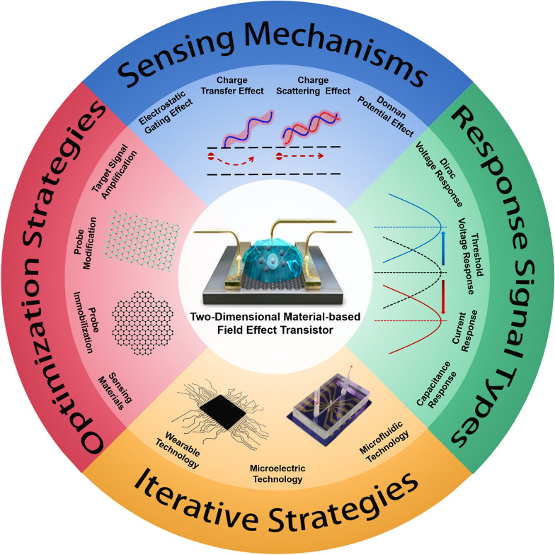
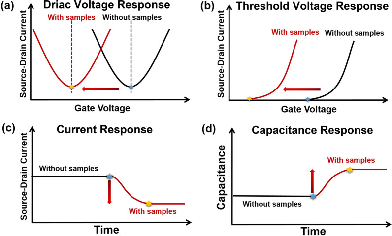
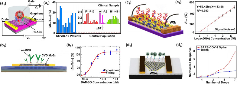
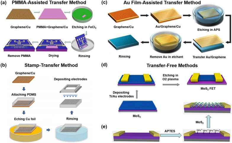
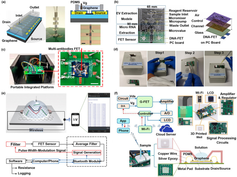
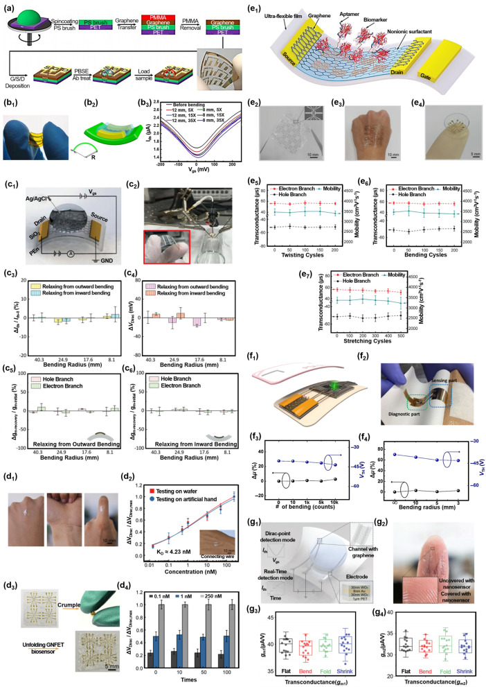
Field-effect transistor (FET) is regarded as the most promising candidate for the next-generation biosensor, benefiting from the advantages of label-free, easy operation, low cost, easy integration, and direct detection of biomarkers in liquid environments. With the burgeoning advances in nanotechnology and biotechnology, researchers are trying to improve the sensitivity of FET biosensors and broaden their application scenarios from multiple strategies. In order to enable researchers to understand and apply FET biosensors deeply, focusing on the multidisciplinary technical details, the iteration and evolution of FET biosensors are reviewed from exploring the sensing mechanism in detecting biomolecules (research direction 1), the response signal type (research direction 2), the sensing performance optimization (research direction 3), and the integration strategy (research direction 4). Aiming at each research direction, forward perspectives and dialectical evaluations are summarized to enlighten rewarding investigations.

Keywords: Biomarker detection; Biosensor; Field-effect transistor; Sensing application; Two-Dimensional Material.


Conflict of interest statement

The authors declare that they have no competing interests.

Figures

Scheme 1
Scheme 1
A brief introduction of two-dimensional material-based FET (2D material-based FET) on the sensing mechanisms, response signal types, optimization strategies, and iterative strategies. Reproduced with permission [30]. Copyright 2020, American Chemical Society. Reproduced with permission [5]. Copyright 2017, Nature Publishing Group
Fig. 1
Fig. 1
Sensing mechanisms in FET biomolecule detection. a and b Electrostatic gating effect and geometrical capacitance model. c Electron transfer between graphene and DNA. d Electrostatic potential simulation of graphene surface after binding double-stranded DNA. e Charge transfer between graphene and DNA induced by MoS2. f Potential modulation mechanism based on charge transfer. g and h Donnan potential-based potential distribution and capacitance model. i Electron scattering effect. j Transmission spectrum simulation of sensing material after binding DNA. k Charge scattering caused by adsorption of DNA (K1) and surface groups (K2) on graphene. (a,e) Reproduced with permission [31]. Copyright 2020, Elsevier Ltd. b Reproduced with permission [5]. Copyright 2017, Nature Publishing Group. c Reproduced with permission [32]. Copyright 2021, Royal Society of Chemistry. d Reproduced with permission [33]. Copyright 2021, Springer Netherlands. f Reproduced with permission [34]. Copyright 2022, American Chemical Society. g, h Reproduced with permission [35]. Copyright 2015, Wiley–VCH Verlag GmbH & Co. KGaA, Weinheim. i Reproduced with permission [36]. Copyright 2020, American Chemical Society. j Reproduced with permission [37]. Copyright 2019, Elsevier Ltd. k Reproduced with permission [38]. Copyright 2021, Wiley–VCH Verlag GmbH & Co. KGaA, Weinheim
Fig. 2
Fig. 2
Response signal types of FET in detecting biomolecules. a Dirac voltage response of graphene FET. b Threshold voltage response of MX2 FET. c Current response. d Capacitance response
Fig. 3
Fig. 3
Optimization strategies from the direction of sensing material types. a1 The graphene-based FET detecting SARS-COV-2 characteristic protein. a2 Dynamic response of this biosensor to SARS-COV-2 characteristic protein almost 100%. b1 The MoS2-based FET detecting polypeptide receptor wsMOR. b2 The detection range of this biosensor to wsMOR from 0.1 nM to 10 uM. c1 The WS2-based FET detecting DNA hybridization. c2 The response range of this biosensor to target DNA from 10−16 M to 10−9 M. d1 The WSe2-based FET detecting SARS-COV-2 characteristic protein. d2 The detection range of this biosensor to SARS-COV-2 characteristic protein from 25 fg/ul to 10 ng/ul. a Reproduced with permission [88]. Copyright 2021, American Chemical Society. b Reproduced with permission [127]. Copyright 2019, Institute of Physics Science. c Reproduced with permission [105]. Copyright 2022, American Chemical Society. d Reproduced with permission [106]. Copyright 2021, American Chemical Society
Fig. 4
Fig. 4
Optimization strategies from the direction of sensing material’s transfer steps. a PMMA-assisted transfer of graphene from the growth substrate to the target substrate. b Stamp-transfer graphene from the growth substrate to the target substrate. c Au film-assisted transfer of graphene from the growth substrate to the target substrate. d Growing MoS2 on the target substrate with transfer-free. e Depositing MoS2 on the APTES-modified target substrate with transfer-free. a Reproduced with permission [143]. Copyright 2015, American Chemical Society. b Reproduced with permission [144]. Copyright 2013, American Institute of Physics. c Reproduced with permission [145]. Copyright 2018, Elsevier Ltd. d Reproduced with permission [15]. Copyright 2019, American Chemical Society. e Reproduced with permission [16]. Copyright 2018, Elsevier Ltd
Fig. 5
Fig. 5
Optimization strategies from the direction of sensing material configurations. a1 The wrinkled graphene-based FET detecting DNA hybridization. a2 The detection range of this biosensor to target DNA from 2 aM to 2 uM. b1 The MoS2/graphene-based FET detecting DNA hybridization. b2 The response signal of this biosensor to probe DNA is four times that of graphene FET. c1 The suspended graphene-based FET detecting HF. c2 The carrier mobility of suspended graphene is two times that of graphene contacted with the substrate. d1 The suspended MoS2-based FET detecting charged ions. d2 The conductance of the suspended MoS2 is 1 − 2 orders of magnitude higher than that of the MoS2 supported with the substrate. e1 The platinum nanoparticles/reduced graphene oxide-based FET detecting DNA hybridization. e2 The response signal of this biosensor to probe DNA is five times that of graphene FET. f1 The gold nanoparticles/graphene-based FET detecting DNA hybridization. f2 The detection range of this biosensor to target DNA from 1 aM to 1 pM. a Reproduced with permission [4]. Copyright 2020, Nature Publishing Group. b Reproduced with permission [31]. Copyright 2020, Elsevier Ltd. c Reproduced with permission [149]. Copyright 2010, American Chemical Society. d Reproduced with permission [150]. Copyright 2015, American Chemical Society. e Reproduced with permission [151]. Copyright 2012, Royal Society of Chemistry. f Reproduced with permission [92]. Copyright 2020, Elsevier Ltd
Fig. 6
Fig. 6
Optimization strategies from the direction of probe immobilization methods. a, b Immobilizing unmodified probes based on physical absorption. c, d Immobilizing unmodified probes based on electrostatic adsorption. e Immobilizing amino-modified probes based on glutaraldehyde cross-linking. f, g Immobilizing thiol-modified probes based on Au–S bond. h Immobilizing amino-modified probes based on EDC + NHS cross-linking. i Immobilizing amino-modified probes based on PBASE cross-linking. j Immobilizing biotin-modified probes based on Biotin-streptavidin cross-linking. k Immobilizing amino-modified probes based on MUA cross-linking. a Reproduced with permission [95]. Copyright 2020, American Chemical Society. b Reproduced with permission [101]. Copyright 2015, Royal Society of Chemistry. c Reproduced with permission [93]. Copyright 2022, American Chemical Society. d Reproduced with permission [94]. Copyright 2021, American Chemical Society. e Reproduced with permission [155]. Copyright 2018, Elsevier Ltd. f Reproduced with permission [15]. Copyright 2019, American Chemical Society. g Reproduced with permission [65]. Copyright 2020, American Chemical Society. h Reproduced with permission [119]. Copyright 2019, Elsevier Ltd. i Reproduced with permission [47]. Copyright 2021, Elsevier Ltd. j Reproduced with permission [156]. Copyright 2014, Nature Publishing Group. k Reproduced with permission [106]. Copyright 2021, American Chemical Society
Fig. 7
Fig. 7
Optimization strategies from the direction of probe types. a Single-stranded nucleic acid probes. b Tetrahedral nucleic acid probes. c Y-shaped nucleic acid probes. d Hairpin-shaped nucleic acid probes. e Nucleic acid-protein composite probes. f Electrically neutral PMO probes. a Reproduced with permission [37]. Copyright 2019, Elsevier Ltd. b Reproduced with permission [50]. Copyright 2021, American Chemical Society. c Reproduced with permission [87]. Copyright 2021, American Chemical Society. d Reproduced with permission [79]. Copyright 2018, Wiley–VCH Verlag GmbH & Co. KGaA, Weinheim. e Reproduced with permission [3]. Copyright 2019, Nature Publishing Group. f Reproduced with permission [99]. Copyright 2021, Elsevier Ltd
Fig. 8
Fig. 8
Optimization strategies from the direction of multiplying target signals. a Multiplying target signals based on the MMP-2-cutting reaction between the probe peptide sequence and MMP-2; b based on the·OH-cutting reaction between the cysteamine and·OH; c based on the VEGF165-catalyzed HCA; d based on the target DNA-catalyzed HCA; e based on the CRISPR-Cas13a system; f based on the CRISPR-Cas13a/Csm6 synergistic system; g based on the CRISPR-Cas12a system. a Reproduced with permission [186]. Copyright 2013, American Chemical Society. b Reproduced with permission [187]. Copyright 2019, Nature Publishing Group. c Reproduced with permission [44]. Copyright 2022, Elsevier Ltd. d Reproduced with permission [68]. Copyright 2018, American Chemical Society. e Reproduced with permission [188]. Copyright 2021, Elsevier Ltd. f Reproduced with permission [189]. Copyright 2021, Nature Publishing Group. g Reproduced with permission [190]. Copyright 2020, Elsevier Ltd
Fig. 9
Fig. 9
Iterative strategies for FET development using microfluidics and microelectronics. a Single-channel microfluidic chip for quantitative DNA hybridization detection. b Multifunctional microfluidic chip for miRNA detection. c Portable integrated platform for COVID-19 antigen detection. d Smart sensing platform for SARS-COV-2 RNA detection. e Intelligent analysis platform for single-base mismatch detection in DNA. f Intelligent sensing platform for cytokine biomarker detection in saliva. a Reproduced with permission [156]. Copyright 2014, Nature Publishing Group. b Reproduced with permission [193]. Copyright 2021, Springer-Verlag GmbH Germany. c Reproduced with permission [88]. Copyright 2021, American Chemical Society. d Reproduced with permission [1]. Copyright 2022, Nature Publishing Group. e Reproduced with permission [79]. Copyright 2018, Wiley–VCH Verlag GmbH & Co. KGaA, Weinheim. f Reproduced with permission [83]. Copyright 2019, Elsevier Ltd
Fig. 10
Fig. 10
Iterative strategies of FET from the direction of wearable technology. a The flexible biosensor detecting the HIV-1 virus and MLV virus. b The wearable biosensor detecting related miRNA of breast cancer. c The flexible wearable sensor attached to the skin surface detecting the TNF-α of sweat. d The flexible wearable sensor attached to the wrist or finger surface detecting the IFN-γ of sweat. e The flexible wearable sensor attached to human tissue or skin surface detecting the TNF-α of sweat. f The epidermal skin-type point-of-care device detected PSA protein. g The wearable biosensor mounted on the eyeball detecting the L-cysteine of tears. a Reproduced with permission [90]. Copyright 2019, Institute of Physics Science. b Reproduced with permission [95]. Copyright 2020, American Chemical Society. c Reproduced with permission [80]. Copyright 2018, Royal Society of Chemistry. d Reproduced with permission [96]. Copyright 2021, Wiley–VCH Verlag GmbH & Co. KGaA, Weinheim. e Reproduced with permission [81]. Copyright 2019, Wiley–VCH Verlag GmbH & Co. KGaA, Weinheim. f Reproduced with permission [196]. Copyright 2017, Tsinghua University Press and Springer-Verlag Berlin Heidelberg. g Reproduced with permission [197]. Copyright 2022, Wiley–VCH GmbH

Similar articles

Cited by

References

    1. Wang L, Wang X, Wu Y, Guo M, Gu C, Dai C, Kong D, Wang Y, Zhang C, Qu D. Rapid and ultrasensitive electromechanical detection of ions, biomolecules and SARS-CoV-2 RNA in unamplified samples. Nat Biomed Eng. 2022;6:276–285. doi: 10.1038/s41551-021-00833-7. - DOI - PubMed
    1. Balderston S, Taulbee JJ, Celaya E, Fung K, Jiao A, Smith K, Hajian R, Gasiunas G, Kutanovas S, Kim D. Discrimination of single-point mutations in unamplified genomic DNA via Cas9 immobilized on a graphene field-effect transistor. Nat Biomed Eng. 2021;5:713–725. doi: 10.1038/s41551-021-00706-z. - DOI - PubMed
    1. Hajian R, Balderston S, Tran T, DeBoer T, Etienne J, Sandhu M, Wauford NA, Chung J-Y, Nokes J, Athaiya M. Detection of unamplified target genes via CRISPR–Cas9 immobilized on a graphene field-effect transistor. Nat Biomed Eng. 2019;3:427–437. doi: 10.1038/s41551-019-0371-x. - DOI - PMC - PubMed
    1. Hwang MT, Heiranian M, Kim Y, You S, Leem J, Taqieddin A, Faramarzi V, Jing Y, Park I, van der Zande AM. Ultrasensitive detection of nucleic acids using deformed graphene channel field effect biosensors. Nat Commun. 2020;11:1–11. doi: 10.1038/s41467-020-15330-9. - DOI - PMC - PubMed
    1. Xu S, Zhan J, Man B, Jiang S, Yue W, Gao S, Guo C, Liu H, Li Z, Wang J. Real-time reliable determination of binding kinetics of DNA hybridization using a multi-channel graphene biosensor. Nat Commun. 2017;8:1–10. - PMC - PubMed

MeSH terms

LinkOut - more resources