. 2023 May 1;44(17):1560-1570.
doi: 10.1093/eurheartj/ehad205.

Big tau aggregation disrupts microtubule tyrosination and causes myocardial diastolic dysfunction: from discovery to therapy

Affiliations

Big tau aggregation disrupts microtubule tyrosination and causes myocardial diastolic dysfunction: from discovery to therapy

Marco Luciani et al. Eur Heart J. .

Erratum in

Abstract

Background: Amyloid plaques and neurofibrillary tangles, the molecular lesions that characterize Alzheimer's disease (AD) and other forms of dementia, are emerging as determinants of proteinopathies 'beyond the brain'. This study aims to establish tau's putative pathophysiological mechanistic roles and potential future therapeutic targeting of tau in heart failure (HF).

Methods and results: A mouse model of tauopathy and human myocardial and brain tissue from patients with HF, AD, and controls was employed in this study. Tau protein expression was examined together with its distribution, and in vitro tau-related pathophysiological mechanisms were identified using a variety of biochemical, imaging, and functional approaches. A novel tau-targeting immunotherapy was tested to explore tau-targeted therapeutic potential in HF. Tau is expressed in normal and diseased human hearts, in contradistinction to the current oft-cited observation that tau is expressed specifically in the brain. Notably, the main cardiac isoform is high-molecular-weight (HMW) tau (also known as big tau), and hyperphosphorylated tau segregates in aggregates in HF and AD hearts. As previously described for amyloid-beta, the tauopathy phenotype in human myocardium is of diastolic dysfunction. Perturbation in the tubulin code, specifically a loss of tyrosinated microtubules, emerged as a potential mechanism of myocardial tauopathy. Monoclonal anti-tau antibody therapy improved myocardial function and clearance of toxic aggregates in mice, supporting tau as a potential target for novel HF immunotherapy.

Conclusion: The study presents new mechanistic evidence and potential treatment for the brain-heart tauopathy axis in myocardial and brain degenerative diseases and ageing.

Keywords: Alzheimer’s; HMW tau; Heart failure; immunotherapy; tau protein.


Conflict of interest statement

Conflict of interest All authors declare no conflict of interest for this contribution.

Figures

Structured Graphical Abstract
Tangle-like aggregates of hyperphosphorylated tau are present in the heart of patients with dilated cardiomyopathy or Alzheimer’s disease. A mouse model of tauopathy recapitulates the pathological findings of the human disease. Disruption of microtubule de/tyrosination causes myocardial diastolic dysfunction observed in the mouse model, and it can be targeted with immunotherapy. Abbreviations: iDCM, idiopathic dilated cardiomyopathy; PAO, pre-amyloid oligomers; TOMA, tau oligomer monoclonal antibody.
Figure 1
Figure 1
Human hearts express HMW tau: SDS–PAGE of tau expression in myocardial tissue. The age of patients is indicated at the bottom of the blots. Soluble (A) and insoluble (B) fractions of iDCM and age/sex/ethnicity-matched controls; (C) quantification of tau expression. Soluble (D) and insoluble fraction (E) of AD and age/sex/ethnicity-matched controls. (F) quantification of tau expression; (G–I) total tau quantification by ELISA using tau5 and phosphor-tau employing p-Ser396/404 antibodies in the heart of patients with iDCM and AD and in the brain of patients with AD and controls. Data are presented as means ± SEM. P-values have been calculated using the non-parametric two-tailed Mann–Whitney test for the SDS–PAGE. Statistical analysis for the ELISA was performed using one-way ANOVA with Tukey post-hoc analysis. When values were normalized by age, the P = value for total tau was 0.870 for Con vs. iDCM and 0.003 for Con vs. AD. Abbreviations: H = heart; B = brain; iDCM = idiopathic dilated cardiomyopathy; AD = Alzheimer’s disease.
Figure 2
Figure 2
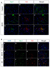
Oligomerized tau accumulates in myocardial tissue in iDCM and AD. (A) Immunofluorescence images of total tau (tau5) and tau oligomers (T22) and merged images in control (A–C) and iDCM (D–I) myocardial tissue; (B) immunofluorescence images of total tau (tau5) and tau oligomers (T22) and merged images in control (A–D), iDCM (E–H), and AD (I–L) myocardial tissue. Abbreviations: iDCM = idiopathic dilated cardiomyopathy; AD = Alzheimer’s disease.
Figure 3
Figure 3
Phospho-tau Ser396 and p-Ser262 in human iDCM heart and AD heart and brain. SDS–PAGE of phospho-tau in myocardial tissue. The age of patients is indicated at the bottom of the blots. Tau is hyperphosphorylated on Ser396 (A) and segregated in the aggregates (B) in human iDCM hearts, while no changes appear in AD hearts (D–F). p-Ser262 in iDCM (G, H) and AD hearts (I, L). AD brain expression of tau p-Ser396 could not be quantified due to signal saturation. Data are presented as means ± SEM. P-values have been calculated using the non-parametric two-tailed Mann–Whitney test for the SDS–PAGE. Abbreviations: H = heart; B = brain; iDCM = idiopathic dilated cardiomyopathy; AD = Alzheimer’s disease.
Figure 4
Figure 4
Oligomerized tau accumulates in tissue and isolated cardiomyocytes in iDCM and AD. (A) Immunofluorescence images of tau oligomers in control, iDCM, and AD myocardial tissue; (B) co-localization and Manders coefficient of co-localization of tau p-Ser396 and tau oligomers in control, iDCM and AD myocardial tissue. The arrow indicates the site of measurement of the co-localization. (C) Tau p-Ser396 and tau oligomers localize inside cardiomyocytes as shown by the staining of isolated cardiomyocytes. Green, phospho-tau; red, tau oligomers; blue DAPI, nuclei. Abbreviations: iDCM = idiopathic dilated cardiomyopathy; AD = Alzheimer’s disease.
Figure 5
Figure 5
Functional effect and mechanisms of tauopathy in the heart in hTau and WT mice. (A) Representative M-Mode echocardiographic images and (B) quantification of the contractile parameter: fractional shortening (FS). Representative echocardiographic images of (C) pulsed-wave Doppler of the mitral valve (MV) and (D) flow tissue Doppler at the level of the mitral valve annulus. Quantification of the diastolic function parameters: (E) MV E/A ratio; (F) MV deceleration time. An insert with the graphical representation of the progression of the MV flow from normal to grade I to II–III diastolic dysfunction is shown between the data panels (E) and (F); (G) E/e′ ratio; (H) left atrium (LA) dimensions; (I) LA calculated pressure. Data are presented with dotted lines since the data were not collected longitudinally from the same mice. Immunostaining of tau oligomers using TOMA antibodies (green): (J) myocardial tissue of all three age groups and (K) quantitative analysis of the signal. Data were analysed in R using t-test with Welch’s correction for unequal variances. Data are presented as untransformed means ± SEM.
Figure 6
Figure 6
(A–E) Sarcomeres and Ca2+ transient parameters indicating diastolic defect in isolated cardiomyocytes from five male/one female WT mice and five male hTau mice. Data were analysed using a linear mixed effects model (LMM) with post-hoc comparison with Holm–Bonferroni correction within outcomes. (F) Increased deposits of tau oligomers stained in green with monoclonal antibody (TOMA) in isolated cardiomyocytes and (G) quantification of the signal. (H) Representative single-plane images of cardiomyocytes stained with a pan-microtubule antibody (cyan) and the nuclear dye Hoechst (grey). The thresholded images showing the microtubule skeleton are displayed below. (I) Quantification of microtubule density (n = 29, 38 non-carrier; n = 3, 4 hTau). (J) Representative single-plane images of cardiomyocytes stained with an antibody specific for detyrosinated (deTyr) microtubules (magenta) and the nuclear dye Hoechst (grey). The thresholded images showing the deTyr microtubule skeleton are displayed below. (K) Quantification of deTyr microtubule density (n = 31, 40 non-carrier n = 3, 4 hTau). (L) Representative single-plane images of the same cardiomyocytes from (C) stained with an antibody specific for tyrosinated (Tyr) microtubules (orange) and the nuclear dye Hoechst (grey). The thresholded images showing the Tyr microtubule skeleton are displayed below. (M) Quantification of Tyr microtubule density (n = 31, 40 non-carrier; n = 3, 4 hTau). Each data point represents the average from a single cell and the box-plot depicts the mean ± SEM. Statistical significance was determined via one-way ANOVA.
Figure 7
Figure 7
Structural and functional myocardial outcomes of TOMA immunotherapy for hTau mice. (A) Injection and readout measurements protocol. (B–D) Representative echocardiographic images of IgG- (six male/six female) or TOMA- (six male/six female) treated 12-month-old hTau mice 1 month after injection. (B) M-mode; (C) pulsed-wave Doppler of the mitral valve flow; (D) tissue Doppler at the level of the mitral valve annulus. (E–G) Diastolic function parameters change by echocardiography: (E) E/e′ ratio; (F) left atrium (LA) dimension; (G) mitral valve deceleration time. Changes from the value at 10 days and 1 month are linked by a straight line since the same mice were followed over time. Data were analysed in R using a linear mixed effects model (LMM) approach. Within- and between-group differences are indicated in each graph. (H) Immunohistochemistry of the IgG- and TOMA-treated hTau mice to visualize total tau (tau5) (blue), tau oligomers (T22) (red); WGA (wheat germ agglutinin; green) defines the cell membrane to localize the intra-/extracellular accumulation of oligomers; (I) quantification of the oligomers; (J) polymerized tubulin (red), and RyRs (green). Data are presented as means ± SEM.

Comment in

References

    1. Iadecola C. Neurovascular regulation in the normal brain and in Alzheimer’s disease. Nat Rev Neurosci 2004;5:347–360. - PubMed
    1. Ruitenberg A, den Heijer T, Bakker SL, van Swieten JC, Koudstaal PJ, Hofman A, et al. . Cerebral hypoperfusion and clinical onset of dementia: the Rotterdam study. Ann Neurol 2005;57:789–794. - PubMed
    1. Troncone L, Luciani M, Coggins M, Wilker EH, Ho CY, Codispoti KE, et al. . Abeta amyloid pathology affects the hearts of patients with Alzheimer’s disease: mind the heart. J Am Coll Cardiol 2016;68:2395–2407. - PMC - PubMed
    1. Stamatelopoulos K, Sibbing D, Rallidis LS, Georgiopoulos G, Stakos D, Braun S, et al. . Amyloid-beta (1–40) and the risk of death from cardiovascular causes in patients with coronary heart disease. J Am Coll Cardiol 2015;65:904–916. - PubMed
    1. Drastichova Z, Skrabalova J, Jedelsky P, Neckar J, Kolar F, Novotny J. Global changes in the rat heart proteome induced by prolonged morphine treatment and withdrawal. PLoS One 2012;7:e47167. 10.1371/journal.pone.0047167 - DOI - PMC - PubMed

Publication types